Clinical Trial
. 2024 Nov 14;16(1):131.
doi: 10.1186/s13073-024-01388-3.

Neoantigen DNA vaccines are safe, feasible, and induce neoantigen-specific immune responses in triple-negative breast cancer patients

Affiliations
Clinical Trial

Neoantigen DNA vaccines are safe, feasible, and induce neoantigen-specific immune responses in triple-negative breast cancer patients

Xiuli Zhang et al. Genome Med. .

Abstract

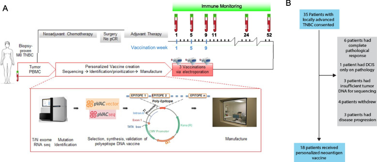
Background: Neoantigen vaccines can induce or enhance highly specific antitumor immune responses with minimal risk of autoimmunity. We have developed a neoantigen DNA vaccine platform capable of efficiently presenting both HLA class I and II epitopes and performed a phase 1 clinical trial in triple-negative breast cancer patients with persistent disease on surgical pathology following neoadjuvant chemotherapy, a patient population at high risk of disease recurrence.

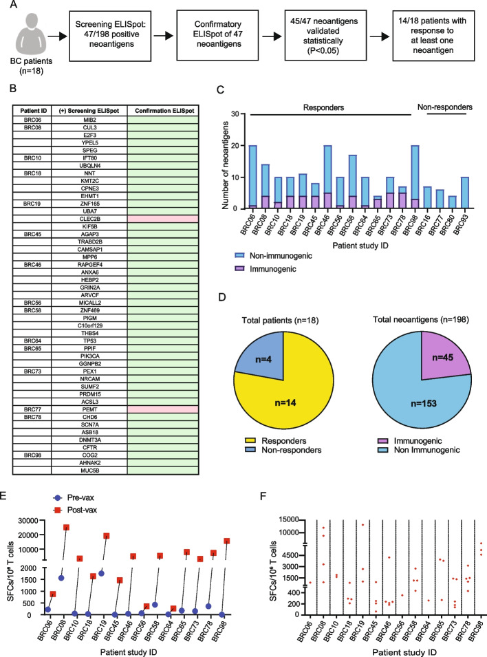
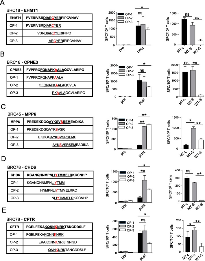
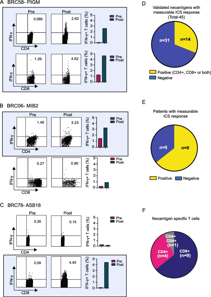
Methods: Expressed somatic mutations were identified by tumor/normal exome sequencing and tumor RNA sequencing. The pVACtools software suite of neoantigen prediction algorithms was used to identify and prioritize cancer neoantigens and facilitate vaccine design for manufacture in an academic GMP facility. Neoantigen DNA vaccines were administered via electroporation in the adjuvant setting (i.e., following surgical removal of the primary tumor and completion of standard of care therapy). Vaccines were monitored for safety and immune responses via ELISpot, intracellular cytokine production via flow cytometry, and TCR sequencing.

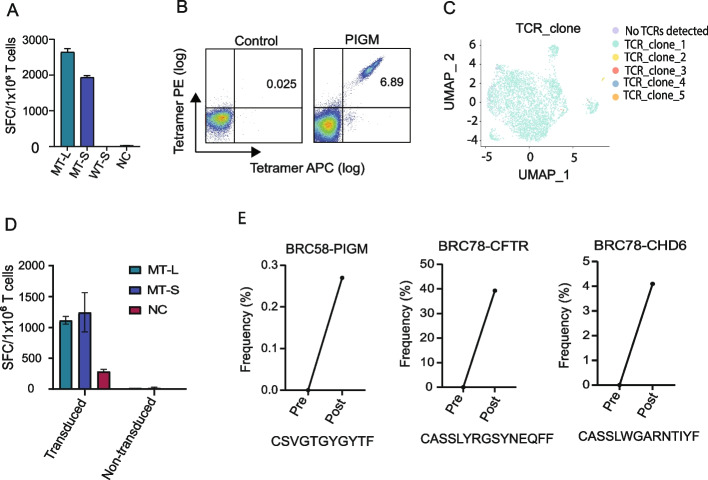
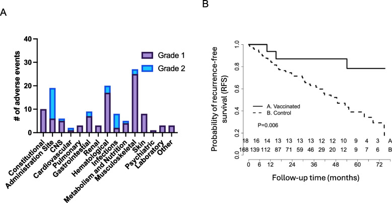
Results: Eighteen subjects received three doses of a neoantigen DNA vaccine encoding on average 11 neoantigens per patient (range 4-20). The vaccinations were well tolerated with relatively few adverse events. Neoantigen-specific T cell responses were induced in 14/18 patients as measured by ELISpot and flow cytometry. At a median follow-up of 36 months, recurrence-free survival was 87.5% (95% CI: 72.7-100%) in the cohort of vaccinated patients.

Conclusion: Our study demonstrates neoantigen DNA vaccines are safe, feasible, and capable of inducing neoantigen-specific immune responses.

Clinical trial registration number: NCT02348320.

Keywords: Clinical trial; DNA Vaccine; Immune response; Neoantigen; Phase I; TNBC.


Conflict of interest statement

Declarations Ethics approval and consent to participate The clinical protocol was reviewed and approved by the Institutional Review Board (ID# 201505074) at Washington University School of Medicine. The research conformed to the principles of the Helsinki/Tokyo/Venice Declaration on experimentation in humans. Consent for publication Written consent for publication was obtained from all participating patients. Competing interests K.S., M.G., and O.G. are consultants for the Jaime Leandro Foundation. All other authors declare that they have no competing interests.

Figures

Fig. 1
Fig. 1
Design, manufacture, and administration of neoantigen DNA vaccines for TNBC patients. A Somatic mutations were identified by whole exome sequencing of tumor and germline DNA. Mutation expression was confirmed by tumor RNA-seq with cDNA capture. Candidate neoantigens were prioritized for inclusion in the vaccines on the basis of HLA binding predictions by pVAC-seq (Methods). Neoantigen DNA vaccines were administered intramuscularly using a TriGrid electroporation device. Peripheral blood was drawn prior at each vaccination timepoint and at selected timepoints after all vaccinations as indicated in A. B 35 patients with locally advanced TNBC were consented. Patients were excluded due to complete pathological response after neoadjuvant chemotherapy (NAC), insufficient tumor, patient withdrawal, and disease recurrence. 18 patients received personalized neoantigen DNA vaccines
Fig. 2
Fig. 2
Neoantigen DNA vaccines induce neoantigen-specific immune responses. A Overview of ELISpot immune monitoring assays including a screening ELISpot assay followed by confirmatory ELISpot assay. The response to 45/47 neoantigens was statistically validated in the “confirmatory ELISpot.” Patients who responded to at least one neoantigen were considered to be responders. B List of all neoantigens conferring immune responses, as assessed by both screening and confirmatory IFN-γ ELISpot assay. Patient-derived PBMCs were stimulated with pooled candidate neoantigens for 12 days. On day 12, cells were harvested and stimulated in IFN-γ ELISpot assays (screening ELISpot) with autologous, irradiated PBMC pulsed with overlapping peptide pools of neoantigens or individual overlapping peptides. ELISpot assays were repeated for those neoantigens that elicited an immune response (confirmatory ELISpot), and confirmatory ELISpot demonstrated that almost all neoantigens were statically validated using Student’s t-test (indicated by the green color), with the exception of two neoantigens (pink color). C Breakdown of the number of immunogenic vs non-immunogenic neoantigens per patient. D The number of patients responding to at least one neoantigen (responders), and the number of immunogenic vs non-immunogenic antigens based on a total of 198 candidate neoantigens for all patients. E The cumulative number of spot-forming cells for all neoantigens per patient comparing pre- and post-vaccination analysis of T cells after 12-days stimulation with neoantigen peptides (P < 0.05 for all patients). F The number of SFC for each immunogenic neoantigen post vaccination and culture
Fig. 3
Fig. 3
Specificity of immune responses to predicted candidate neoantigens before and after vaccination. PBMC at baseline (pre-vax) and after vaccination (2 weeks post vaccination) were stimulated with pooled OP encoding two candidate neoantigens for 12 days. PBMCs were selected based on the relative strength of the immune response (as assessed by ELISpot), the predicted binding of the corresponding minimal epitope, and the availability of PBMC. For each patient, T cell IFN-γ ELISpot assays against pooled (MT-L) and individual OP (OP1–3), as well as the minimal predicted neoantigen (MT-S) and matching wild type peptide (WT-S) were performed on day 12 by co-culturing stimulated T cells overnight with autologous, irradiated PBMC pulsed with peptide. The sequence of individual OP from representative patients is listed with the minimal predicted MHC class I epitope underlined and the missense mutation indicated in red. Panels AE show IFN-γ secretion ELISpot assays for patients BRC18 (A, B), patient BRC45 (C), and patient BRC78 (D, E). Different OPs are indicated in color in the bar graphs (black: OP-1; gray: OP-2; white: OP-3). The negative controls in the ELISpot assays included responder T cells cultured with no peptide (the number of spot-forming cells per 106 cells was 10–120). The background without peptide was subtracted from the experimental condition in each case. Data are presented as means ± SEM (n = 2–3 wells per peptide in ELISpot assay) and are representative of three independent experiments. Samples were compared using unpaired, Student’s t-test (*, P < 0.05; **, P < 0.01; ns, no significant difference); SFC, spot-forming cells. ELISpot experiments were performed in duplicate or triplicate wells per condition
Fig. 4
Fig. 4
Neoantigen vaccines elicit both CD4 and CD8 T cell responses. (AC Examples of increased intracellular IFN-γ production after vaccination and in vitro culture with neoantigen peptide in three patients. Bar graphs reflect the percent IFN-γ + cells per T cell subset and blood draw. D Breakdown of positive vs negative neoantigens by ICS. E Breakdown of ICS data by patients and F T cell subset with regard to immunogenicity. Increases between pre- and post-vaccination are considered positive when a twofold or greater increase is observed in percent positive cells, with a minimum percent positive of ≥ 1% in the post-vaccination sample. All positive neoantigen responses are indicated by blue shading
Fig. 5
Fig. 5
T cell receptor sequencing analysis confirms neoantigen vaccination expands neoantigen-specific T cells. A PBMCs from patient BRC58 were stimulated with pooled PIGM OPs for 12 days. T cell IFN-γ ELISpot assay against pooled OPs (MT-L), as well as the minimal predicted neoantigen (MT-S) and matching wild-type peptide (WT-S) were performed on day 12 by co-culturing stimulated T cells overnight with autologous, irradiated PBMC pulsed with peptide. Cells were rescued from ELISpot plates and were continuously cultured for another 12 days for subsequent tetramer-based TCR analysis. B Tetramer staining of cells. Tetramer + cells were sorted and subjected to scTCR-seq. C UMAP plot of tetramer-sorted cells, showing an almost monoclonal population. TCR genes of the dominant TCR clone were transduced into naïve PBMC of BRC58, activated with PHA for 48 h. Transduced cells were expanded for 96 h and tested for recognition of the PIGM neoantigen by ELISpot. D IFN-γ ELISpot data showing TCR-transduced but not untransduced cells recognize the PIGM neoantigen. NC refers to unpulsed PBMC. E Bulk TCR-seq was performed on cells collected before and 2 weeks post vaccination that were cultured with neoantigen as listed above for three patients. The CDR3 of the dominant TCRVβ is shown for each patient
Fig. 6
Fig. 6
Neoantigen vaccine treatment is safe and prolongs recurrence-free survival compared to historical controls. A Adverse events (grade 1 and 2 toxicity) are depicted by type and number among all vaccinated patients. B Kaplan–Meier survival curve of patients vaccinated with a personalized neoantigen DNA vaccine compared to non-vaccinated historical control patients

References

    1. Matsushita H, Vesely MD, Koboldt DC, Rickert CG, Uppaluri R, Magrini VJ, et al. Cancer exome analysis reveals a T-cell-dependent mechanism of cancer immunoediting. Nature. 2012;482(7385):400–4. http://www.ncbi.nlm.nih.gov/pubmed/22318521:10.1038/nature10755. - DOI - PMC - PubMed
    1. Gubin MM, Zhang X, Schuster H, Caron E, Ward JP, Noguchi T, et al. Checkpoint blockade cancer immunotherapy targets tumour-specific mutant antigens. Nature. 2014;515(7528):577–81. - PMC - PubMed
    1. Alspach E, Lussier DM, Miceli AP, Kizhvatov I, DuPage M, Luoma AM, et al. MHC-II neoantigens shape tumour immunity and response to immunotherapy. Nature. 2019;574(7780):696–701. https://www.ncbi.nlm.nih.gov/pubmed/31645760:10.1038/s41586-019-1671-8. - DOI - PMC - PubMed
    1. Zhang X, Kim S, Hundal J, Herndon JM, Li S, Petti AA, et al. Breast Cancer Neoantigens Can Induce CD8(+) T-Cell Responses and Antitumor Immunity. Cancer Immunol Res. 2017;5(7):516–23. https://www.ncbi.nlm.nih.gov/pubmed/28619968:10.1158/2326-6066.CIR-16-0264. - DOI - PMC - PubMed
    1. Li L, Zhang X, Wang X, Kim SW, Herndon JM, Becker-Hapak MK, et al. Optimized polyepitope neoantigen DNA vaccines elicit neoantigen-specific immune responses in preclinical models and in clinical translation. Genome Med. 2021;13(1):56. https://www.ncbi.nlm.nih.gov/pubmed/33879241:10.1186/s13073-021-00872-4. - DOI - PMC - PubMed

Publication types

Associated data

LinkOut - more resources