

Mini-LET: A Technique Note
- PMID: 39539321
- PMCID: PMC11554595
- DOI: 10.1007/s43465-024-01269-2
Mini-LET: A Technique Note
Abstract
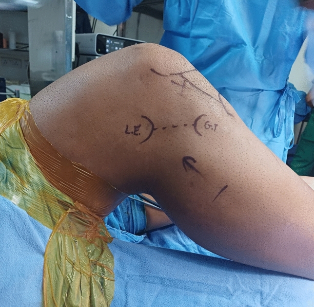
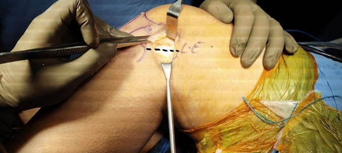
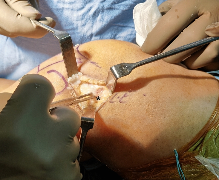
Lateral extra-articular tenodesis (LET) has shown an upswing in popularity since the role of antero-lateral rotatory instability (ALRI) in ACL tears has become better understood. Its primary aim is to restore antero-lateral rotatory stability and hence reduce stresses placed on the ACL graft in high-risk individuals which should reduce graft failure rates. Many techniques have been described for LET using a variety of fixation methods (screws, anchors, etc.). Most of these techniques rely on a large incision laterally. We describe a novel technique ('mini-LET') which uses a 3-4 cm incision, to improve cosmesis.
Keywords: Lateral extra-articular tenodesis (LET); Surgical technique; Technique.
© Indian Orthopaedics Association 2024. Springer Nature or its licensor (e.g. a society or other partner) holds exclusive rights to this article under a publishing agreement with the author(s) or other rightsholder(s); author self-archiving of the accepted manuscript version of this article is solely governed by the terms of such publishing agreement and applicable law.
Conflict of interest statement
Conflict of interestThe author has no conflict of interest to disclose.
Figures


References
-
- Hewison, C. E., Tran, M. N., Kaniki, N., Remtulla, A., Bryant, D., & Getgood, A. M. (2015). Lateral extra-articular tenodesis reduces rotational laxity when combined with anterior cruciate ligament reconstruction: a systematic review of the literature. Arthrosc J Arthrosc Relat Surg,31(10), 2022–2034. - PubMed
-
- Devitt, B. M., Bell, S. W., Ardern, C. L., Hartwig, T., Porter, T. J., Feller, J. A., et al. (2017). The role of lateral extra-articular tenodesis in primary anterior cruciate ligament reconstruction: a systematic review with meta-analysis and best-evidence synthesis. Orthopaedic Journal of Sports Medicine,5(10), 2325967117731767. - PMC - PubMed
LinkOut - more resources
Full Text Sources