. 2024 Oct 30:15:1393017.
doi: 10.3389/fimmu.2024.1393017. eCollection 2024.

A cost-effective protocol for single-cell RNA sequencing of human skin

Affiliations

A cost-effective protocol for single-cell RNA sequencing of human skin

Saba Khoshbakht et al. Front Immunol. .

Abstract

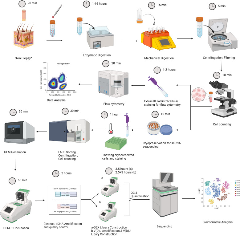
Introduction: Single-cell RNA sequencing (scRNAseq) and flow cytometry studies in skin are methodologically complex and costly, limiting their accessibility to researchers worldwide. Ideally, RNA and protein-based analyses should be performed on the same lesion to obtain more comprehensive data. However, current protocols generally focus on either scRNAseq or flow cytometry of healthy skin.

Methods: We present a novel label-free sample multiplexing strategy, building on the souporcell algorithm, which enables scRNAseq analysis of paired blood and skin samples. Additionally, we provide detailed instructions for simultaneous flow cytometry analysis from the same sample, with necessary adaptations for both healthy and inflamed skin specimens.

Results: This tissue multiplexing strategy mitigates technical batch effects and reduces costs by 2-4 times compared to existing protocols. We also demonstrate the effects of varying enzymatic incubation durations (1, 3, and 16 hours, with and without enzyme P) on flow cytometry outcomes. Comprehensive explanations of bioinformatic demultiplexing steps and a detailed step-by-step protocol of the entire experimental procedure are included.

Discussion: The protocol outlined in this article will make scRNAseq and flow cytometry analysis of skin samples more accessible to researchers, especially those new to these techniques.

Keywords: flow cytometry; inflammation; multiplexing; single cell RNA sequencing (scRNA); skin; skin dissociation; souporcell.


Conflict of interest statement

The authors declare that the research was conducted in the absence of any commercial or financial relationships that could be construed as a potential conflict of interest.

Figures

Figure 1
Figure 1
Graphic representation of our experimental workflow that involves skin biopsy, tissue dissociation using the Whole Skin Dissociation Kit (Miltenyi Biotec, Germany) in 2 steps of enzymatic and mechanical dissociation, staining part of the dissociated cells for flow cytometry analysis, and cryopreservation part of cells for subsequent scRNAseq analysis. Second part includes thawing of cell suspensions, sample preparation by Fluorescence-activated cell sorting (FACS), cell counting by trypan blue exclusion method, Gel Beads-in-emulsion (GEM) generation, library preparation, sequencing and data analysis. The figure was created with BioRender.com. *For lesional skin, one 4mm punch biopsy was obtained. For healthy skin samples, two 6 mm specimens were obtained.
Figure 2
Figure 2
Representative flow cytometry graphs demonstrating the effect of enzymatic treatment for 1 hour, 3 hours, 16 hours; and 3 hours of incubation using enzyme P on all cells in healthy skin (A), live CD19-CD14- cells (B), CD45+ and CD45- cells (C), CD3+ T cells and CD56+ NK cells (D), CD69+ resident memory T cells (TRMs) (E) CD4+ T helper cells (THLs) and CD8+ cytotoxic T cells (CTLs) (F), Granzyme K+ (GrK) CTLs (G), and Granzyme B+ (GrB) CTLs (H). It is demonstrated that, due to the cleavage of CD69 and CD8 antigens by enzyme P, the population of TRMs (E) and CTLs (F) decreased dramatically.
Figure 3
Figure 3
The impact of various enzymatic incubation periods (1 hour, 3 hours, 16 hours) on cell percentages (A) and mean fluorescence intensity (MFI) measurements (B) is shown. A) Live cell counts (determined by trypan blue exclusion method) increased with longer incubation times, indicating enhanced cell yield. The percentage of viable CD14-CD19- leukocytes, assessed by flow cytometry, showed no significant difference across the three incubation periods. However, the percentage of CD45- cells decreased, leading to a significant increase in CD45+ percentage after 16 hours compared to 3 hours of incubation. There was no notable difference in the percentage of lymphocyte subtypes across different incubation times, except for a decrease in CD8+Granzyme B+ cytotoxic T lymphocytes (CTLs) after 16 hours compared to 1 hour incubation. B) MFI values of CD3, CD8, CD56, CD69, and Granzyme K (GrK) showed no significant difference following enzymatic incubation for different durations. However, CD4 and Granzyme B (GrB) MFI decreased significantly after 16 hours of incubation.
Figure 4
Figure 4
The impact of enzyme P on all cell percentages (A) and mean fluorescence intensity (MFI) measurements (B) is shown. A) Live cell counts demonstrated increased cell yield with the use of enzyme P. However, the percentage of NK cells, CD69+ cells, and CD8+ cytotoxic T lymphocytes (CTLs) significantly declined when enzyme P was used. B) The MFI of CD8, CD56, and CD69 significantly decreased when enzyme P was used in the enzymatic incubation. Statistical analyses were performed using Repeated Measures ANOVA (to compare different time points) and paired ratio T-test (for comparison of the effect of enzyme P).
Figure 5
Figure 5
Schematic representation of our label-free triplet pooling strategy. First, we segregate paired PBMC (S3a) and dissociated skin samples (S3b) from a selected individual into two distinct tubes. Additionally, two genetically unrelated samples, each from subjects of different sexes (Female is demonstrated as a circle and Male as a square), are introduced to each tube, resulting in three samples per tube. Following scRNA-seq and sample demultiplexing using souporcell, we first identify the clusters exhibiting identical genetic single nucleotide polymorphism (SNP) patterns in dual reactions and then annotate their sample identity. Subsequently, Y chromosome genes are utilized to ascertain the sex information of the remaining two samples in each tube. By using this information the sample identities of the remaining clusters can be determined readily. S, sample. Different colors denote samples from separate subjects.
Figure 6
Figure 6
Bioinformatic workflow for analyzing single-cell RNA sequencing results that include alignment, demultiplexing, preprocessing, and downstream analysis of sequencing data.
Figure 7
Figure 7
Representative results of the single cell RNAseq analysis of skin samples (n=7) obtained from patients with Behçet’s disease (n=4) and healthy controls (n=3). (A) Demultiplexed skin samples are highlighted on UMAP embedding. (B) Identified high-hierarchy cell types are visualized. (C) Cell type proportion for skin samples from each donor is illustrated with a stacked bar plot. (D) Cell type proportions for PBMC samples from 4 Behçet’s disease patients are illustrated with a stacked bar plot. (E) The cell counts and proportions of cell types in skin samples are shown. The ratio of fibroblasts is less than 1%, indicating the high purity of the sorted CD45+ cells. (F) The cell counts and percentages of cell types in PBMC samples are illustrated. (G) Marker genes to characteristic for each cell type are shown as a dot plot.

References

    1. He H, Suryawanshi H, Morozov P, Gay-Mimbrera J, Del Duca E, Kim HJ, et al. . Single-cell transcriptome analysis of human skin identifies novel fibroblast subpopulation and enrichment of immune subsets in atopic dermatitis. J Allergy Clin Immunol. (2020) 145:1615–28. doi: 10.1016/j.jaci.2020.01.042 - DOI - PubMed
    1. Rojahn TB, Vorstandlechner V, Krausgruber T, Bauer WM, Alkon N, Bangert C, et al. . Single-cell transcriptomics combined with interstitial fluid proteomics defines cell type–specific immune regulation in atopic dermatitis. J Allergy Clin Immunol. (2020) 146:1056–69. doi: 10.1016/j.jaci.2020.03.041 - DOI - PubMed
    1. Liu J, Chang HW, Huang ZM, Nakamura M, Sekhon S, Ahn R, et al. . Single-cell RNA sequencing of psoriatic skin identifies pathogenic TC17 cell subsets and reveals distinctions between CD8+ T cells in autoimmunity and cancer. J Allergy Clin Immunol. (2021) 147:2370–80. doi: 10.1016/j.jaci.2020.11.028 - DOI - PMC - PubMed
    1. Kim J, Lee J, Kim HJ, Kameyama N, Nazarian R, Der E, et al. . Single-cell transcriptomics applied to emigrating cells from psoriasis elucidate pathogenic versus regulatory immune cell subsets. J Allergy Clin Immunol. (2021) 148:1281–92. doi: 10.1016/j.jaci.2021.04.021 - DOI - PMC - PubMed
    1. Burja B, Paul D, Tastanova A, Edalat SG, Gerber R, Houtman M, et al. . An optimized tissue dissociation protocol for single-cell RNA sequencing analysis of fresh and cultured human skin biopsies. Front Cell Dev Biol. (2022) 10:872688. doi: 10.3389/fcell.2022.872688 - DOI - PMC - PubMed

MeSH terms

LinkOut - more resources