

Reduced p63 expression is linked to unfavourable prognosis in muscle-invasive urothelial carcinoma of the bladder
- PMID: 39539567
- PMCID: PMC11557268
- DOI: 10.1002/bco2.431
Reduced p63 expression is linked to unfavourable prognosis in muscle-invasive urothelial carcinoma of the bladder
Erratum in
-
Erratum.BJUI Compass. 2024 Dec 30;5(12):1324-1329. doi: 10.1002/bco2.482. eCollection 2024 Dec. BJUI Compass. 2024. PMID: 39744071 Free PMC article.
Abstract
Objective: There is a shortage of established prognostic biomarkers in bladder cancer. One candidate is tumour protein 63 (p63), a transcription factor of the p53 gene family that is expressed in the normal urothelium. Recently proposed RNA expression-based molecular classifiers of bladder cancer identified high p63 expression as a component of a basal/squamous subtype linked to poor patient prognosis.
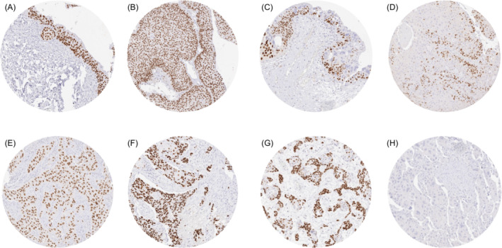
Methods: In this study, p63 protein expression was analysed by immunohistochemistry on more than 2500 urothelial bladder carcinomas in a tissue microarray format to determine its relationship with clinicopathological parameters of disease progression and patient outcome.
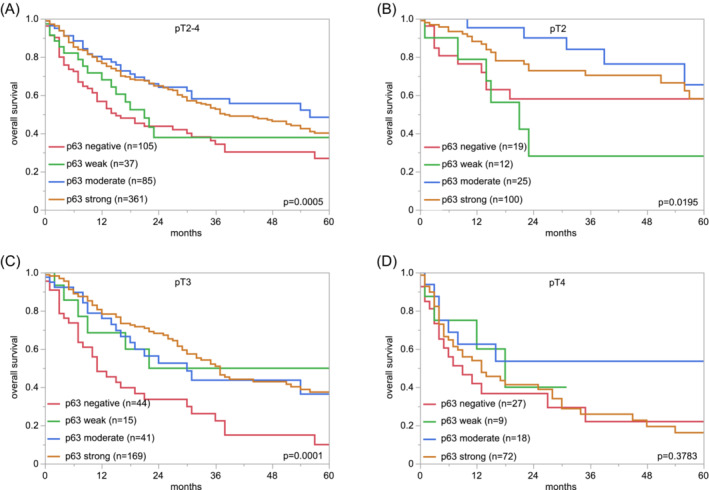
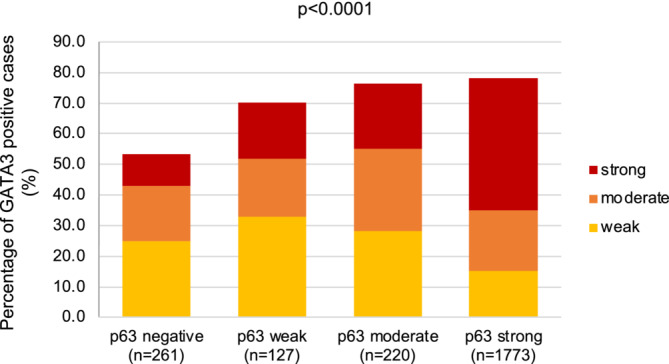
Results: Nuclear p63 staining was seen in all cells of normal urothelium and at elevated levels in pTaG2 tumours. The rate of p63 positive cases and the staining intensity was lower in pTaG3 tumours (93.2%, p < 0.0001 for pTaG3 vs. pTaG2) and markedly lower in pT2-4 carcinomas (83.5%, p = 0.0120 for pT2-4 vs. pTaG3). Within 1018 pT2-4 carcinomas treated by cystectomy, low p63 expression was linked to nodal metastasis (p = 0.0028) and overall survival (p = 0.0005). The association with survival was independent of pT and pN (p = 0.0081). p63 expression was associated with GATA3 expression (p < 0.0001), a luminal cell type marker associated with favourable disease. A joint analysis of p63 and GATA3 did not suggest that GATA3 could provide additional prognostic information.
Conclusion: The independent prognostic role of reduced p63 expression in advanced urothelial carcinomas suggests that p63 could be a useful biomarker to distinguish pT2-4 urothelial carcinomas.
Keywords: biomarker; immunohistochemistry; p63; tissue microarray; urothelial carcinoma.
© 2024 The Author(s). BJUI Compass published by John Wiley & Sons Ltd on behalf of BJU International Company.
Conflict of interest statement
The recombinant rabbit monoclonal p63 antibody, clone MSVA‐063R, was obtained from MS Validated Antibodies GmbH, Hamburg, Germany (owned by a family member of GS).
Figures

References
-
- di Como CJ, Urist MJ, Babayan I, Drobnjak M, Hedvat CV, Teruya‐Feldstein J, et al. p63 expression profiles in human normal and tumor tissues. Clin Cancer Res. 2002;8(2):494–501. - PubMed
LinkOut - more resources
Full Text Sources
Research Materials
Miscellaneous