Review
. 2024 Oct 30:12:1498032.
doi: 10.3389/fcell.2024.1498032. eCollection 2024.

Unveiling the dynamics of circulating tumor cells in colorectal cancer: from biology to clinical applications

Affiliations
Review

Unveiling the dynamics of circulating tumor cells in colorectal cancer: from biology to clinical applications

Claudia Dompé et al. Front Cell Dev Biol. .

Abstract

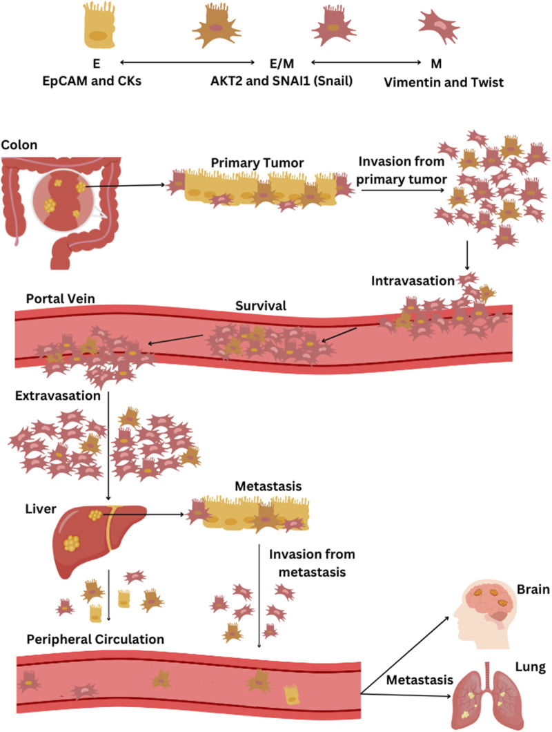
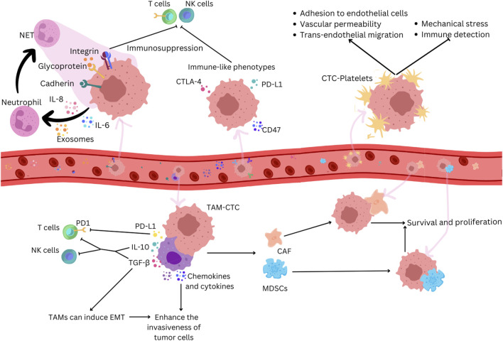
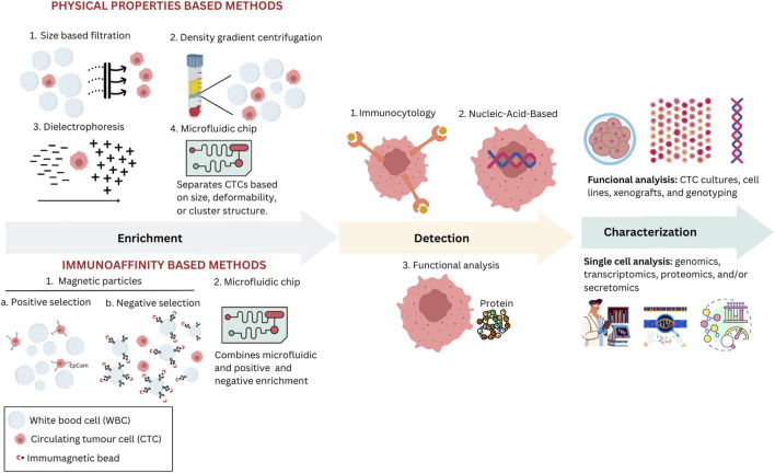
This review delves into the pivotal role of circulating tumor cells (CTCs) in colorectal cancer (CRC) metastasis, focusing on their biological properties, interactions with the immune system, advanced detection techniques, and clinical implications. We explored how metastasis-competent CTCs evade immune surveillance and proliferate, utilizing cutting-edge detection and isolation technologies, such as microfluidic devices and immunological assays, to enhance sensitivity and specificity. The review highlights the significant impact of CTC interactions with immune cells on tumor progression and patient outcomes. It discusses the application of these findings in clinical settings, including non-invasive liquid biopsies for early diagnosis, prognosis, and treatment monitoring. Despite advancements, challenges remain, such as the need for standardized methods to consistently capture and analyze CTCs. Addressing these challenges through further molecular and cellular research on CTCs could lead to improved interventions and outcomes for CRC patients, underscoring the importance of unraveling the complex dynamics of CTCs in cancer progression.

Keywords: cancer cell biology; circulating tumor cells; colorectal cancer; liquid biopsy; metastasis.


Conflict of interest statement

The authors declare that the research was conducted in the absence of any commercial or financial relationships that could be construed as a potential conflict of interest.

Figures

FIGURE 1
FIGURE 1
The process of EMT and the dissemination of CTCs from CRC through the bloodstream. CTC phenotypes are characterized by specific markers: epithelial (E) phenotype by EpCAM and cytokeratins (CKs), epithelial-mesenchymal hybrid (E/M) phenotype by AKT2 and SNAI1 (Snail), and the mesenchymal (M) phenotype by vimentin and twist. The progression of CRC and the spread of CTCs via the bloodstream begins with the primary tumor in the colon, primarily composed of epithelial cells, followed by tumor cell invasion into surrounding tissues and intravasation into blood vessels. CTCs that detach from the tumor, predominantly of the M- and some E/M phenotypes, then circulate in the portal vein. Most CTCs that survive in the portal vein are of the M-phenotype and eventually extravasate in the liver, leading to potential liver metastasis. Some CTCs enter the peripheral circulation; they predominantly present weaker E/M and E-phenotypes and may initiate a further metastatic cascade that can potentially spread to other organs, including the lungs and brain. If liver metastasis occurs, cells may detach from the secondary tumor, undergo once more EMT transition, and contribute to the further metastatic cascade towards other organs.
FIGURE 2
FIGURE 2
CTCs adopt immune-like phenotypes to evade immune recognition in cancer. Neutrophils can promote tumor growth by suppressing anti-tumor immune responses and facilitating tumor cell proliferation and metastasis. CTCs secrete chemokines that attract neutrophils, which, when activated, promote the formation of NETs which trap CTCs by creating a physical mesh that immobilizes them in the bloodstream and by facilitating adhesion through interactions involving a series of cell adhesion proteins, such as cadherin, integrin, and surface glycoprotein. NETs can suppress immune surveillance by inhibiting the activation of NK cells and effector T cells. CTCs express immune antigens like CTLA-4 and CD47 to evade immune responses. Platelets bind to CTCs, protecting them from mechanical stress and immune surveillance and facilitating their adhesion to endothelial cells, increasing vascular permeability and supporting trans-endothelial migration. TAMs interact with CTCs, secreting factors that help them evade immune detection by T cells and NK cells and induce EMT. TAMs produce chemokines and cytokines that enhance the invasiveness of tumor cells, and their interaction with CTCs attracts MDSCs and CAFs. These cells enhance CTC survival and proliferation through the secretion of chemokines and cytokines that further suppress immune responses.
FIGURE 3
FIGURE 3
Various methodologies are used to enrich, detect, and characterize CTCs in clinical and research settings. Physical properties-based methods include size-based filtration, density gradient centrifugation, dielectrophoresis, and microfluidic chips, each utilizing unique physical characteristics such as size and electric properties to isolate CTCs. In immunoaffinity-based methods, magnetic particles are employed either for positive selection using specific antibodies or for negative selection to remove unwanted cells, enhancing the purity of CTCs; microfluidic chips can also employ one of these selection strategies. After enrichment, isolated CTCs are identified through various methods, including immunocytological, molecular, or functional assays. Immunocytological methods involve staining CTCs using antibodies targeting specific cellular markers. Molecular methods focus on identifying CTCs through nucleic-acid-based assays. Functional assays allow viable CTCs to be detected based on their biological activities, such as assays that identify specific proteins secreted by CTCs. Characterization techniques, such as functional analysis through CTC cultures and single-cell analysis encompassing genomics and proteomics, are used to assess the biological and functional properties of isolated CTCs.

Similar articles

Cited by

References

    1. Abdalla T. S. A., Meiners J., Riethdorf S., König A., Melling N., Gorges T., et al. (2021). Prognostic value of preoperative circulating tumor cells counts in patients with UICC stage I-IV colorectal cancer. PLoS One 16 (6 June), e0252897. 10.1371/journal.pone.0252897 - DOI - PMC - PubMed
    1. Agarwal A., Balic M., El-Ashry D., Cote R. J. (2018). Circulating tumor cells: strategies for capture, analyses, and propagation. Cancer J. U. S., 70–77. 10.1097/PPO.0000000000000310 - DOI - PMC - PubMed
    1. Aktar S., Hamid F. B., Gamage S. M. K., Cheng T., Parkneshan N., Lu C. T., et al. (2023a). Gene expression analysis of immune regulatory genes in circulating tumour cells and peripheral blood mononuclear cells in patients with colorectal carcinoma. Int. J. Mol. Sci. 24, 5051. 10.3390/ijms24055051 - DOI - PMC - PubMed
    1. Aktar S., Islam F., Cheng T., Gamage S. M. K., Choudhury I. N., Islam M. S., et al. (2023b). Correlation between KRAS mutation and CTLA-4 mRNA expression in circulating tumour cells: clinical implications in colorectal cancer. Genes 14, 1808. 10.3390/genes14091808 - DOI - PMC - PubMed
    1. Alix-Panabières C., Mader S., Pantel K. (2017). Epithelial-mesenchymal plasticity in circulating tumor cells. J. Mol. Med. 95, 133–142. 10.1007/s00109-016-1500-6 - DOI - PubMed

LinkOut - more resources