

A Clinical Retrospective Study on the Qualitative Value of Multimodal Ultrasonography for ACR-TIRADS 4 Thyroid Nodules Ranging from 1 cm to 1.5 cm
- PMID: 39540218
- PMCID: PMC11561986
- DOI: 10.1177/15330338241297599
A Clinical Retrospective Study on the Qualitative Value of Multimodal Ultrasonography for ACR-TIRADS 4 Thyroid Nodules Ranging from 1 cm to 1.5 cm
Abstract
Introduction: This study explored the clinical value and application of ultrasound contrast imaging technology in the American College of Radiology Thyroid Imaging Reporting and Data System (ACR TI-RADS) TR4 benign and malignant thyroid nodules.
Methods: We retrospectively analyzed data from the medical records of 40 patients who met the inclusion criteria between January 2020 and December 2023. Each patient was evaluated using the ACR TI-RADS classification and contrast-enhanced ultrasound (CEUS). The agreement between pathological outcomes and ultrasound indicators and the diagnostic value and significance of each parameter were assessed.
Results: The diameters did not differ between benign and malignant nodules (P = 0.324). Ring enhancement was closely associated with benign thyroid nodules, with a negative predictive value of 100%. Homogeneous enhancement and enhancement intensity showed good diagnostic value for pathological results, with an area under the curve (AUC) > 0.8. This parameter showed a high diagnostic value for serial and parallel combinations of homogeneous enhancement and enhancement intensity, with a sensitivity of 77.8% and specificity of 85.7% for the serial combination and 100% and 71.4%, respectively. for the parallel combination.
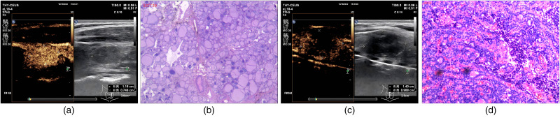
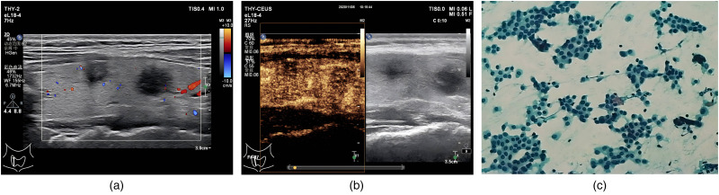
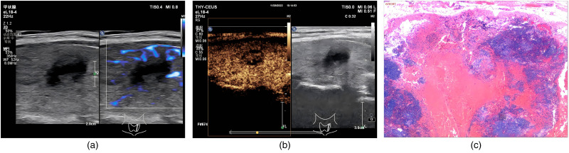
Conclusion: Among ACR TI-RADS TR4 nodules, diameter 1.0-1.5 cm was not significantly correlated with a benign or malignant nature. Nodules featuring ring enhancement with ring-enhancing features should be considered benign. Similarly, nodules showing no, homogeneous, or high enhancement with clear borders on CEUS imaging may be benign. However, nodules with uneven low enhancement or unclear borders may be malignant. Therefore, uneven and low enhancement on CEUS imaging may have a high diagnostic value for malignant nodules. Moreover, the combination of these features may have even higher specificity.
Keywords: TI-RADS TR4; contrast-enhanced ultrasound imaging; diagnosis; multimodal ultrasonography; thyroid nodule.
Conflict of interest statement
Declaration of Conflicting InterestsThe authors declared no potential conflicts of interest with respect to the research, authorship, and/or publication of this article.
Figures
Similar articles
-
Building radiomics models based on ACR TI-RADS combining clinical features for discriminating benign and malignant thyroid nodules.Front Endocrinol (Lausanne). 2025 Jul 21;16:1486920. doi: 10.3389/fendo.2025.1486920. eCollection 2025. Front Endocrinol (Lausanne). 2025. PMID: 40761820 Free PMC article.
-
A Practical CEUS TI-RADS Refer to Score and Grading of ACR TI-RADS (2017) for Thyroid Nodules.Ultrasound Med Biol. 2025 Aug;51(8):1352-1360. doi: 10.1016/j.ultrasmedbio.2025.05.005. Epub 2025 May 24. Ultrasound Med Biol. 2025. PMID: 40414781
-
Efficacy Assessment and Influencing Factors on Superb Microvascular Imaging (SMI) Microflow Patterns in Solid Thyroid Nodules: What Matters?Ultrasound Med Biol. 2025 Sep;51(9):1389-1398. doi: 10.1016/j.ultrasmedbio.2025.04.002. Epub 2025 Jun 14. Ultrasound Med Biol. 2025. PMID: 40518320
-
Diagnostic Performance of Six Ultrasound Risk Stratification Systems for Thyroid Nodules: A Systematic Review and Network Meta-Analysis.AJR Am J Roentgenol. 2023 Jun;220(6):791-803. doi: 10.2214/AJR.22.28556. Epub 2023 Feb 8. AJR Am J Roentgenol. 2023. PMID: 36752367
-
Contrast-enhanced ultrasound using SonoVue® (sulphur hexafluoride microbubbles) compared with contrast-enhanced computed tomography and contrast-enhanced magnetic resonance imaging for the characterisation of focal liver lesions and detection of liver metastases: a systematic review and cost-effectiveness analysis.Health Technol Assess. 2013 Apr;17(16):1-243. doi: 10.3310/hta17160. Health Technol Assess. 2013. PMID: 23611316 Free PMC article.
References
-
- Gharib H, Papini E, Paschke R, et al. American Association of clinical endocrinologists, associazione medici endocrinologi, and European thyroid association medical guidelines for clinical practice for the diagnosis and management of thyroid nodules. J Endocrinol Invest. 2010;33(S5):1-50. doi:10.1007/BF03346587 - DOI - PubMed
Publication types
MeSH terms
Substances
LinkOut - more resources
Full Text Sources
Miscellaneous
