. 2024 Dec 11;15(12):e0150524.
doi: 10.1128/mbio.01505-24. Epub 2024 Nov 14.

Whole-blood model reveals granulocytes as key sites of dengue virus propagation, expanding understanding of disease pathogenesis

Affiliations

Whole-blood model reveals granulocytes as key sites of dengue virus propagation, expanding understanding of disease pathogenesis

Hansa Praneechit et al. mBio. .

Abstract

Dengue virus (DENV) infection poses a significant global health threat, yet our understanding of its immunopathogenesis remains incomplete due to limitations of existing models. Here, we establish an in vitro whole-blood model using hirudin, an anticoagulant that preserves complement activity and cellular interactions, to study DENV infection. Our model reveals the susceptibility of all major leukocyte populations to DENV infection, with monocytes and granulocytes demonstrating high permissiveness and production of infectious virus progeny. Notably, granulocytes emerge as previously unrecognized targets of DENV infection, highlighting the importance of studying viral tropism within a physiologically relevant context. We also observed efficient DENV binding to B cells, but limited production of infectious virus, suggesting a potential role in viral sequestration or immune dysregulation. Interestingly, both NK and T cells, while less permissive, were also found to be susceptible to DENV infection. Our ex vivo analysis of whole blood from DENV-infected patients confirms the susceptibility of granulocytes, monocytes, B cells, natural killer cells, and T cells to infection, further validating the clinical relevance of our model. Additionally, we observed dynamic changes in circulating blood cell populations during acute dengue, potentially reflecting both direct virus-mediated effects and immune responses. This whole-blood model offers a valuable tool for investigating the complex interplay between DENV and host factors, facilitating a deeper understanding of dengue pathogenesis and ultimately contributing to the development of novel therapeutic strategies.IMPORTANCEDengue virus (DENV) infection is a significant global health threat, with increasing incidence in endemic regions and expanding geographic range due to factors like global warming. Current models for studying DENV pathogenesis often lack the complexity of the human immune system, hindering the development of effective therapies and vaccines. To address this, we have established the first in vitro whole-blood model using hirudin, preserving critical immune components and cellular interactions. This model reveals granulocytes as previously unrecognized targets of productive DENV infection, challenging existing paradigms of viral tropism. Our ex vivo analysis of patient blood samples confirms the clinical relevance of this finding and validates our model's utility. This unique model offers a powerful platform for future studies to dissect the complex interactions between DENV and the host immune system, including the roles of different leukocyte populations, ultimately informing the development of novel therapeutic strategies to combat this devastating disease.

Keywords: dengue target cells; dengue virus; granulocytes; in vitro whole-blood infection model; neutrophils.


Conflict of interest statement

The authors declare no conflict of interest.

Figures
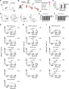
Fig 1
Fig 1
Hirudin compatibility with a whole-blood DENV infection model. (A) Hirudin preserves complement function. Plasma was separated from hirudinized whole blood or EDTA-treated hirudinized blood after 2 h, then incubated with yeast cells. C3b deposition on yeast was detected by flow cytometry. (B) Diagram of the in vitro whole-blood DENV infection model (18 h post-infection). At 2 and 18 h post-infection, each cell population was analyzed based on their size, granularity, and CD markers for cell viability and dengue antigens. (C) Hirudin does not interfere with DENV infectivity. Hirudinized blood from DENV-naïve donors was incubated with mock or DENV (10⁷ or 10⁸ genome copies/mL). Infectious DENV titers in plasma were determined by focus-forming unit (FFU) assay at 2 h (white bars) and 18 h (gray bars) post-infection. (D) Representative gating strategy for analyzing DENV-infected whole blood. Cells were gated based on forward scatter (FSC), side scatter (SSC), and established CD markers to distinguish platelets (CD41a+), granulocytes (CD66+), monocytes (CD14+), B (CD19+) and T (CD3+) lymphocytes, and NK cells (CD56+). (E, F) Hirudin maintains leukocyte viability in a whole-blood infection model. Cell viability in each white blood cell (WBC) population was determined by live/dead staining and flow cytometry after incubation with mock (white bars), DENV 10⁷ genome copies/mL (gray bars), or 10⁸ genome copies/mL (black bars) at 2 h (E) and 18 h (F) post-infection. (G–I) DENV infection in each cell population in a whole-blood infection model. Percentages of DENV-positive cells, as determined by surface DENV envelope (E), protein (G), intracellular NS1 (H), and intracellular NS3 (I) in each WBC cell population were determined at 2 h (white circles) and 18 h (gray circles) post-infection. Data are presented as individual dot plots from 8 to 10 independent experiments. Student’s t-test was used to compare expression levels of DENV antigens among each time-point and each condition in each cell population. Asterisks (*, **, and ***) indicate statistical significance (P < 0.05, P < 0.01, and P < 0.005, respectively).
Fig 2
Fig 2
DENV infection and replication efficiency in different blood cell populations. (A) Schematic diagram of the experimental setup for analyzing DENV infection and replication in sorted blood cell population 18 h post-infection with DENV at 10⁸ genome copies/mL. (B) Purity assessment of each sorted WBC population from mock-infected (white bars) or DENV-infected (black bars) whole blood. (C) Confocal microscopy images of DENV-infected WBCs. Intracellular DENV NS1 and NS5 proteins are shown in green, and nuclei are stained with Hoechst dye (blue). (D) Quantification of cell-associated DENV RNA in each sorted cell population by qRT-PCR. (E) Infectious DENV titers (FFU/10³ cells) in supernatants from co-cultures of sorted blood cell populations with permissive BHK cells for 2 days. (F) DENV genome copies (per 10³ cells) in supernatants from co-cultures as in panel E. (G) Specific infectivity (si) of DENV in each cell population, calculated as the ratio of infectious DENV titers (FFU) to viral genome copies. Data are representative of three independent experiments. N.D. indicates "not detected."
Fig 3
Fig 3
DENV antigen detection in whole blood from adult and pediatric patients. (A) Representative gating strategy for flow cytometry analysis of DENV-infected patient whole blood. Cell populations were identified based on forward scatter (FSC), side scatter (SSC), and the following CD markers: platelets (CD41a+), granulocytes (CD66+), monocytes (CD14+), B cells (CD19+) and T cells (CD3+), and NK cells (CD56+). (B–D) Percentages of cells positive for surface DENV envelope (E), protein (B), intracellular NS1 (C), or intracellular NS3 (D) in the indicated cell populations from adult DENV-infected patients (n = 24). Whole blood was collected at the following two time points: acute (1–6 days before defervescence) and convalescent (7–24 days after defervescence). Each symbol represents an individual patient. The X/X number in each plot indicates the number of patients with detectable positive cells out of the total number of analyzed for that cell population. (E) Kinetics of NS1-positive cells in the indicated cell populations from pediatric DENV-infected patients (n = 8; four with dengue fever [DF], four with dengue hemorrhagic fever [DHF]). Whole blood was collected daily during the acute phase, with one convalescent sample. Lines represent individual patients.
Fig 4
Fig 4
CBC and cell differential count in adult and pediatric dengue patients. Total cell counts and differential counts of white blood cell (WBC) populations (lymphocytes, monocytes, neutrophils, eosinophils, basophils, atypical lymphocytes, band neutrophils) and platelets were determined by standard CBC for whole-blood samples collected from adult (A) and pediatric (B) dengue patients at the same time points as in Fig. 3. Circles and squares represent DF and DHF patients, respectively. Data are presented as dot plots with unique colored symbols representing individual patients. A Student’s t-test was used to analyze the differences in cell numbers between acute (days −2 to 2) and convalescent (days 12 to 24) specimens. P values are labeled on each graph.

References

    1. Bhatt S, Gething PW, Brady OJ, Messina JP, Farlow AW, Moyes CL, Drake JM, Brownstein JS, Hoen AG, Sankoh O, Myers MF, George DB, Jaenisch T, Wint GRW, Simmons CP, Scott TW, Farrar JJ, Hay SI. 2013. The global distribution and burden of dengue. Nature New Biol 496:504–507. doi:10.1038/nature12060 - DOI - PMC - PubMed
    1. Stanaway JD, Shepard DS, Undurraga EA, Halasa YA, Coffeng LE, Brady OJ, Hay SI, Bedi N, Bensenor IM, Castañeda-Orjuela CA, Chuang T-W, Gibney KB, Memish ZA, Rafay A, Ukwaja KN, Yonemoto N, Murray CJL. 2016. The global burden of dengue: an analysis from the global burden of disease study 2013. Lancet Infect Dis 16:712–723. doi:10.1016/S1473-3099(16)00026-8 - DOI - PMC - PubMed
    1. Yang X, Quam MBM, Zhang T, Sang S. 2021. Global burden for dengue and the evolving pattern in the past 30 years. J Travel Med 28:taab146. doi:10.1093/jtm/taab146 - DOI - PubMed
    1. Monath TP. 1994. Dengue: the risk to developed and developing countries. Proc Natl Acad Sci U S A 91:2395–2400. doi:10.1073/pnas.91.7.2395 - DOI - PMC - PubMed
    1. Halstead SB, O’Rourke EJ. 1977. Dengue viruses and mononuclear phagocytes. I. Infection enhancement by non-neutralizing antibody. J Exp Med 146:201–217. doi:10.1084/jem.146.1.201 - DOI - PMC - PubMed

LinkOut - more resources