Comparative Study
. 2024 Nov 14;19(11):e0313697.
doi: 10.1371/journal.pone.0313697. eCollection 2024.

Manufacturing exosomes for wound healing: Comparative analysis of culture media

Affiliations
Comparative Study

Manufacturing exosomes for wound healing: Comparative analysis of culture media

Huy Hoang Dao et al. PLoS One. .

Abstract

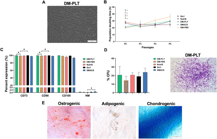
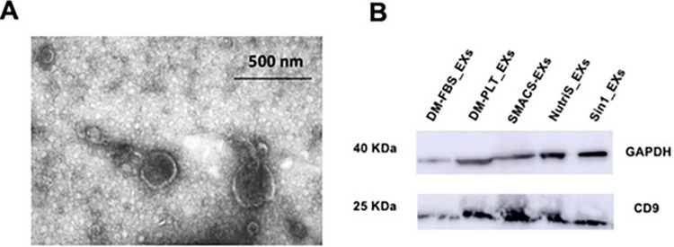
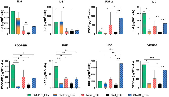
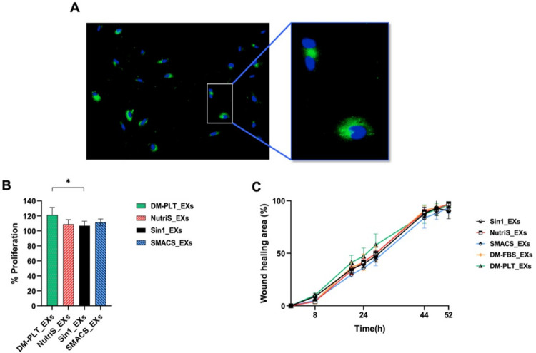
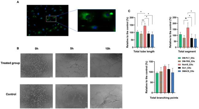
Mesenchymal stem cell (MSC)-derived exosomes (EXs) have emerged as promising therapeutic agents for wound healing. However, the optimal conditions for manufacturing MSC-derived EXs that maximize their wound-healing potential have yet to be established. Hence, we compared the efficacy of five different MSC culture media, including three different serum-free, a platelet-supplemented, and a fetal bovine serum-supplemented media, in exosome manufacturing for wound healing applications. Although umbilical cord-derived MSCs (UCMSCs) cultured in these media exhibited similar proliferation, morphology, MSC surface marker expression, and stemness, EXs derived from UCMSCs cultured in different culture media displayed varying levels of growth factors and cytokines. Notably, EXs derived from platelet-supplemented media (DM-PLT_EXs) exhibited significantly higher concentrations of keratinocyte growth factor (KGF), vascular endothelial growth factor (VEGF-A), platelet-derived growth factor (PDGF-BB), interleukin 6 (IL-6), interleukin 7 (IL-7), and interleukin 8 (IL-8) than EXs from other media. These differences correlated with the superior capability of DM-PLT_EXs to promote human skin fibroblast proliferation and stimulate angiogenesis of human umbilical vein endothelial cells, making them a more suitable choice for wound healing applications. Our findings emphasize the significance of the culture medium selection in tailoring the therapeutic potential of UCMSC-derived EXs for wound healing.


Conflict of interest statement

The authors have declared that no competing interests exist.

Figures

Fig 1
Fig 1. Characteristic of UCMSCs cultured in five different media.
(A) A representative morphology of UCMSCs cultured in DM-PLT medium at P5. Magnification 10X. Scale bar 50 μm. (B) A comparative population doubling time of three UCMSC lines cultured in 10% FBS supplemented (DM-FBS), a 5% PLT supplemented (DM-PLT), and three serum-free media (STEMin1, NutriStem, and StemMACS) (n = 3). (C) Expression of MSC surface markers (n = 3) and (D) colony-forming potential of UCMSCs cultured in five different culture media (n = 3). A representative image from DM-PLT culture medium independent experiments is shown. Magnification 4X. Scale bar 200 μm. (E) A representative trilineage differentiation of UCMSCs cultured in DM-PLT medium. Magnification 10X. Scale bar 50 μm. Sin1, NutriS, DM-PLT, SMACS, DM-FBS are UCMSCs cultured in STEMin1, NutriStem, DMEM/F12 medium supplemented with 5% PLT, StemMACS and DMEM/F12 medium supplemented with 10% FBS, respectively. P: Passage, CFU: Colonies forming unit.
Fig 2
Fig 2. Exosomes derived from UCMSCs displayed typical exosome morphology and surface marker.
(A) Representative morphology of EXs derived from UCMSCs cultured in DM-PLT medium observed under transmission electron microscopy. (B) Internal control GAPDH and CD9 were detected in all EX samples with 15 μg total exosomal protein loaded into each lane. DM-FBS_EXs, DM-PLT_EXs, SMACS_EXs, NutriS_EXs, Sin1_EXs are EXs derived from UCMSCs cultured in DMEM/12 + 10% FBS, DMEM/F12 + 5% PLT, StemMACS, NutriStem, and STEMin1, respectively.
Fig 3
Fig 3. Quantity of growth factors and cytokines in exosomes purified from 108 UCMSCs cultured in different media (n = 3).
DM-FBS_EXs, DM-PLT_EXs, SMACS_EXs, NutriS_EXs, Sin1_EXs are EXs from UCMSCs cultured in DMEM/12 + 10% FBS, DMEM/F12 + 5% PLT, StemMACS, NutriStem, and STEMin1, respectively. Statistical significance was determined by ANOVA and post-hoc Tukey HSD tests and is indicated by: * where p < 0.05; ** where p < 0.01; *** where p < 0.001; **** where p < 0.0001.
Fig 4
Fig 4. Effects of EXs on fibroblast proliferation and migration (n = 3).
(A) Confocal images of fibroblasts incubated for two hours with ExoGlow (green) labeled UCMSC-derived EXs. DAPI was used to stain nuclei (B) Fibroblast proliferation rates induced by EXs from different media. The percentage of cell proliferation was normalized to medium supplement with DM-FBS_EXs. (C) EXs-mediated fibroblast migration as determined by a scratch assay. DM-FBS_EXs, DM-PLT_EXs, SMACS_EXs, NutriS_EXs, Sin1_EXs are EXs from UCMSCs cultured in DMEM/12 + 10% FBS, DMEM/F12 + 5% PLT, StemMACS, NutriStem, and STEMin1, respectively. Statistical significance was determined by ANOVA and post-hoc Tukey HSD tests, two-way ANOVA, and is indicated by: * where p < 0.05; ** where p < 0.01; *** where p < 0.001; **** where p < 0.0001.
Fig 5
Fig 5. Influence of EX treatment on the tube formation (n = 3).
(A) Uptake of ExoGlow-labeled EXs (green) in endothelial cells after two hours. The nucleus was stained with DAPI. (B) The representative tube structures of hUVECs under an inverted microscope. (C) Quantitative analysis of hUVEC tube formation normalized to the control group. DM-FBS_EXs, DM-PLT_EXs, SMACS_EXs, NutriS_EXs, Sin1_EXs are EXs from UCMSCs cultured in DMEM/12 + 10% FBS, DMEM/F12 + 5% PLT, StemMACS, NutriStem, and STEMin1, respectively. Statistical significance was determined by ANOVA and post-hoc Tukey HSD tests and is indicated by: * where p < 0.05; ** where p < 0.01; *** where p < 0.001; **** where p < 0.0001.

References

    1. Than UTT, Guanzon D, Leavesley D, Parker T. Association of Extracellular Membrane Vesicles with Cutaneous Wound Healing. Int J Mol Sci. 2017;18: 956. doi: 10.3390/ijms18050956 - DOI - PMC - PubMed
    1. Frykberg RG, Banks J. Challenges in the Treatment of Chronic Wounds. Adv Wound Care. 2015;4: 560–582. doi: 10.1089/wound.2015.0635 - DOI - PMC - PubMed
    1. Nagamura-Inoue T, He H. Umbilical cord-derived mesenchymal stem cells: Their advantages and potential clinical utility. World J Stem Cells. 2014;6: 195–202. doi: 10.4252/wjsc.v6.i2.195 - DOI - PMC - PubMed
    1. Lee CH, Moioli EK, Mao JJ. Fibroblastic differentiation of human mesenchymal stem cells using connective tissue growth factor. Conf Proc Annu Int Conf IEEE Eng Med Biol Soc IEEE Eng Med Biol Soc Annu Conf. 2006;2006: 775–778. doi: 10.1109/IEMBS.2006.259866 - DOI - PMC - PubMed
    1. De Ugarte DA, Morizono K, Elbarbary A, Alfonso Z, Zuk PA, Zhu M, et al.. Comparison of multi-lineage cells from human adipose tissue and bone marrow. Cells Tissues Organs. 2003;174: 101–109. doi: 10.1159/000071150 - DOI - PubMed

Publication types

MeSH terms

LinkOut - more resources