. 2025 Jan:59:101070.
doi: 10.1016/j.neo.2024.101070. Epub 2024 Nov 14.

A novel regimen for pancreatic ductal adenocarcinoma targeting MEK, BCL-xL, and EGFR

Affiliations

A novel regimen for pancreatic ductal adenocarcinoma targeting MEK, BCL-xL, and EGFR

Song Han et al. Neoplasia. 2025 Jan.

Abstract

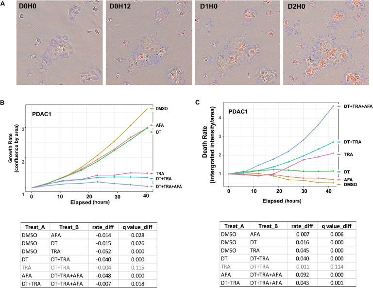
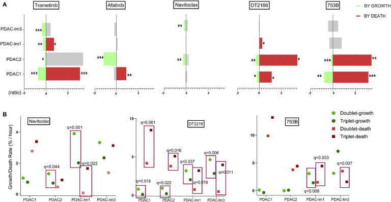
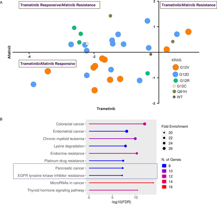
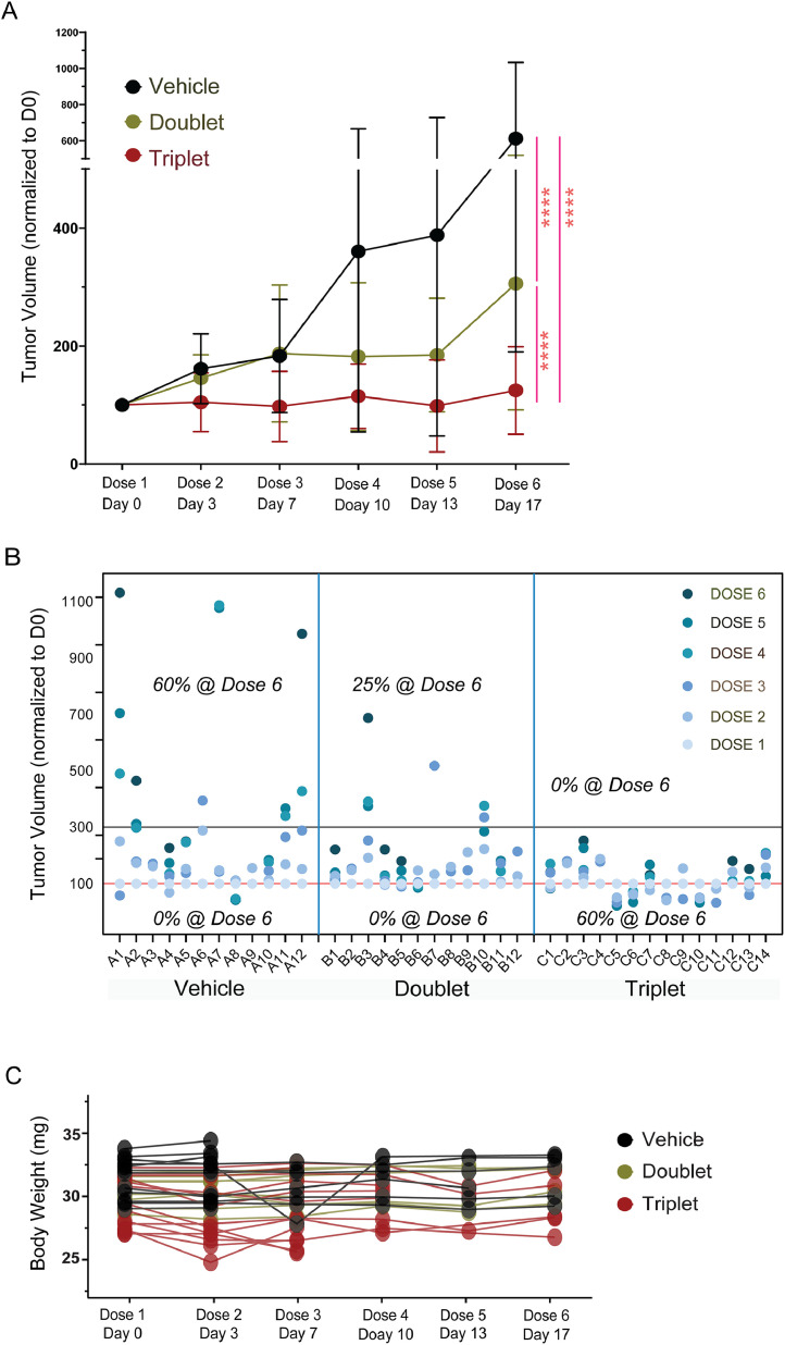
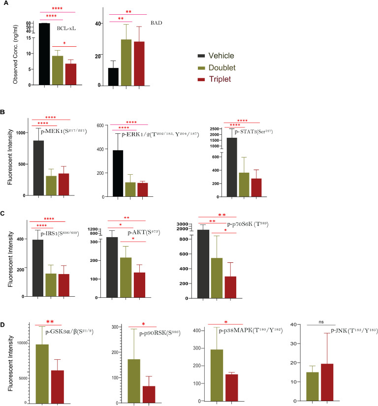
Oncogenic KRAS signaling plays a critical role in pancreatic ductal adenocarcinoma (PDAC) biology. Recent studies indicate that the combination of MEK and BCL-xL inhibition is synthetically lethal and holds promise for some types of solid cancers, however, patient response was poorly observed in PDAC predominantly due to amplified EGFR signaling. Here, we leverage the advantage of the combinational treatment strategy and designed a triplet regimen targeting the comprehensive RAS activation networks through simultaneously blocking MEK/BCL-xL/EGFR. The cytotoxicity of trametinib (MEK inhibitor), DT2216 (BCL-xL degrader) and afatinib (pan-EGFR inhibitor) and their combination was tested in patient-derived, primary PDAC cells using a live cell imaging system. Patient-derived xenograft (PDX) model was employed for the evaluation of the therapeutic efficacy and safety of the combinational regimen. Targeted pathway cascades activities were analyzed using multiplex phosphor-immune assays. In vitro comparisons showed the addition of afatinib as a third agent was statistically superior compared to a doublet of trametinib+DT2216 in suppressing cell growth and inducing cell death in all cell lines tested. This triplet similarly demonstrated significant superiority over the doublet of MEK/BCL-xL inhibition in the in vivo murine model. The triplet regimen was well tolerated in vivo. Overall tumor growth rates were significantly reduced in doublet treatment compared to controls, and further reduced in the triplet treatment group. Pathway analysis revealed the addition of afatinib in triplet regimen further inhibited PI3K/AKT effectors of p90RSK, p70S6K, and GSK3α/β along with a secondary pathway of P38 MAPK. Our study identifies an important contribution of EGFR inhibition to elevate the response of PDAC, supporting a clinical assessment of this triplet combination in patients.

Keywords: Combinational targeting therapy; EGFR, Epidermal Growth Factor Receptor; KRAS, Kirsten Rat Sarcoma Viral Oncogene Homolog; MEK, Mitogen-Activated Protein Kinase Kinase; PDAC, Pancreatic Ductal Adenocarcinoma.


Conflict of interest statement

Declaration of competing interest The authors declare the following financial interests/personal relationships which may be considered as potential competing interests: P.Z., G.Z., and D.Z. are inventors of the following patent and pending patent application for use of BCL-xL PROTACs as senolytic and antitumor agents: 1. COMPOUNDS THAT INDUCE DEGRADATION OF ANTI-APOPTOTIC BCL-2 FAMILY PROTEINS AND THE USES THEREOF (Patent number: 10807977, Status: Granted, Applicant/Assignee: BioVentures, LLC, Inventors: Guangrong Zheng, Daohong Zhou, Xuan Zhang, Yingying Wang, Jianhui Chang). 2. THERAPEUTIC AGENTS AND METHODS OF TREATMENT (Patent number: 2020218367, Status: Pending, Applicant/Assignee: University of Florida Research Foundation, Inventors: Guangrong Zheng, Daohong Zhou, Pratik Pal, Xingui Liu, Dinesh Thummuri, Wenyi Hu, Peiyi Zhang, Dongwen Lyu, Yaxia Yuan, and Xuan Zhang). G.Z., and D.Z. are co-founders of and have equity in Dialectic Therapeutics, which develops BCL-xL/2 PROTACs to treat cancer. A.C. is the scientific advisory board for Dialectic that is developing DT2216 PROTAC.

Figures

Fig 1
Fig. 1
Live cell imaging assessment of treatment responses. (A) Phase contrast images of live and dead cells. A set of four representative images of PDAC1 cells were taken at 20x magnification from time points of day 0, 12 hours, day 1, and day 2 as indicated. Cytotox NIR Dye (red) staining indicates dead cells. (B) Quantification of cell proliferation in cultures was assessed by confluence (area covered by cells), and (C) Quantification of dead cells was measured by Cytotox NIR Dye staining (ratio of integrated intensity of red staining over total cell area). Growth rate and death rates were plotted with y-axis reporting rate change against x-axis in elapsed hours. Experiments were performed in quintuplicates and four images were taken from each well. A q-value < 0.05 was considered statistically significant.
Fig 2
Fig. 2
Treatment effectiveness comparison of various combinations. (A) Mono-treatment of the five compounds on cell growth and cell death assessed on four PDAC patient-derived cell lines. x-axis is scaled by ratio of rate change between the individual compounds and the control treatment of DMSO. Ratio of changes comparing to DMSO control for decreased cell growth was scaled towards the left and increased cell death was scaled towards the right. Green and red bars indicate significant changes and gray bars indicate changes without statistical significance. * q < 0.05, ** q < 0.01, *** q < 0.001; and (B) Comparisons between triplet treatment of MEK, BCL-xL, and EGFR inhibition vs doublet inhibition of MEK and BCL-xL. Growth rates are indicated by green dots (light green for doublet and dark green for triplet treatments), and cell death rates are indicated by red dots (light red for doublet and dark red for triplet treatment). Difference between rates that met statistical significance (q < 0.05) were boxed and the associated q value is indicated.
Fig 3
Fig. 3
In silico analysis of molecular basis of PDAC cell lines in response to trametinib and afatinib. (A) DepMap data of cell lines with KRAS mutation in response to trametinib and afatinib. Both x- and y-axis are log2 fold changes. Each dot represents one cell lines and KRAS mutation statues are colored as illustrated; (B) KEGG enriched pathways analysis. Genes with mutations in 14 PDAC cell lines that are sensitive to both trametinib and afatinib were subjected to ShinyGO 0.80 [ShinyGO 0.80 (sdstate.edu)]. FDR cutoff was set at <0.0001 with minimum 10 genes per pathway. Lollipop dots indicate fold enrichment.
Fig 4
Fig. 4
Impact of the triplet regimen on tumor growthin vivoas compared to controls. Mice bearing PDAC-lm1 subcutaneous xenograft tumors were randomly divided into three groups and treated with vehicle (n = 12), doublet of trametinib and DT2216 (n = 12), and triplet of trametinib, DT2216, and afatinib (n = 14). Tumor volumes were normalized to their volume on dose 1 (day 0) for comparisons. Tumor growth kinetics were plotted as indicated with mean ±/ SEM (A) and individually (B), and mice body weight (mg) was plotted in (C) against treatment periods. **** q < 0.0001.
Fig 5
Fig. 5
Key signaling molecules in the targeted pathways. Cascades in EGFR/RAS regulated pathways are arranged and connected based upon IPA canonical/established signaling networks, including EGF, Insulin Receptor, STAT3 Pathway, PI3K/AKT Signaling, and Pancreatic adenocarcinoma Signaling. Molecules tested in the immune-plexes are colored nodes and uncolored nodes represent untested molecules. Phosphorylation of tested molecules are illustrated with a “p” in a circle.
Fig 6
Fig. 6
Doublet and triplet treatments affecting molecules in MAPK and PI3K pathways. PDAC-lm1 subcutaneous xenograft tumors were treated with vehicle, a doublet regimen of trametinib/DT2216, or a triplet regimen of trametinib/DT2216/afatinib. Three mice from each group were sacrificed 4 hours after dose 3 administration and tumors were collected and analyzed using the three immune-plex kits, (A) Expression levels of BCL-xL and BAD by concentration of ng/ml, (B) Phosphorylation status (p-) of p-MEK1, p-ERK1/2, and the substrate of p-STAT3, (C) Phosphorylation status (p-) of p-IRS1, p-AKT, and p-p70S6K, and (D) Phosphorylation status (p-) of p-GSK3α/β, p-p90RSK, p-P38 MAPK, and p-JNK. All phosphorylation sites are as labeled and fluorescent intensity (y-axis) indicate levels of phosphorylation. Bar graphs for mean ±/ SEM. *q < 0.05, **q < 0.01, ****q < 0.0001, ns = not statistically significant.

Similar articles

Cited by

References

    1. Siegel RL, Giaquinto AN, Jemal A. Cancer statistics, 2024. CA Cancer J. Clin. 2024;74(1):12–49. doi: 10.3322/caac.21820. - DOI - PubMed
    1. Conroy T, Desseigne F, Ychou M, Bouche O, Guimbaud R, Becouarn Y, et al. FOLFIRINOX versus gemcitabine for metastatic pancreatic cancer. N. Engl. J. Med. 2011;364(19):1817–1825. doi: 10.1056/NEJMoa1011923. - DOI - PubMed
    1. Long SA, Amparo AM, Goodhart G, Ahmad SA, Waters AM. Evaluation of KRAS inhibitor-directed therapies for pancreatic cancer treatment. Front. Oncol. 2024;14 doi: 10.3389/fonc.2024.1402128. - DOI - PMC - PubMed
    1. Corcoran RB, Cheng KA, Hata AN, Faber AC, Ebi H, Coffee EM, et al. Synthetic lethal interaction of combined BCL-XL and MEK inhibition promotes tumor regressions in KRAS mutant cancer models. Cancer Cell. 2013;23(1):121–128. doi: 10.1016/j.ccr.2012.11.007. - DOI - PMC - PubMed
    1. Corcoran RB, Do KT, Kim JE, Cleary JM, Parikh AR, Yeku OO, et al. Phase 1/2 study of combined BCL-xL and MEK inhibition with navitoclax and trametinib in KRAS or NRAS mutant advanced solid tumors. Clin. Cancer Res. 2024 doi: 10.1158/1078-0432.CCR-23-3135. - DOI - PMC - PubMed

Publication types

MeSH terms