. 2025 Aug;21(4):577-592.
doi: 10.1007/s11302-024-10065-4. Epub 2024 Nov 15.

CD39 activities in the treated acupoints contributed to the analgesic mechanism of acupuncture on arthritis rats

Affiliations

CD39 activities in the treated acupoints contributed to the analgesic mechanism of acupuncture on arthritis rats

Yu-Jia Li et al. Purinergic Signal. 2025 Aug.

Abstract

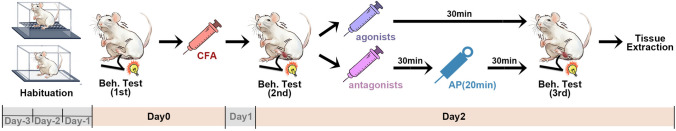
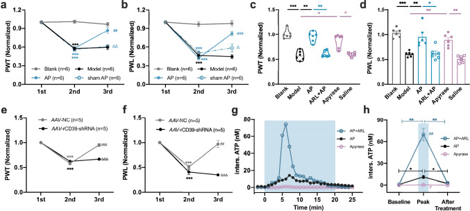
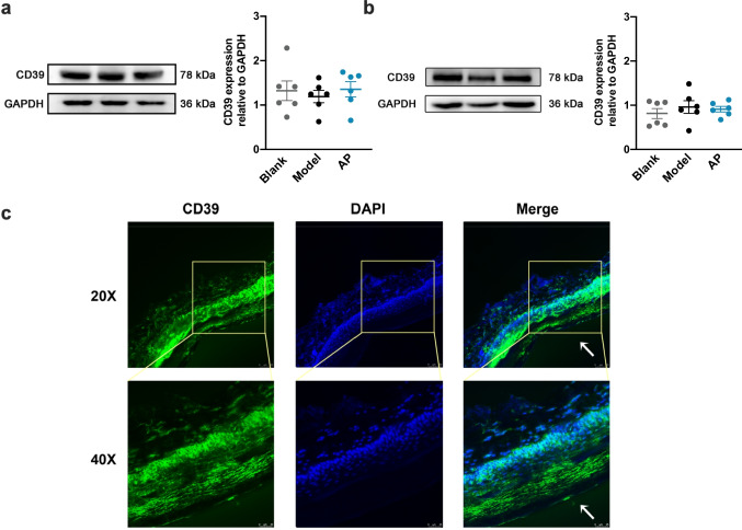
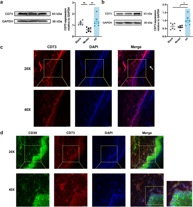
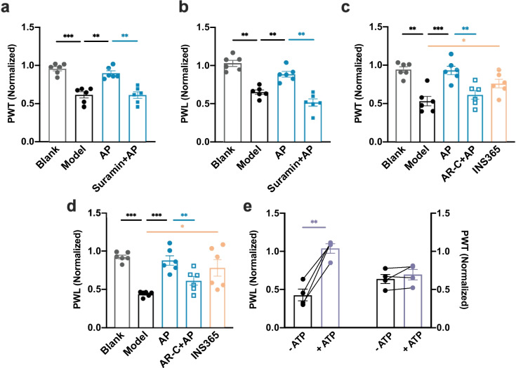
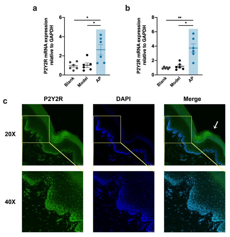
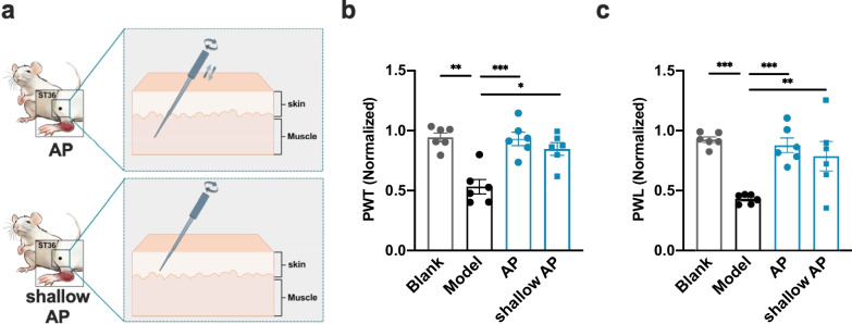
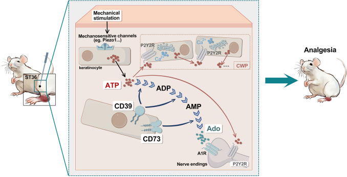
Our previous work had identified that at the acupuncture point (acupoint), acupuncture-induced ATP release was a pivotal event in the initiation of analgesia. We aimed to further elucidate the degradation of ATP by CD39. Acupuncture was administered at Zusanli acupoint on arthritis rats, and pain thresholds of the hindpaws were determined. Pharmacological tools or adeno-associated viruses were administered at the acupoints to interfere with targeting signals. Protein expression was determined with qRT-PCR, WB, or immunofluorescent labeling. Cultured keratinocytes, HaCaT line, were subjected to hypotonic shock to simulate needling stimulation. Extracellular ATP and adenosine levels were quantified using luciferase-luciferin assay and ELISA, respectively. Acupuncture-induced prompt analgesia was impaired by inhibiting CD39 activities to prevent the degradation of ATP to AMP but was mimicked by using CD39 agonists. Acupuncture-induced ATP accumulation exhibited synchronous changes. Similarly, acupuncture analgesia was hindered by suppressing CD73 to prevent the conversion of AMP to adenosine. Furthermore, the acupuncture effect was replicated by agonism at P2Y2Rs but inhibited by antagonism at them. Acupuncture upregulated CD73 and P2Y2Rs but not CD39. Immunofluorescent labeling demonstrated that keratinocytes were a primary site for these proteins. Shallow acupuncture also demonstrated antinociception. In vitro tests showed that hypotonic shock induced HaCaT cells to release ATP and adenosine, which was impaired by suppressing CD39 and CD73, respectively. Finally, agonism at P2Y2Rs promoted ATP release and [Ca2+]i rise. CD39 at the acupoints contributes to the analgesic mechanism of acupuncture. It may facilitate adenosine signaling in conjunction with CD73 or provide an appropriate ATP milieu for P2Y2Rs. Skin tissue may be one of the scenes for these signalings.

Keywords: ATP; Acupuncture analgesia; Acupuncture points; CD39; CD73; P2Y2.


Conflict of interest statement

Declarations. Ethical approval: All experimental protocols were approved by the Animal Care Committee of Shanghai University of Traditional Chinese Medicine (Shanghai, China) (protocol code: PZSHUTCM2212270003). Conflict of interest: The authors declare no competing interests.

Figures

Fig. 1
Fig. 1
Rats grouping and experimental procedures. AP, acupuncture; P2YRs, P2Y receptors; P2Y2Rs, P2Y2 receptors; ARL, ARL67156; AR–C, AR-C118925xx; act., active; inh., inhibit
Fig. 2
Fig. 2
Timeline of the behavioral assays. Pain thresholds were determined before CFA injection (1st), before (2nd), and after treatment (3rd). Beh. Test, behavioral test; AP, acupuncture
Fig. 3
Fig. 3
The effects of CD39 activities at the treated acupoint on AP analgesia and mobilization of inters. ATP (mean ± SEM or median (Q1, Q3), n = 3–6). a, b. The analgesia effect of needling at ST36. Data show the levels of tactile allodynia (PWT) (a) and thermal hyperalgesia (PWL) (b) of injured-side hindpaws (n = 6). Data are expressed as normalized values against their respective baselines. Comparisons were carried out among groups at the same time point: ***p < 0.001 vs. blank; ##p < 0.01, ###p < 0.001 vs. model. Δp < 0.05, ΔΔ p < 0.01 vs. AP. c, d. The responses of pain levels to modulating CD39 at ST36 (n = 6). Data show the last PWT (c) and PWL (d) of each group. ARL67156 (ARL), the antagonist of CD39, was injected into ST36 before needling. Apyrase, the analog of CD39, was introduced to ST36 to mimic AP intervention. *p < 0.05, **p < 0.01, ***p < 0.001. e, f. The prevention effect of AAV-rCD39-shRNA on AP analgesia in PWT (e) and PWL (f) (n = 5). AAV-NC was taken as the negative control. ***p < 0.001 vs. 1st and ##p < 0.01, ###p < 0.001 vs. 2nd, comparison within group. ΔΔΔp < 0.001 vs. AAV-NC in 3rd, comparison among groups. g Representative time courses of inters. ATP level at ST36 of AA rats during AP (0–20 min, highlighted by the blue box) in response to modulation of CD39 activities. Each trace was from 3–4 tests. h. Comparison of inters. ATP levels among each group at three representative time points (n = 3–4). Data were extracted from the time-course recordings. *p < 0.05 **p < 0.01, comparison within group. #p < 0.05, ##p < 0.01 vs. peak in AP, comparison among groups
Fig. 4
Fig. 4
Expression of CD39 at ST36 and in rats’ glab skin. a, b. WB intensity and quantification of CD39 in the skin (a) and muscle (b) at the treated ST36 from different groups (mean ± SEM, n = 6). c Confocal images showing immunofluorescence staining of CD39 (green) and nuclei (blue) in glab skin excised from normal rats. The white arrow points to the surface layer of the skin
Fig. 5
Fig. 5
Responses of AP-analgesia to downregulating CD73 at ST36 (mean ± SEM, n = 6). a, b. The suppression effects of antagonism at CD73 in ST36 on AP analgesia (n = 6). a and b are changes in PWT and PWL, respectively. Data show the last behavioral tests of each group. AMP-CP, the specific antagonist of CD73, was injected into ST36 30 min before needling. **p < 0.01, ***p < 0.001. c, d. The prevention of AP analgesia on PWT (c) and PWL (d) by AAV-rCD73-shRNA interference (n = 6). AAV-NC rats were taken as the control group. ***p < 0.001 vs. 1st and #p < 0.05, ##p < 0.01 vs. 2nd, comparison within group. Δp < 0.05, ΔΔ p < 0.01 vs. AAV-NC in 3rd, comparison among groups
Fig. 6
Fig. 6
Expression of CD73 at ST36 and in rats’ glab skin. a, b WB intensity and quantification of CD73 in the skin (a) and muscle (b) at ST36 from different groups (mean ± SEM, n = 6). *p < 0.05 **p < 0.01. c Confocal images showing immunofluorescence staining of CD73 (red) and nuclei (blue) in glab skin excised from normal rats. d Co-staining with immunofluorescence for CD39 (green) and CD73 (red) in glab skin. Orange represents double-labeling. The white arrows in c and d point to the surface layer of the skin
Fig. 7
Fig. 7
The involvement of P2Y2Rs in AP analgesia (mean ± SEM, n = 4–6). a, b The suppression effects of inhibiting P2YRs on AP analgesia on PWT (a) and PWL (b). Suramin, an antagonist of P2Yrs, was injected into ST36 before needling. **p < 0.01, ***p < 0.001. c, d Effects of antagonism or agonism at P2Y2Rs in ST36 on AP analgesia. AR-C118925xx (AR–C) and INS365 were the antagonists and agonists of P2Y2Rs, respectively. AR–C was injected 30 min before needling, and INS365 was injected without needling. Data show the last behavioral tests of each group. e Effects of 1 µM ATP in ST36 on AP analgesia. ATP was injected without needling. Data illustrates the within-group comparison of pain thresholds before and after ATP injection. *p < 0.05, **p < 0.01, ***p < 0.001
Fig. 8
Fig. 8
Expression of P2Y2Rs at ST36 and in rats’ glab skin. a-b Expression of P2Y2Rs in the skin (a) and muscle (b) from different groups at the treated ST36 by qRT-PCR (mean ± SEM, n = 6). *p < 0.05 **p < 0.01. c Immunofluorescence staining for P2Y2Rs (green) and nuclei (blue) in glab skin. The white arrow points to the surface layer of the skin
Fig. 9
Fig. 9
The analgesic effect of shallow AP (mean ± SEM, n = 6). a Schematic diagrams of AP and shallow AP. The AP intervention involved a 20-min session of lifting-thrusting and twisting, and shallow AP was only penetrating the skin layer (0.5–0.6 mm in depth) without lifting-thrusting. b, c The effects of shallow AP on tactile allodynia (PWT, b) and thermal hyperalgesia (PWL, c). *p < 0.05, **p < 0.01, ***p < 0.001. Data show the last behavioral tests of each group
Fig. 10
Fig. 10
Mechanical triggering of the cascade purinergic signaling in HaCaT (mean ± SEM or median (Q1, Q3), n = 3–8). a Expression of CD39, CD73, and P2Y2Rs in HaCaT by qRt-PCR (n = 5). b Comparison of 10 min HS-induced ATP release in the presence or absence of ARL (n = 8). c Comparison of HaCaT extracellular Ado levels in response to IS or 10 min-HS with or without AMP-CP (n = 4–6). d Left: the representative curve reflecting responses of [Ca.2+]i to 1 µM ATP in HaCaT. Right: the peak of the ratio of F340/F380 with or without antagonism at P2Y2Rs (n = 3). e Effects of activating P2Y2Rs by INS (INS365) on ATP release in HaCaT (n = 6). *p < 0.05, **p < 0.01, ***p < 0.001
Fig. 11
Fig. 11
CD39 activities in the treated acupoints contributed to the analgesic mechanism of acupuncture on arthritis rats. Manual acupuncture generates mechanical stimulation and subsequently triggers the release and accumulation of ATP at acupoints via activating mechanosensitive channels. The released ATP is then degraded to AMP by CD39. AMP is further converted into adenosine (Ado) by CD73, which activates A1 receptors on adjacent nerve endings. Additionally, an appropriate low level of ATP created by CD39 activates P2Y2Rs, either directly ascending analgesic signals or facilitating the spread of acupuncture signals through the formation of calcium wave propagation (CWP)

References

    1. Tan H, Tumilty S, Chapple C, Liu L, McDonough S, Yin H (2019) Understanding acupoint sensitization: a narrative review on phenomena, potential mechanism, and clinical application. Evid Based Complement Alternat Med 2019:6064358. 10.1155/2019/6064358 - PMC - PubMed
    1. Ding G, Zhang D, Huang M, Wang L, Yao W (2012) The function of collagen and mast cells at acupoints. In: Xia Y, Ding G, Wu G (eds) The function of collagen and mast cells at acupoints, Eds. Springer, Springer Location, pp 53–87
    1. Burnstock G (2009) Acupuncture: a novel hypothesis for the involvement of purinergic signalling. Med Hypotheses 73:470–472. 10.1016/j.mehy.2009.05.031 - PubMed
    1. Vincenzi F, Pasquini S, Borea PA, Varani K (2020) Targeting adenosine receptors: a potential pharmacological avenue for acute and chronic pain. Int J Mol Sci 21. 10.3390/ijms21228710 - PMC - PubMed
    1. Huang M, Wang X, Xing B, Yang H, Sa Z, Zhang D (2018) Critical roles of TRPV2 channels, histamine h1 and adenosine A1 receptors in the initiation of acupoint signals for acupuncture analgesia. Sci Rep 8:6523–6533. 10.1038/s41598-018-24654-y - PMC - PubMed