. 2024 Jan-Dec;16(1):2426609.
doi: 10.1080/19490976.2024.2426609. Epub 2024 Nov 14.

Induction of intercrypt goblet cells upon bacterial infection: a promising therapeutic target to restore the mucosal barrier

Affiliations

Induction of intercrypt goblet cells upon bacterial infection: a promising therapeutic target to restore the mucosal barrier

Renaud Léonard et al. Gut Microbes. 2024 Jan-Dec.

Abstract

Intestinal mucins play a crucial role in the mucosal barrier, serving as the body's initial defense against microorganisms. However, how the host regulates the secretion and glycosylation of these mucins in response to bacterial invasion remains unclear. Our study demonstrates that when exposed to Streptococcus gallolyticus (SGG), a gut pathobiont, the host mucosa promptly adjusts the behavior of specialized goblet cells (GCs) located in the middle of the crypts. A subset of these cells undergoes a remodeling, becoming intercrypt goblet cells (icGCs), which do not detach from the surface but instead migrate along intercrypt spaces while secreting a mucus impermeable to bacterial pathogens. Significantly, a non-piliated SGG mutant unable to bind to mucus fails to induce icGCs, allowing its translocation through the mucosa and submucosa. Interestingly, a closely related nonpathogenic bacterium, SGM, able to bind to mucus, also triggers the differentiation of GCs into icGCs. This discovery opens new avenues for treating patients with a "leaky gut" as observed in intestinal diseases such as inflammatory bowel diseases and metabolic disorders, but also patients with a history of repeated antibiotic use. Utilizing mucus-adherent probiotics to induce icGCs represents a promising strategy for reinforcing the mucosal barrier.

Keywords: Intercrypt goblet cells; Streptococcus gallolyticus; infection; intestinal mucosal barrier; mucin sialylation; mucus-binding bacteria; pili.


Conflict of interest statement

No potential conflict of interest was reported by the author(s).

Figures

None
Graphical abstract
Figure 1.
Figure 1.
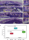
Induction of intercrypt goblet cells (icGCs) after infection of mouse distal colon by SGG. Representative distal colonic sections stained with Alcian Blue (AB) and periodic acid Schiff (PAS) to visualize acidic and neutral mucins from non-infected A/J mice (a), mice infected orally with SGG WT (b), Pil3+ (c) and ΔPil1/Pil3 (d). Only few icGCs are indicated (yellow arrows) but others are easily distinguishable from crypt goblet cells due to their small, typical rectangular shape. Quantification of the number of icGCs per mm, in colon of non-infected mice and mice infected by SGG WT, Pil3+ or ∆Pil1/Pil3 or SGM (e).
Figure 2.
Figure 2.
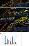
Formation of intercrypt goblet cells (icGCs) from differentiated goblet cells of the crypts. Representative distal colonic sections from mice infected by SGG Pil3+ (a, b) or ΔPil1/Pil3 (e) and from non-infected A/J mice (c, d), stained with Alcian Blue and Periodic Acid Schiff. The yellow panels are enlarged views of the boxed regions within the cross-sections. The rectangular shape of the icGCs is highlighted by green rectangular boxes whereas the globular shape of GCs is highlighted by orange circles.
Figure 3.
Figure 3.
Production of a dense, fully glycosylated, mucus layer by intercrypt goblet cells (icGCs). Representative immunofluorescence staining of mouse distal colonic tissue infected by SGG. Colon cross-sections were stained with DAPI to detect DNA (blue), anti Muc2 to visualize colonic mucus (red), and fluorescently conjugated lectins UEA-1 (a, c, e, g) or SNA (b, d, f, h) to visualize fucosylation and sialylation of mucus, respectively (green). Distal colonic sections from non-infected A/J mice (a-b), mice infected orally with SGG strains WT (c-d), Pil3+ (e-f) and ΔPil1/Pil3 (g-h). The yellow panels are enlarged views of the boxed regions within the cross-sections. Only few icGCs are indicated (white arrows) but others are easily distinguishable from crypt goblet cells due to their small, typical rectangular shape. FC: fecal content; PDM: partially degraded mucus; NSM: newly synthesized mucus. Relative percentage of neutral, sialylated, and sulfated O-glycans carried by mucins purified from distal colon of non-infected A/J mice, mice infected by SGG WT, Pil3+, ΔPil1/Pil3, and SGM, showing a modulation of mucin glycosylation upon bacterial infection (i). Data are expressed as mean ± SD; n = 6 per group. Differences in the level of expression of mucin O-glycosylation were analyzed using the student’s t-test, by comparing the non-infected group to the other ones. A P-value <0.01 was considered statistically significant.
Figure 4.
Figure 4.
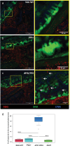
Production of a mucus impenetrable to bacteria by intercrypt goblet cells (icGcs). Representative immunofluorescence staining of mouse distal colonic tissue infected by SGG. Colon cross-section was stained with DAPI to detect DNA (blue), anti SGG (red) to visualize bacteria, and fluorescently FITC labeled SNA lectin to visualize mucus (green). Distal colonic sections from mice infected by SGG WT (a), Pil3+ (b) and ΔPil1/Pil3 (c) strains of SGG. The yellow panels are enlarged views of the boxed regions within the cross-sections. The enlarged view of Figure 4c indicates a subpopulation of ΔPil1/Pil3 mutant strains that have translocated and are localized in the mucosa and submucosa (white arrows). Quantification of the percentage of bacteria in the mucosa/submucosa compared to the percentage of bacteria in the mucus layer, in the distal colon of mice infected by SGG WT, Pil3+ or ∆Pil1/Pil3 (e) strains of SGG and SGM.

References

    1. Flint HJ, Scott KP, Louis P, Duncan SH.. The role of the gut microbiota in nutrition and health. Nat Rev Gastroenterol Hepatol. 2012;9(10):577–12. doi:10.1038/nrgastro.2012.156. - DOI - PubMed
    1. Fan Y, Pedersen O. Gut microbiota in human metabolic health and disease. Nat Rev Microbiol. 2021;19(1):55–71. doi:10.1038/s41579-020-0433-9. - DOI - PubMed
    1. Bergstrom K, Xia L. The barrier and beyond: roles of intestinal mucus and mucin-type O-glycosylation in resistance and tolerance defense strategies guiding host-microbe symbiosis. Gut Microbes. 2022;14(1):2052699. doi:10.1080/19490976.2022.2052699. - DOI - PMC - PubMed
    1. Zhao Q, Maynard CL. Mucus, commensals, and the immune system. Gut Microbes. 2022;14(1):2041342. doi:10.1080/19490976.2022.2041342. - DOI - PMC - PubMed
    1. Pothuraju R, Chaudhary S, Rachagani S, Kaur S, Roy HK, Bouvet M, Batra SK. Mucins, gut microbiota, and postbiotics role in colorectal cancer. Gut Microbes. 2021;13(1):1974795. doi:10.1080/19490976.2021.1974795. - DOI - PMC - PubMed

LinkOut - more resources