. 2024 Dec;36(52):e2408307.
doi: 10.1002/adma.202408307. Epub 2024 Nov 14.

Charge-Stabilized Nanodiscs as a New Class of Lipid Nanoparticles

Affiliations

Charge-Stabilized Nanodiscs as a New Class of Lipid Nanoparticles

Ivan S Pires et al. Adv Mater. 2024 Dec.

Abstract

Nanoparticles have the potential to improve disease treatment and diagnosis due to their ability to incorporate drugs, alter pharmacokinetics, and enable tissue targeting. While considerable effort is placed on developing spherical lipid-based nanocarriers, recent evidence suggests that high aspect ratio lipid nanocarriers can exhibit enhanced disease site targeting and altered cellular interactions. However, the assembly of lipid-based nanoparticles into non-spherical morphologies has typically required incorporating additional agents such as synthetic polymers, proteins, lipid-polymer conjugates, or detergents. Here, charged lipid headgroups are used to generate stable discoidal lipid nanoparticles from mixed micelles, which are termed charge-stabilized nanodiscs (CNDs). The ability to generate CNDs in buffers with physiological ionic strength is restricted to lipids with more than one anionic group, whereas monovalent lipids only generate small nanoliposomal assemblies. In mice, the smaller size and anisotropic shape of CNDs promote higher accumulation in subcutaneous tumors than spherical liposomes. Further, the surface chemistry of CNDs can be modified via layer-by-layer (LbL) assembly to improve their tumor-targeting properties over state-of-the-art LbL-liposomes when tested using a metastatic model of ovarian cancer. The application of charge-mediated anisotropy in lipid-based assemblies can aid in the future design of biomaterials and cell-membrane mimetic structures.

Keywords: anisotropy; layer‐by‐layer; lipid nanoparticles; nanodiscs; self‐assembly; tumor targeting.


Conflict of interest statement

I.S.P., P.T.H., and D.J.I. are inventors on a provisional patent filed by the Massachusetts Institute of Technology related to this work.

Figures

Figure 1
Figure 1
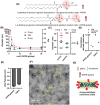
Charge‐stabilized nanodiscs (CND) with monovalent lipids are unstable in physiological ionic strength media. A) Schematic process for dilution of mixed micelles to generate discoidal lipid assemblies or liposomes. B) Lipid/detergent micelles (10 mg mL−1 of 6:3:1 molar mixture of DSPC:cholesterol:POPG in 10% MEGA‐10) in 10 mm HEPES buffer with different concentration of NaCl were diluted to indicated final MEGA‐10 concentrations and particle sizes (hydrodynamic Z‐avg) were assessed by DLS. C,D) CryoTEM micrographs of purified samples from dilution of lipid/detergent micelles using C) 200 mm NaCl and D) 0 mm NaCl. E) Intensity‐weighted diameter (Z‐avg) of PBS‐diluted lipid/detergent micelles containing 10 mg mL−1 of lipids in 10% MEGA‐10 composed of either 5:3:2, 4:3:3, or 3:3:4 molar ratios of DSPC:cholesterol:POPG. F) Representative cryoTEM micrograph of a 3:3:4 molar composition (DSPC:cholesterol:POPG) of mixed micelles allowed to equilibrate at 0.05% MEGA‐10 overnight then purified via TFF to remove MEGA‐10.
Figure 2
Figure 2
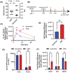
Charge density of anionic lipid DOPE‐glutaryl enables synthesis of CNDs stable at physiological ionic strength buffers. A) Chemical structure of POPG and DOPE‐glutaryl. B) DLS intensity weighted size (Z‐avg), number‐weighted average size (#‐avg), and polydispersity index (PDI) of particles assembled from varying compositions of DPSC, cholesterol, and DOPE‐glutaryl diluted in PBS to 0.1% MEGA‐10 concentrations (all samples contained 30 mol% cholesterol and the mol% indicated of DOPE‐glutaryl with the remainder being DSPC). C) DLS Z‐avg, #‐avg, and PDI of 10 mol% DOPE‐glutaryl CNDs before and after TFF purification. D) DLS count rate with or without polarized light filters. E) Zeta potential of CNDs composed with 10 mol% DOPE‐glutaryl compared to extrusion‐based liposomes with 10 mol% POPG (both samples contained 30% cholesterol and 60% DSPC). F) Representative negative stain TEM (NS‐TEM) micrograph of purified CNDs from (C). G) Proposed structure of CNDs composed of DOPE‐glutaryl, DSPC, and cholesterol.
Figure 3
Figure 3
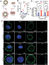
CNDs show greater tumor accumulation than liposomes in solid tumors. A) DLS Z‐avg, #‐avg, and PDI of purified CNDs composed with 10 mol% DOPE‐glutaryl compared to extrusion‐based liposomes with 10 mol% POPG (both samples contained 30% cholesterol and 60% DSPC). B) In vivo study timeline in which mice were inoculated subcutaneously with 106 MC38 cells and then dosed intravenously on day 7 with 1 nmol of cyanine‐5 labeled NPs (1 mol%). C) Tumor radiant efficiency measured in vivo via IVIS. D) Area under the curve (AUC) or data from (H). E) Serum fluorescence of CNDs and liposome‐dosed animals at 4 and 16 h after dosing. F) Recovered radiant efficiency from tumor, liver, and spleen 24 h after dosing mice with either CNDs or liposomes. Error bars represent SEM (n = 3). Statistical comparisons in (C,E,F) were performed using two‐way analysis of variance (ANOVA), with Tukey's multiple‐comparisons test and an unpaired two‐tailed t‐test was performed for D. Asterisks denote p‐values: **** p < 0.0001, *** p < 0.001, ** p < 0.01, * p < 0.05.
Figure 4
Figure 4
Deposition of polyelectrolyte layers composed of PLR and PLE onto CNDs enables improved association of CNDs with ovarian cancer cells in vitro. A) Schematic of LbL technique to generate LbL‐liposomes and LbL‐CNDs. B) Size and PDI of CND and liposomes before and after LbL modification. C) In vitro measurement of total HM‐1‐associated NP fluorescence relative to liposomes using a plate reader after 4 or 24 h of incubation. D) Confocal images of HM‐1 cells after 4 h of incubation with NPs. Error bars represent SEM. Statistical comparisons in C was performed using two‐way analysis of variance (ANOVA), with Tukey's multiple‐comparisons. Asterisks denote p‐values: **** p < 0.0001, *** p < 0.001, ** p < 0.01, * p < 0.05.
Figure 5
Figure 5
LbL‐CNDs NPs efficiently target metastatic ovarian cancer in vivo. A) In vivo timeline for treatment of fluorescently‐labeled NPs i.p. in ovarian cancer model. Mice were inoculated with 106 HM‐1‐luc cells i.p. and dosed with NPs 14 days later. B) Total radiant efficiency of NP fluorescence from peritoneum. C) AUC of peritoneal fluorescence readings from (B). D) Ex vivo weight normalized NP fluorescence in liver, spleen, urogenital tract (UGT), and omentum. E) Spearman's correlation coefficient between weight‐normalized NP fluorescence and weight‐normalized BLI readings. Error bars (s.e.m.) derived from parameter estimates for each group. F) Slope of linear fit between weight‐normalized NP fluorescence and weight‐normalized BLI readings. Error bars (s.e.m.) represent variation between each animal in respective treatment groups (n = 4 mice/group). Statistical comparisons in B and D were performed using two‐way analysis of variance (ANOVA) one‐way ANOVA was used in C and F with Tukey's multiple‐comparisons test. Spearman's correlation significance for E was performed based on a t‐test analysis with the null hypothesis of no (r = 0) correlation. Asterisks denote p‐values: **** p < 0.0001, *** p < 0.001, ** p < 0.01, * p < 0.05.

References

    1. Mundekkad D., Cho W. C., Int. J. Mol. Sci. 2022, 23, 1685. - PMC - PubMed
    1. Durymanov M., Kamaletdinova T., Lehmann S. E., Reineke J., J. Controlled Rel. 2017, 261, 10. - PubMed
    1. Albanese A., Tang P. S., Chan W. C. W., Annu. Rev. Biomed. Eng. 2012, 14, 1. - PubMed
    1. Zhu X., Vo C., Taylor M., Smith B. R., Mater. Horiz. 2019, 6, 1094.
    1. Ding J., Chen J., Gao L., Jiang Z., Zhang Y., Li M., Xiao Q., Lee S. S., Chen X., Nano Today 2019, 29, 100800.

LinkOut - more resources