Review
. 2024 Nov 14;22(1):710.
doi: 10.1186/s12951-024-02972-w.

Navigating the intricate in-vivo journey of lipid nanoparticles tailored for the targeted delivery of RNA therapeutics: a quality-by-design approach

Affiliations
Review

Navigating the intricate in-vivo journey of lipid nanoparticles tailored for the targeted delivery of RNA therapeutics: a quality-by-design approach

Elahe Haghighi et al. J Nanobiotechnology. .

Abstract

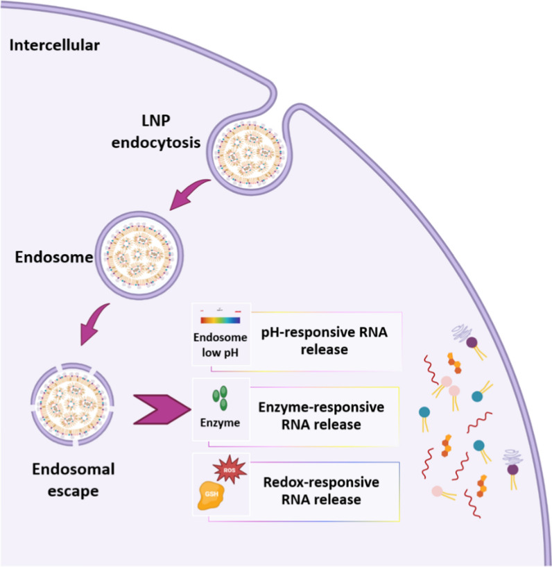
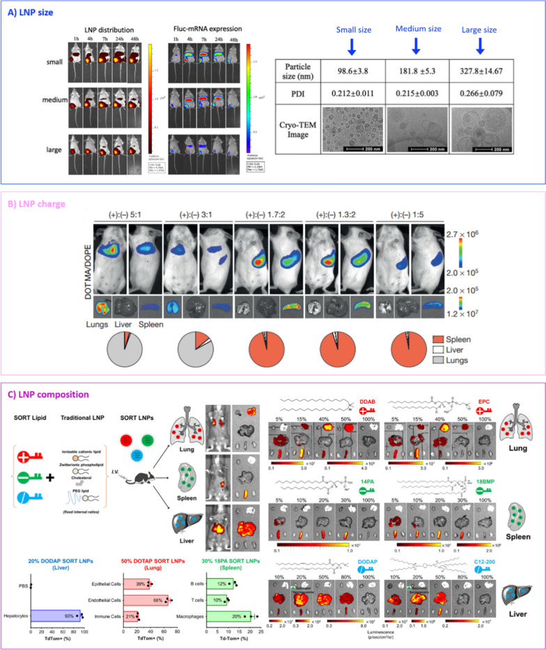
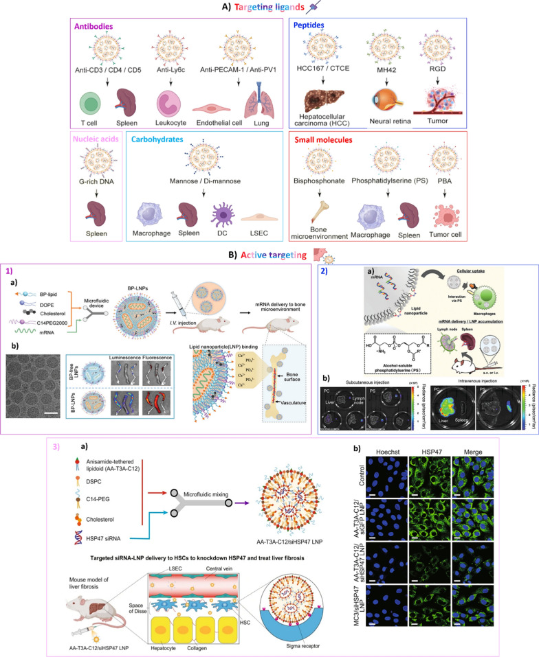
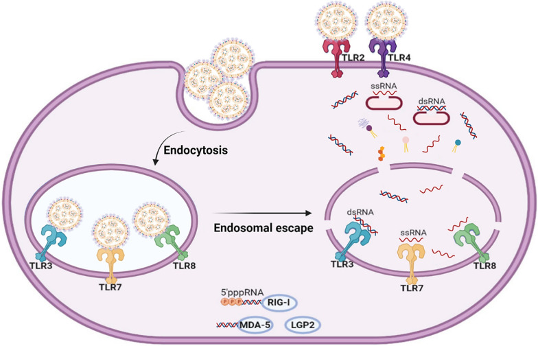
RNA therapeutics, such as mRNA, siRNA, and CRISPR-Cas9, present exciting avenues for treating diverse diseases. However, their potential is commonly hindered by vulnerability to degradation and poor cellular uptake, requiring effective delivery systems. Lipid nanoparticles (LNPs) have emerged as a leading choice for in vivo RNA delivery, offering protection against degradation, enhanced cellular uptake, and facilitation of endosomal escape. However, LNPs encounter numerous challenges for targeted RNA delivery in vivo, demanding advanced particle engineering, surface functionalization with targeting ligands, and a profound comprehension of the biological milieu in which they function. This review explores the structural and physicochemical characteristics of LNPs, in-vivo fate, and customization for RNA therapeutics. We highlight the quality-by-design (QbD) approach for targeted delivery beyond the liver, focusing on biodistribution, immunogenicity, and toxicity. In addition, we explored the current challenges and strategies associated with LNPs for in-vivo RNA delivery, such as ensuring repeated-dose efficacy, safety, and tissue-specific gene delivery. Furthermore, we provide insights into the current clinical applications in various classes of diseases and finally prospects of LNPs in RNA therapeutics.


Conflict of interest statement

Declarations Ethics approval and consent to participate Not applicable. Consent for publication All authors confirm that this work is original, has not been previously published, and is not under consideration for publication elsewhere. They have all consented to submitting this manuscript to the Journal of Nanobiotechnology. Competing interests The authors declare no competing interests.

Figures

Fig. 1
Fig. 1
Obstacles in the development of RNA therapeutics: A chemical modifications including alterations at the 5′ and 3′ ends (5′-capping, 3′-tail modifications, and 5′ and 3′-end conjugations), nucleotide modifications, ribose sugar substitutions at the 2′ position, and alterations in the phosphate backbone. B Nanocarrier delivery systems including lipid nanoparticle (LNP), lipoplex, polyplex, lipopolyplex, polymersome, polymeric micelle, exosome, and DNA nanostructures
Fig. 2
Fig. 2
Structure of lipid nanoparticle (LNP) encapsulating nucleic acids. A Compact nanostructured core. The LNPs exhibit a compact, electron-rich core where the siRNA is tangled with positively charged lipids forming inverted micelle structures, enclosed by cholesterol and helper lipids; B the multilamellar assembly features an external bilayer and a dense inner core; C ‘Bleb’ configuration. The cationic lipid predominantly interacts with the mRNA, creating a solid core, while the helper lipid is mainly partitioned into a ‘bleb’ bilayer (Adopted from [38])
Fig. 3
Fig. 3
Chemical compositions of lipid nanoparticles (LNPs): A ionizable cationic lipids (ICLs); B helper lipids; C cholesterol and its derivatives; D PEG-lipids
Fig. 4
Fig. 4
Schematic representation of various lipid nanoparticle (LNP) fabrication techniques: A bulk mixing methods including thin-film hydration, solvent injection, bulk nanoprecipitation, and melting emulsification; B microfluidic methods utilizing distinct mixer designs such as T-junction, HFF, staggered herringbone, ring-type, Tesla, and baffle mixer
Fig. 5
Fig. 5
Fundamental procedures for applying the quality-by-design (QbD) approach in developing lipid nanoparticles (LNPs) which includes quality target product profile (QTPP), critical quality attributes (CQAs), critical process parameters (CPPs) and critical material attributes (CMAs). In the QbD approach by using statistical, analytical, and risk-management methodologies optimized products can be designed and developed
Fig. 6
Fig. 6
Summary of common labeling and identification techniques for LNPs, RNAs, and protein products. Lipid nanoparticles (LNPs) are labeled with fluorescent and radioisotope tags for imaging purposes (1 and 2). LNP components are analyzed using mass spectroscopy (MS) (3). Specific nucleic acid sequences are detected using reverse transcription-quantitative PCR (RT-qPCR) (4). Reporter gene expressions, such as enhanced green fluorescent protein (eGFP) and luciferase (LUC), are visualized through fluorescent and bioluminescent imaging (5). Immunohistochemistry (IHC) techniques identify proteins produced by mRNA-LNP (6) or suppressed by siRNA-LNP. Fluorescence in situ hybridization (FISH) probes bind to specific nucleic acid sequences within cells, enabling their visualization (7)
Fig. 7
Fig. 7
Natural targeting of various cells by LNP-RNA therapeutics influenced by PEG-lipid shedding and protein corona composition. ApoE, high-density lipoprotein (HDL), vitronectin, and albumin can naturally target LNPs to the liver parenchyma, lymphatic vessel, and tumor and breast cancer cells, respectively
Fig. 8
Fig. 8
Illustration of endosomal escape and stimuli-responsive RNA release mechanisms; pH-, enzyme-, and redox-responsive RNA release can be achieved following LNP endocytosis
Fig. 9
Fig. 9
Factors affecting lipid nanoparticle (LNP) passive targeting: A Fluorescence and bioluminescence biodistribution patterns of 1,1′-dioctadecyl-3,3,3′,3′-tetramethylindotricarbocyanine iodide (DiR)-tagged Fluc-mRNA LNPs varying in size and the corresponding luciferase expression after intramuscular administration [235]; B biodistribution of Luc-LNP at various charge ratios in BALB/c mice 24 h after intravenous administration [238]; C incorporating an additional element, known as a selective organ targeting (SORT) molecule, into conventional LNPs modifies their distribution within the body and facilitates targeted delivery to specific tissues [128]
Fig. 10
Fig. 10
A Schematic representation of common targeting ligands used for specific LNP-RNA delivery; B examples of recent LNP surface modification for active targeting: 1 bisphosphonate (BP) lipid-like component for mRNA delivery to the bone microenvironment: a illustration depicting the construction of BP-LNPs for mRNA delivery to the bone environment, leveraging the interaction of BP-LNPs with Ca2+; b Cryo-TEM image showing BP-LNP morphology (100 nm scale bar) and ex-vivo imaging of bones post-LNP delivery. LNPs encapsulate luciferase (Luc) mRNA and are labeled with 1′-dioctadecyl-3,3,3′,3′-tetramethylindotricarbocyanine iodide (DiR) dye [280]; 2 phosphatidylserine-loaded LNP for delivering mRNA to secondary lymphoid tissues: a illustration outlining the preparation of phosphatidylserine LNPs and strategies for mRNA delivery to macrophages in secondary lymphoid organs; b images of Luc activity in tissues from mice treated with subcutaneous and intravenous injections of phosphatidylserine-LNPs encapsulating Luc mRNA [302]; 3 ligand-tethered lipid nanoparticles for RNA delivery to treat liver fibrosis: a fabrication of AA-T3A-C12/siHSP47 LNP by microfluidic mixing and specific delivery to activated hepatic stellate cells (HSCs) for silencing heat shock protein 47 (HSP47) in liver fibrosis; b immunofluorescence staining of HSP47 in LNP-treated activated 3T3 fibroblasts [295]
Fig. 11
Fig. 11
Pattern recognition receptors (PRRs) identify both mRNA and lipid nanoparticles (LNPs) extracellularly and intracellularly via toll-like receptors (TLRs), RIG-I-like receptors (RLRs), and NOD-like receptors (NLRs)
Fig. 12
Fig. 12
Activation of innate and adaptive immune cells upon mRNA-LNP administration: A intramuscular injection of mRNA-LNP vaccines leads to local inflammation, recruiting neutrophils, monocytes, and dendritic cells (DCs) from the bloodstream to the site of injection through the release of chemokines and cytokines, facilitating the migration of other immune cells; B mRNA-LNPs alone, or along with antigen-presenting cells (APCs), are localized in the nearby lymph nodes; C DCs, monocytes or macrophages present antigens and initiate the activation of T cells; D Tfh cells assist B cells during germinal center (GC) reactions, alongside follicular DCs, to refine antibody affinity. In mouse models, LNP-induced IL-6 is crucial for developing T follicular helper (Tfh) and GC B cell responses, while type I IFNs are known to enhance cytotoxic T lymphocyte (CTL) reactions Adopted from [419]

Similar articles

Cited by

References

    1. Paunovska K, Loughrey D, Dahlman JE. Drug delivery systems for RNA therapeutics. Nat Rev Genet. 2022;23:265–80. 10.1038/s41576-021-00439-4. - PMC - PubMed
    1. Feng R, Patil S, Zhao X, Miao Z, Qian A. RNA therapeutics—research and clinical advancements. Front Mol Biosci. 2021;8:1–13. 10.3389/fmolb.2021.710738. - PMC - PubMed
    1. Zhang C, Zhang B. RNA therapeutics: updates and future potential. Sci China Life Sci. 2023;66:12–30. 10.1007/S11427-022-2171-2. - PMC - PubMed
    1. Burrows C, Helm M, Zhou X. Modulating RNA with chemical modifications. Acc Chem Res. 2024;57:175–6. 10.1021/acs.accounts.3c00770. - PubMed
    1. Jiao L, Sun Z, Sun Z, Liu J, Deng G, Wang X. Nanotechnology-based non-viral vectors for gene delivery in cardiovascular diseases. Front Bioeng Biotechnol. 2024;12:1349077. 10.3389/fbioe.2024.1349077. - PMC - PubMed

LinkOut - more resources