Review
. 2024 Nov 14;23(1):254.
doi: 10.1186/s12943-024-02171-z.

Immune dynamics shaping pre-metastatic and metastatic niches in liver metastases: from molecular mechanisms to therapeutic strategies

Affiliations
Review

Immune dynamics shaping pre-metastatic and metastatic niches in liver metastases: from molecular mechanisms to therapeutic strategies

Chang Zhu et al. Mol Cancer. .

Abstract

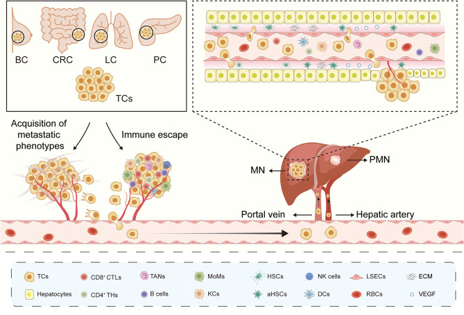
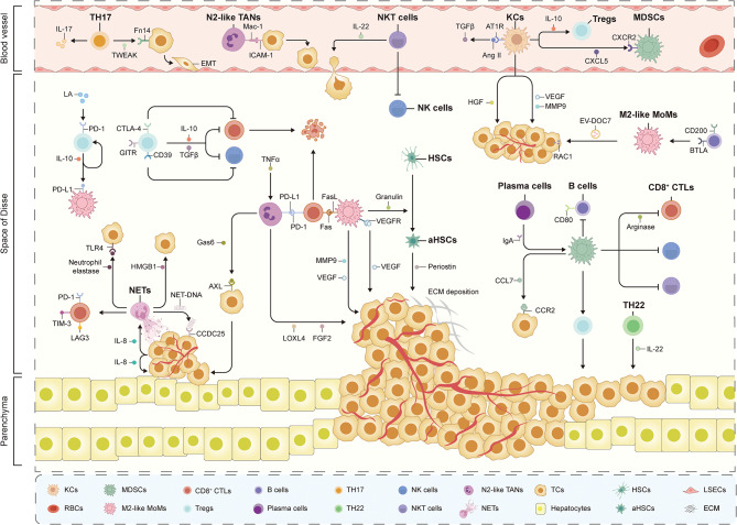
Liver metastases are commonly detected in the advanced stages of various malignant tumors, representing a significant clinical challenge. Throughout the process of liver metastases formation, immune cells play a pivotal role, particularly in the pre-metastatic and metastatic niches within the liver. Immune cells establish extensive and intricate interactions with tumor cells and other components in the liver, collectively promoting and sustaining the growth of liver metastases. Despite the limited efficacy of existing therapeutic modalities against some advanced liver metastases, novel immune-based treatment approaches are continuously being explored and validated. Building on the systematic elucidation of the immunosuppressive characteristics of liver metastases, we explored the potential of novel immunotherapies applicable to patients with liver metastases from multiple dimensions.

Keywords: Immunotherapy; Liver metastases; Tumor immunology; Tumor microenvironment.


Conflict of interest statement

Declarations Ethical approval Not applicable. Competing interests The authors declare no competing interests.

Figures

Fig. 1
Fig. 1
Process of liver metastases formation. The schematic diagram illustrates the general process of metastasis of tumors from primary sites to the liver. Prior to metastasis, essential preparatory steps include the acquisition of the metastatic phenotypes and the achievement of immune escape of in situ tumor cells (TCs), as well as the formation of a pre-metastatic niche (PMN) in the liver. Subsequently, TCs enter the circulation and access the rich bloodstream of the liver through major vessels such as the portal vein and hepatic artery. TCs adhere to and traverse through liver sinusoidal endothelial cells (LSECs) upon entering the liver sinusoids, finally settling and growing based on the hepatic parenchyma, forming liver metastases (LM) in the liver metastatic niche (MN). Immune cells participate extensively throughout the whole dynamic process. BC, breast cancer; CRC, colorectal cancer; LC, lung cancer; PC, pancreatic cancer; CTLs, cytotoxic T cells; THs, helper T cells; TANs, tumor-associated neutrophils; MoMs, monocyte-derived macrophages; KCs, Kupffer cells; HSCs, hepatic stellate cells; aHSCs, activated hepatic stellate cells; NK cells, natural killer cells; DCs, dendritic cells; RBCs, red blood cells; ECM, extracellular matrix; VEGF, vascular endothelial growth factor
Fig. 2
Fig. 2
Immune involvement in liver pre-metastatic niche formation. Prior to the development of liver metastases (LM), exosomes and secretory factors derived from primary tumors exert a distant influence on the liver, modifying its immune characteristics in anticipation of the arrival of tumor cells (TCs). As illustrated in the diagram, MIF (migration inhibitory factor), lipopolysaccharide binding protein (LBP) and exosomal microRNA-21-5p (miR-21-5p), miR-151a-3p, miR-135a-5p, and integrin αvβ5 from TCs act on liver resident Kupffer cells (KCs), promoting high secretion of transforming growth factor beta (TGFβ) and interleukin-6 (IL-6), environmental fibrosis, and subsequent changes in the stemness and adhesion characteristics of TCs upon arrival. Exosomal angiopoietin-like protein 1 (ANGPTL1) expression, which has the potential to inhibit vascular leakage by reducing matrix metalloproteinase 9 (MMP9) expression in KCs, is observed to decrease prior to the onset of metastasis. The M2-like polarization of recruited monocyte-derived macrophages (MoMs) in the liver is activated by exosomal miR-934, miR-519a-3p, circ-0034880 (circular RNA-0034880) and secretory glucose-regulated protein 78 (sGRP78). Molecules such as tissue inhibitor of metalloproteinases-1 (TIMP-1), tRF-GluCTC-0005 (transfer RNA-derived fragment), TGFβ, IL-6, and vascular endothelial growth factor A (VEGFA) from the primary tumors are crucial for initiating the pre-recruitment of neutrophils and myeloid-derived suppressor cells (MDSCs) from the bone marrow. PMN, pre-metastatic niche; TLR, Toll-like receptor; NF-κB, nuclear factor kappa B; SMAD, mothers against decapentaplegic; HSCs, hepatic stellate cells; aHSCs, activated hepatic stellate cells; S1PR1, sphingosine-1-phosphate receptor 1; STAT3, signal transducer and activator of transcription 3; DCs, dendritic cells; HSCs, hepatic stellate cells; aHSCs, activated hepatic stellate cells; CXCL1, CXC chemokine ligand 1
Fig. 3
Fig. 3
Prometastatic immune landscape in the liver metastatic niche. As illustrated in the diagram, a multitude of pro-metastatic immune cells collectively facilitate the growth of tumor cells (TCs) in the liver metastatic niche (MN). M2-like recruited monocyte-derived macrophages (MoMs) play a pivotal role as pro-metastatic members. In addition to activating hepatic stellate cells (HSCs) to create a fibrotic microenvironment for TCs, they also induce apoptosis of cytotoxic T cells (CTLs) through high expression of FAS ligand (FasL). Furthermore, M2-like MoMs promote angiogenesis by secreting vascular endothelial growth factor (VEGF) and matrix metalloproteinase 9 (MMP9). Additionally, M2-like MoMs secrete DOCK7-enriched extracellular vesicles (EVs), which activate the oncogene RAC1 in tumor cells (TCs), directly exerting pro-tumor effects. In the late stages of metastasis, Kupffer cells (KCs) promote the expansion of regulatory T cells (Tregs) and myeloid-derived suppressor cells (MDSCs) respectively by producing interleukin-10 (IL-10) and CXC chemokine ligand 5 (CXCL5) and promote TCs proliferation by secreting transforming growth factor beta (TGFβ), hepatocyte growth factor (HGF), VEGF and MMP9. N2-like tumor-associated neutrophils (TANs) express high levels of programmed cell death 1 ligand 1 (PD-L1), which directly inhibits CTLs activation. Additionally, they secrete growth arrest specific 6 (Gas6) and Mac-1, which respectively promote TCs regeneration and adhesion to liver sinusoidal endothelial cells (LSECs). Neutrophil extracellular traps (NETs) derived from N2-like TANs capture TCs to promote their settlement. The effective protein components within NETs, such as high mobility group box 1 (HMGB1), and DNA components, such as coiled-coil domain containing 25 (CCDC25), each exert specific pro-carcinogenic effects. MDSCs have been reported to interact extensively with various lymphocytes including Tregs, CTLs, natural killer (NK) cells, natural killer T (NKT) cells, and B cells, participating in the shaping of the immunosuppressive microenvironment at multiple levels. Tregs inhibit the cytolytic activity of CTLs and NK cells by expressing IL-10, TGFβ, cytotoxic T-lymphocyte-associated protein 4 (CTLA-4), programmed cell death protein 1 (PD-1) and CD39. Furthermore, helper T cells 17 (TH17), TH22, B cells and NKT cells have also been shown to possess immunosuppressive phenotypes that promote liver metastases (LM). Plasma cells differentiated from B cells secrete specific IgA antibodies, which induce MDSCs activation. RBCs, red blood cells; ECM, extracellular matrix; ICAM-1, intercellular adhesion molecule 1; Ang II, angiotensin II; AT1R, angiotensin II receptor type I; LA, lactic acid; GITR, glucocorticoid-induced tumor necrosis factor receptor; TLR, Toll-like receptor; TIM-3, T-cell immunoglobulin and mucin domain-3; LAG3, lymphocyte-activation gene 3; TNFα, tumor necrosis factor α; LOXL4, lysyl oxidase-like 4; FGF2, fibroblast growth factor 2; VEGFR, vascular endothelial growth factor receptor; CCL, CC chemokine ligand; CCR, CC chemokine receptor
Fig. 4
Fig. 4
Antimetastatic immune landscape in the liver metastatic niche. Metastatic tumor cells (TCs) arriving in the liver are subjected to extensive immune surveillance, coordinated by both innate and adaptive immune cells. M1-like recruited monocyte-derived macrophages (MoMs) enhance the cytolytic ability of natural killer (NK) cells by secreting interleukin-18 (IL-18) and IL-1β. Kupffer cells (KCs) express Dectin-2 and IgG-binding Fc receptor (FcγR) in the early stages of metastasis, which, in combination with IgG, exert antibody-dependent phagocytosis (ADPh). Additionally, KCs can directly kill TCs by releasing nitric oxide (NO) and tumor necrosis factor α (TNFα), or activate NK cells by releasing IL-12. N1-like tumor-associated neutrophils (TANs) can also promote TCs death by releasing NO, TNFα and reactive oxygen species (ROS). Cytotoxic T cells (CTLs) and NK cells are the main executors of adaptive immune responses against liver metastases (LM). They exert anti-tumor effects directly or indirectly by releasing perforin or granzyme to induce tumor cell lysis or apoptosis, or by releasing cytokines such as interferon-gamma (IFN-γ). The CD4 + helper T cells (THs) subset TH1 promotes the activation of CTLs and KCs, respectively, by secreting IL-2 and IFN-γ. Another THs subset, follicular helper T cells (Tfh), contributes to the formation of tertiary lymphoid structures (TLSs), which have been preliminarily shown to have anti-LM potential. Furthermore, intra-tumoral CD45RO + CD103 + CD8 + memory T cells (Tms) may also contribute to LM suppression. Natural killer T (NKT) cells present in the hepatic sinusoids can enhance the immune activity of NK cells and CTLs, and reduce the infiltration of M2-like MoMs. BRCA1, breast cancer type 1 susceptibility protein; DCs, dendritic cells; MHC-I, major histocompatibility complex class I molecules; FasL, FAS ligand; CTSB, cathepsin B; aHSCs, activated hepatic stellate cells; LSECs, liver sinusoidal endothelial cells; RBCs, red blood cells
Fig. 5
Fig. 5
Factors mediating immunosuppression in liver metastases. Multiple factors from tumor cells (TCs) and the liver metastatic niche (MN) contribute to the remodelling of the immune microenvironment in the liver, rendering it more conducive to the survival and sustained growth of liver metastases (LM). The recruitment and polarization of myeloid cells towards a pro-carcinogenic phenotype represent pivotal steps in reshaping the hepatic immune landscape. TCs secrete molecules including CC chemokine ligand 2 (CCL2), complement 5a (C5a) and interleukin-1α (IL-1α) to promote the recruitment of CCR2 + C5aR + monocyte-derived macrophages (MoMs) and secrete molecules including transcription factor 4 (TCF4) and CXC chemokine ligand 12 (CXCL12) to polarize them into M2-like MoMs. Notably, MoMs are also recruited in response to MLKL (mixed lineage kinase domain-like pseudo-kinase)-driven necroptosis of TCs. Similarly, the recruitment of tumor-associated neutrophils (TANs) is regulated by TCs-derived cytokines such as CXCL1, CXCL2 and granulocyte colony-stimulating factor (G-CSF), as well as environmental factors such as KIAA1199 and androgens. Transforming growth factor beta (TGFβ) and cyclic adenosine monophosphate (cAMP) are involved in the N2-like polarization of TANs. The high expression of CD47 on the surface of TCs leads to impaired late-stage phagocytic function of Kupffer cells (KCs), which is characterized by high expression of intracellular MAF bZIP transcription factor B (MafB) and c-Maf. The aggregation and reprogramming of myeloid-derived suppressor cells (MDSCs) are regulated by phosphorylated signal transducer and activator of transcription 3 (pSTAT3) and estrogen. At the level of lymphocytes, the liver MN extensively suppresses the density and activation levels of natural killer (NK) cells and CD8 + cytotoxic T cells (CTLs), and hinders the acquisition of TH1-related phenotypes by helper T cells (THs). Additionally, TCs secrete IL-2 and IL-8 to attract the aggregation and activation of regulatory T cells (Tregs), secrete SDF-1 to promote the migration of activated B cells, and secrete CCL28 to recruit IgA + plasma cells. CCR2, CC chemokine receptor 2; C5aR, complement 5a receptor; uPA, urokinase-type plasminogen activator; CARD9, caspase recruitment domain-containing protein 9; CXCR, CXC chemokine receptor; MRC1, mannose receptor C-type 1; LCFA, long-chain fatty acids; CTHRC1, collagen triple helix repeat containing 1; TNFR2, tumor necrosis factor receptor 2; AR, androgen receptor; LA, lactic acid; HLA-E, major histocompatibility complex class I E; PD-1, programmed cell death protein 1; PD-L1, programmed cell death 1 ligand 1; CHSY1, chondroitin sulfate synthase 1; MGP, matrix Gla protein; TIM-3, T-cell immunoglobulin and mucin domain-3
Fig. 6
Fig. 6
Immune-based therapies for liver metastases. Shown are several major options of immune-based therapies for liver metastases (LM). CCL, CC chemokine ligand; CCR, CC chemokine receptor; M-CSF, macrophage colony-stimulating factor; CSF-1R, colony stimulating factor-1 receptor; CTLA-4, cytotoxic T-lymphocyte-associated protein 4; LAG3, lymphocyte-activation gene 3; TGFβ, transforming growth factor beta; Tregs, regulatory T cells; CAR-T cells, chimeric antigen receptor T cells; TC, tumor cell; DC, dendritic cell

References

    1. Tsilimigras DI, Brodt P, Clavien PA, Muschel RJ, D’Angelica MI, Endo I, Parks RW, Doyle M, de Santibanes E, Pawlik TM. Liver metastases. Nat Rev Dis Primers. 2021;7:27. - PubMed
    1. Ciner AT, Jones K, Muschel RJ, Brodt P. The unique immune microenvironment of liver metastases: challenges and opportunities. Semin Cancer Biol. 2021;71:143–56. - PubMed
    1. Gerstberger S, Jiang Q, Ganesh K. Metastasis. Cell. 2023;186:1564–79. - PMC - PubMed
    1. Shin AE, Giancotti FG, Rustgi AK. Metastatic colorectal cancer: mechanisms and emerging therapeutics. Trends Pharmacol Sci. 2023;44:222–36. - PMC - PubMed
    1. Cervantes A, Adam R, Roselló S, Arnold D, Normanno N, Taïeb J, Seligmann J, De Baere T, Osterlund P, Yoshino T, Martinelli E. Metastatic colorectal cancer: ESMO Clinical Practice Guideline for diagnosis, treatment and follow-up. Ann Oncol. 2023;34:10–32. - PubMed

LinkOut - more resources