. 2024 Nov 14;15(1):428.
doi: 10.1186/s13287-024-04015-5.

Endothelial progenitor cell-derived conditioned medium mitigates chronic cerebral ischemic injury through macrophage migration inhibitory factor-activated AKT pathway

Affiliations

Endothelial progenitor cell-derived conditioned medium mitigates chronic cerebral ischemic injury through macrophage migration inhibitory factor-activated AKT pathway

Ya-Wen Cheng et al. Stem Cell Res Ther. .

Abstract

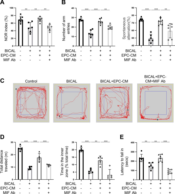
Background: Chronic cerebral ischemia (CCI) is a significant health issue characterized by hypoperfusion due to damage or occlusion of the cerebral or carotid arteries. CCI may lead to progressive cognitive impairment that is considered as a prelude to neurodegenerative diseases, including dementia and Alzheimer's disease (AD). Endothelial progenitor cells (EPCs) have been implicated in vascular repair in ischemic cerebrovascular diseases, primarily by differentiating into endothelial cells (ECs) or through paracrine effects. However, the clinical transplantation of stem cell therapies remains limited. In this study, we investigated the effects of EPC-derived conditioned medium (EPC-CM) on the impaired vasculature and neurological function in a rodent model of CCI and the mechanism involved.

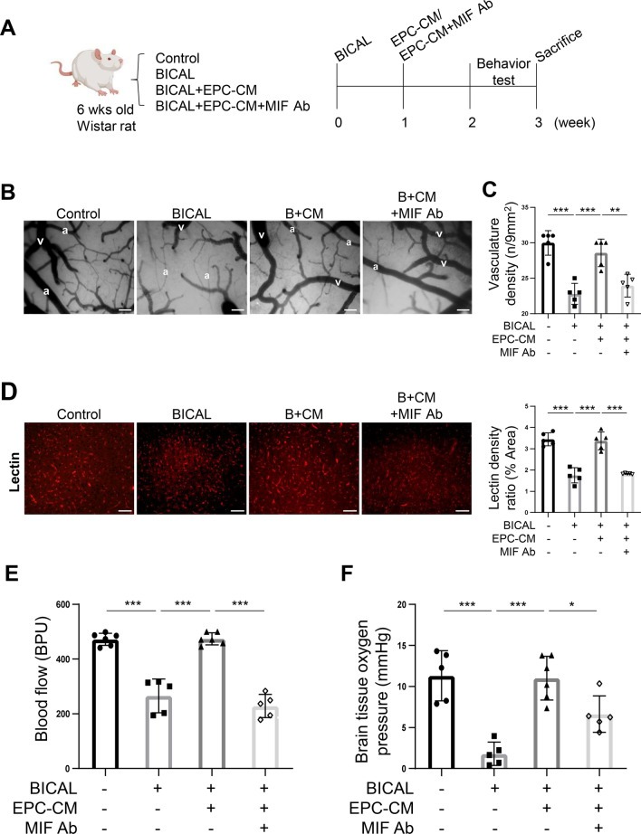
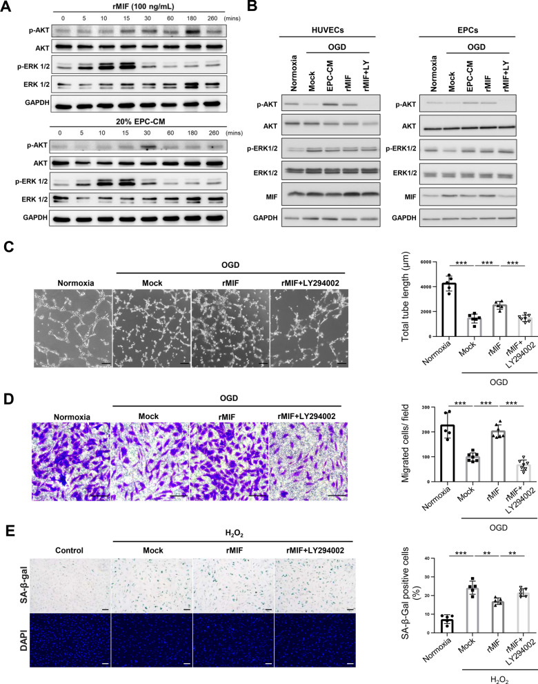
Methods: EPC-CM was analyzed by cytokine array to identify key factors involved in angiogenesis and cellular senescence. The effects and mechanism of the candidate factors in the EPC-CM were validated in vitro using oxygen-glucose deprivation (OGD)-injured ECs and EPCs. The therapeutic effects of EPC-CM and the identified key factor were further examined in a rat model of CCI, which was induced by bilateral internal carotid artery ligation (BICAL). EPC-CM was administered via intracisternal injection one week post BICAL. The cerebral microvasculature and neurobehavior of the rats were examined three weeks after BICAL.

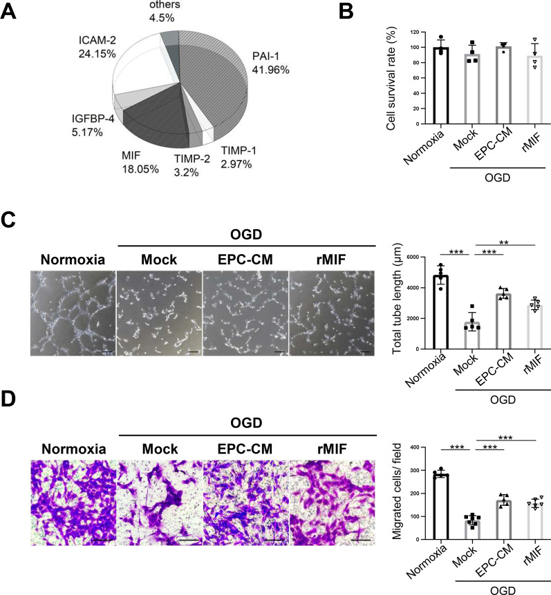
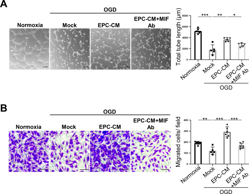
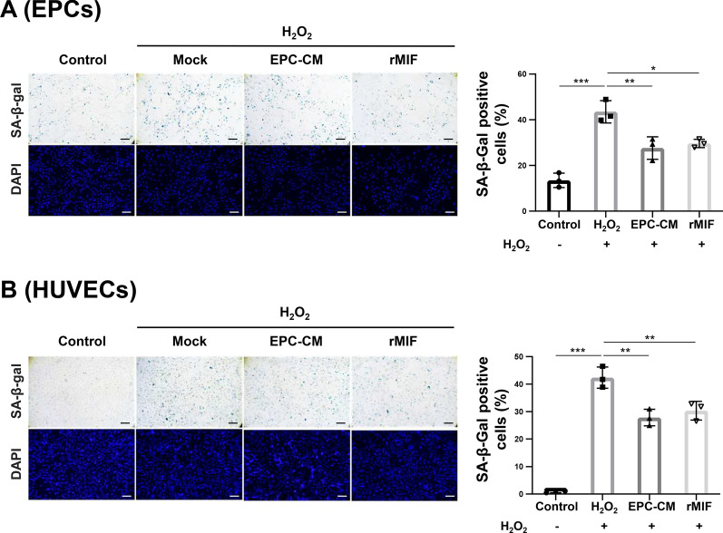
Results: Macrophage migration inhibitory factor (MIF) was identified as a key factor in the EPC-CM. Recombinant MIF protein promoted angiogenesis and prevented senescence in the injured EPCs and ECs. The effect was similar to that of the EPC-CM. These therapeutic effects were diminished when the EPC-CM was co-treated with MIF-specific antibody (Ab). Additionally, the vascular, motor, and cognitive improvements observed in the BICAL rats treated with EPC-CM were abolished by co-treated with MIF Ab. Furthermore, we found MIF promoted angiogenesis and anti-senescence via activating the AKT pathway. Inhibition of the AKT pathway diminished the protective effects of MIF in the in vitro study.

Conclusions: We demonstrated that EPC-CM protected the brain from chronic ischemic injury and promoted functional recovery through MIF-mediated AKT pathway. These findings suggest EPC-CM holds potential as a novel cell-free therapeutic approach for treating CCI through the actions of MIF.

Keywords: AKT pathway; Cerebral ischemia; Endothelial progenitor cell-derived conditioned medium; Macrophage migration inhibitory factor.


Conflict of interest statement

Declarations Ethics approval and consent to participate This animal experiments were conducted in accordance with the ARRIVE guidelines (Animal Research: Reporting of In Vivo Experiments) and was approved by the Institutional Animal Care and Use Committee of National Taiwan University (Approval no. IACUC-20180430, date of approval: 08/01/2019). The EPCs were collected from fresh human umbilical cord blood and was approved by the Institutional Review Board of National Taiwan University Hospital, Taipei, Taiwan (Study title: Platform establishment, validation, and biomarkers study in cerebrovascular diseases: using pediatric moyamoya disease as a pilot. Approval no. IRB-201204074RIC, date of approval: 07/04/2012; Study title: The pathophysiology of indirect revascularization in chronic cerebral ischemia-induced tauopathy: an in vitro, in vivo and clinical study. Approval no. IRB-202012174RIND, date of approval: 01/19/2021). All patients gave written informed consent for participation in the study and the use of samples. Consent for publication All the authors approved this manuscript to be published in the Journal Stem Cell Research & Therapy. Competing interests The authors declare no competing interests.

Figures

Fig. 1
Fig. 1
EPC-CM and MIF protein promote the angiogenesis ability in OGD-treated ECs. A Cytokine profile in the EPC-CM was determined using Quantitative Cytokine Quantibody Human Array 4000 (RayBiotech, Norcross, GA). B The cell survival ratio of the OGD-treated HUVECs having treated with different compounds (Normoxia: 20% basal medium, OGD/Mock: 20% basal medium, EPC-CM: 20% EPC-CM, rMIF: 20% basal medium with 100 ng/ml rMIF) was detected by CCK-8 assay and normalized with normoxic group. C Left panel, representative images of the tube formation in HUVECs with different treatments described in (B). Right panel, the average total length of tubes was quantitatively analyzed by 5 fields randomly. Scale bar = 100 μm. D Representative images of the crystal violet staining of the cells passed through the transwell membrane after different treatments described in (B) (left panel). Scale bar = 100 μm. The migrated cell numbers in each group were quantitatively analyzed by 6 fields randomly (right panel). The data was shown as the mean ± SD and analyzed by One-way ANOVA. ** p < 0.01, *** p < 0.001
Fig. 2
Fig. 2
MIF Ab abolishes the enhancement of angiogenesis by EPC-CM in vitro. A Representative images of tube formation affected by each condition was examined by Matrigel assay (Normoxia: 20% basal medium, OGD/Mock: 20% basal medium, EPC-CM: 20% EPC-CM, MIF Ab: 20% basal medium with 1 µg/ml MIF Ab) (left panel). The average of total tube length in 5 random fields was quantitatively analyzed by MetaMorph software and shown as bar graph (right panel). Scale bar = 100 μm. B Determination of the HUVECs treated with different conditions described in (A) passed through the transwell membrane stained with the crystal violet (left panel). The migrated cell numbers were quantitatively analyzed by 6 fields randomly (right panel). Scale bar = 100 μm. The data was shown as the mean ± SD and analyzed by One-way ANOVA. * p < 0.05, ** p < 0.01, *** p < 0.001
Fig. 3
Fig. 3
EPC-CM and MIF protein protect EPCs and ECs against the H2O2-induced senescence. A and B Representative micrographs of SA-β-gal staining and DAPI staining of H2O2-treated young EPCs (A) or the HUVECs (B) (passage < 10) that treated with/without EPC-CM or recombinant MIF protein (Control: 20% basal medium, H2O2/Mock: 20% basal medium, EPC-CM: 20% EPC-CM, rMIF: 20% basal medium with 100 ng/ml rMIF) (left panel). The senescence ratio was quantitatively analyzed of the SA-β-gal positive cells by 5 independent fields and normalized with DAPI staining. The data was compared with H2O2 group (right panel). Scale bar = 100 μm. The data was shown as the mean ± SD and analyzed by One-way ANOVA. * p < 0.05, ** p < 0.01, *** p < 0.001
Fig. 4
Fig. 4
MIF is the key factor in the EPC-CM to promote the vascular repair after BICAL. A The schematic representation of the experimental process. B The representative micrographs of microcirculation on the brain surface recorded by the videoscope (arteriole marketed as “a” and vein labeled as “v”; B + CM: BICAL + EPC-CM). Scale bar = 50 µm. C The effects of EPC-CM and MIF Ab on the cerebral microvasculature density of BICAL rats were quantified by using a CAM1 capillary anemometer (n = 5). D Representative images of immunostaining of LE-lectin in cortex of different treatments of Wistar rats (left panel, B + CM: BICAL + EPC-CM). Scale bar = 50 μm. The density of LE-lectin was quantified by using ImageJ (right panel). Each symbol represents the average density of 5 independent cortical fields per rat (n = 5). E and F On BICAL rats, the vascular repair effects of EPC-CM and MIF Ab were evaluated by regional blood flow (E) and the partial pressure of brain tissue oxygen (PbtO2) determination (n = 5) (F). The data are presented as mean ± SD, and analyzed using One-way ANOVA. * p < 0.05, ** p < 0.01, *** p < 0.001
Fig. 5
Fig. 5
MIF plays an important role in EPC-CM-mediated repair of the cognitive and motor impairments after BICAL. A The memory function of BICAL rats was evaluated by the NOR test. The duration of time spent exploring the novel object was normalized with total time spent exploring both objects (n = 6). B The spatial working memory of BICAL rats was evaluated by Y-maze, including the numbers of total arm entries (left panel) and the percentage of spontaneous alternation (%) (right panel) (n = 6). C Representative track plots showed the paths of control and BICAL rats treated with/without EPC-CM or MIF Ab in the OFT over a 5 min duration. D The total distance traveled indicated the locomotor activity of each group (left panel). The anxiety-related behaviors were evaluated by the time spent in the center area (% of total time, right panel) in each group (n = 5). E The effects of EPC-CM or EPC-CM + MIF Ab treatment on the motor function after BICAL were evaluated by rotarod test (n = 6). The data was shown as the mean ± SD and analyzed by One-way ANOVA. * p < 0.05, ** p < 0.01, *** p < 0.001
Fig. 6
Fig. 6
MIF promotes the angiogenesis and anti-senescence via activating the AKT pathway. A Determination the AKT and ERK1/2 activity upon the treatment of EPC-CM or recombinant MIF protein in different time points in the EPCs. The EPCs were treated with rMIF (100 ng/ml) or EPC-CM (20%) for the indicated time and harvested the cell lysates for Western blotting analysis. Full-length blots/gels are presented in Additional file 1: Fig. S4. B Detection of the AKT and ERK1/2 activity in OGD-treated cells incubated with EPC-CM, or rMIF, or rMIF + LY (LY294002, 5 µM). 90% confluency HUVECs (left panel) or EPCs (right panel) were incubated with 5% FBS EGM-2 with/without EPC-CM (20%), or rMIF (100 ng/ml), or the rMIF + LY294002 (5 µM) at 1% O2 incubator for 24 h. The cell lysates were collected to detect the AKT and ERK1/2 activity by using the Western analysis. Full-length blots/gels are presented in Additional file 1: Fig. S5. C Representative images of the tube formation of the OGD-treated cells incubated with different chemicals mentioned in (B) (left panel). The average total tube length of five random fields was quantified and showed as bar graph (right panel). Scale bar = 100 μm. D Representative images of the HUVECs passed through the transwell membrane stained with crystal violet (left panel). The migrated cell numbers were quantitative analyzed by 6 fields randomly (right panel). Scale bar = 100 μm. E Images of the SA-β-gal staining of H2O2-induced senescent cells treated with different chemicals in HUVEC cells. Young HUVECs (passage < 10) treated H2O2 (200 µM) for two days, followed with rMIF, or rMIF + LY294002 (5 µM) treatment for three days (left panel). The senescence ratio was the SA-β-gal positive cells normalized with the DAPI in 6 independent fields. The data were compared with the H2O2 group (right panel). Scale bar = 100 μm. The data was showed as the mean ± SD, and analyzed using One-way ANOVA. ** p < 0.01, *** p < 0.001

References

    1. Y.X. He, X. Chen, M. Wu, X.H. Hou, Z.H. Zhou. What type of cell death occurs in chronic cerebral hypoperfusion? A review focusing on pyroptosis and its potential therapeutic implications. Front Cell Neurosci. 2023, 17. - PMC - PubMed
    1. Ciacciarelli A, Sette G, Giubilei F, Orzi F. Chronic cerebral hypoperfusion: an undefined, relevant entity. J Clin Neurosci. 2020;73:8–12. - PubMed
    1. Pappas BA, Davidson CM, Bennett SA, de la Torre JC, Fortin T, Tenniswood MP. Chronic ischemia: memory impairment and neural pathology in the rat. Ann N Y Acad Sci. 1997;826:498–501. - PubMed
    1. Damodaran T, Hassan Z, Navaratnam V, Muzaimi M, Ng G, Müller CP, Liao P, Dringenberg HC. Time course of motor and cognitive functions after chronic cerebral ischemia in rats. Behav Brain Res. 2014;275:252–8. - PubMed
    1. Cechetti F, Pagnussat AS, Worm PV, Elsner VR, Ben J, da Costa MS, Mestriner R, Weis SN, Netto CA. Chronic brain hypoperfusion causes early glial activation and neuronal death, and subsequent long-term memory impairment. Brain Res Bull. 2012;87:109–16. - PubMed

MeSH terms

Substances

LinkOut - more resources