. 2024 Nov 14;21(1):80.
doi: 10.1186/s12979-024-00482-9.

Age-related STING suppression in macrophages contributes to increased viral load during influenza a virus infection

Affiliations

Age-related STING suppression in macrophages contributes to increased viral load during influenza a virus infection

Thurid Lauf et al. Immun Ageing. .

Abstract

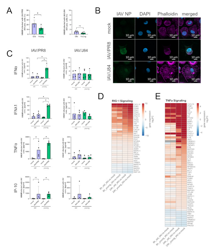
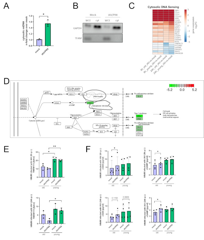
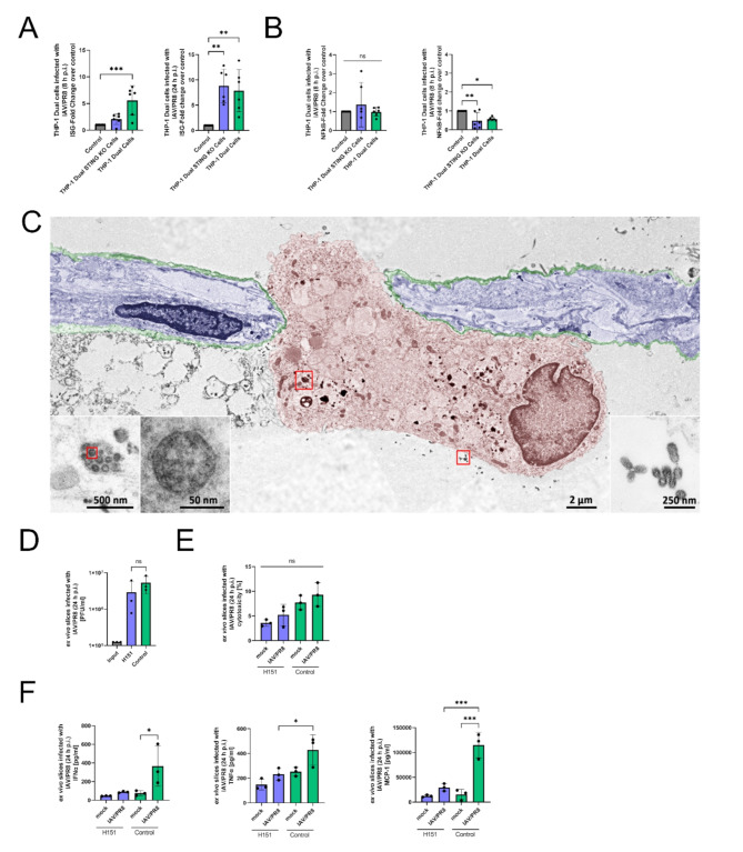
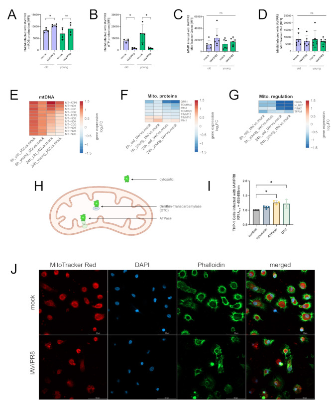
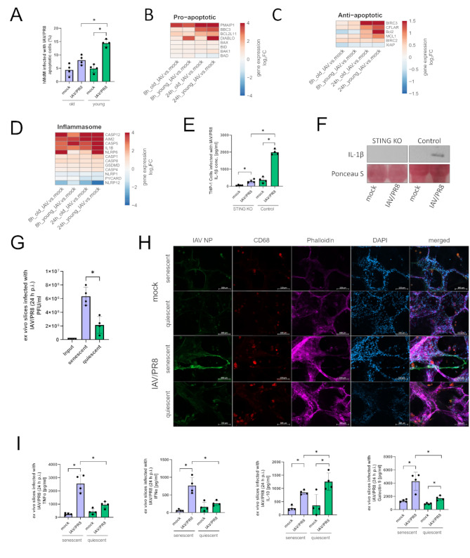
Ageing is a major risk factor that contributes to increased mortality and morbidity rates during influenza A virus (IAV) infections. Macrophages are crucial players in the defense against viral infections and display impaired function during ageing. However, the impact of ageing on macrophage function in response to an IAV infection remains unclear and offers potential insight for underlying mechanisms. In this study, we investigated the immune response of young and aged human monocyte-derived macrophages to two different H1N1 IAV strains. Interestingly, macrophages of aged individuals showed a lower interferon response to IAV infection, resulting in increased viral load. Transcriptomic data revealed a reduced expression of stimulator of interferon genes (STING) in aged macrophages albeit the cGAS-STING pathway was upregulated. Our data clearly indicate the importance of STING signaling for interferon production by applying a THP-1 STING knockout model. Evaluation of mitochondrial function during IAV infection revealed the release of mitochondrial DNA to be the activator of cGAS-STING pathway. The subsequent induction of apoptosis was attenuated in aged macrophages due to decreased STING signaling. Our study provides new insights into molecular mechanisms underlying age-related immune impairment. To our best knowledge, we are the first to discover an age-dependent difference in gene expression of STING on a transcriptional level in human monocyte-derived macrophages possibly leading to a diminished interferon production.

Keywords: Ageing; Influenza A virus; Macrophages; Mitochondria; cGAS-STING pathway.


Conflict of interest statement

Declarations Ethics approval and consent to participate The study was approved by the local ethics committee of the Jena University Hospital (no. 2018 − 1263, 2020 − 1894, and no. 2020 − 1773). Competing interests The authors declare no competing interests.

Figures

Fig. 1
Fig. 1
Suppression of interferon response and increased susceptibility to infection in macrophages of elderly individuals. (A) Measurement of virus-positive hMdM 24 h p.i. after infection with IAV (MOI 1). The experiment was conducted with five young and old donors each. Each data point represents a biological replicate in this and all following figures. Significance was calculated with the Mann-Whitney U test (* p ≤ 0.05). (B) Immunofluorescence staining of infected hMdM (24 h p.i.) with antibody against IAV NP, phalloidin and DAPI. (C) Cytokine concentrations of IFNα, IFNλ1, TNFα and IP-10 were measured in the supernatant of infected hMdM (24 h p.i.). The results of four young and old donors each are shown. Significance was calculated with the Mann-Whitney U test (* p ≤ 0.05). (D) DEGs are shown in heatmaps based on KEGG pathways regarding RIG-I signaling, and (E) TNFα signaling at timepoint 8 h and 24 h p.i. Gene expression in heatmaps over log2 fold change. Red signals upregulation, blue downregulation. The DEG of at least one condition per row had a log2 fold change > 1 with a significance of p ≤ 0.05
Fig. 2
Fig. 2
Reduced expression of STING in aged macrophages albeit the cGAS-STING pathway was upregulated during infection. (A) Abundance of cytosolic mtDNA 8 h p.i. was measured in three biological replicates of hMdM with quantitative RT-PCR. After infection with IAV/PR8 hMdM show elevated levels of cytosolic mtDNA. Significance was evaluated with paired t-test (* p ≤ 0.05). (B) Whole cell extracts (WCE) and cytosolic fractions were analyzed by western blot using the indicated antibodies. (C) Heatmap with DEGs related to cytosolic DNA sensing pathways. Gene expression is presented over log2 fold change. The DEG of at least one condition per row had a log2 fold change > 1 with a significance of p ≤ 0.05. (D) KEGG-Enrichment pathway of cytosolic DNA sensing pathway comparing aged hMdM to young hMdM infected with IAV/PR8 24 h p.i. showing reduced expression of STING in aged hMdM. Red signals upregulation, green signal downregulation. (E) Gene expression of TMEM173 was shown as a bar plot. Significance was calculated with Kruskal-Wallis test (* p ≤ 0.05). (F) Concentration of cGAS and cGAMP were measured with ELISA in four biological replicates of hMdM 8 h and 24 h p.i. Significance was evaluated with paired t-test (* p ≤ 0.05)
Fig. 3
Fig. 3
STING is necessary to induce interferon response in macrophages. (A) IFN stimulator gene (ISG) reporter fold change of THP-1 Dual and THP-1 Dual STING KO cells infected with IAV/PR8 at timepoints 8 h p.i. and 24 h p.i. The results of six biological replicates of each cell line is shown. Significance was calculated with One-way ANOVA (* p ≤ 0.05, ** p ≤ 0.01, *** p ≤ 0.001). (B) Transcriptional activity of NFκB pathway in THP-1 Dual and THP-1 Dual STING KO cells infected with IAV/PR8. Significance was calculated with One-way ANOVA (* p ≤ 0.05, ** p ≤ 0.01). (C) SEMpicture showing IAV on the surface and intracellular of an alveolar macrophage. Colors were added manually. (D) Viral titer of ex vivo lung slices infected with IAV/PR8 24 h p.i. was detected by standard plaque assay shown with logarithmic scaling. The results of three biological replicates are shown. Significance was calculated with One-way ANOVA (ns p ≥ 0.05). (E) LDH was measured in the supernatants of ex vivo slices and presented as percentage of the positive control. Significance was calculated with One-way ANOVA (ns p ≥ 0.05). (F) Cytokines IFNα, TNFα and MCP-1 were measured in the supernatants. Significance was calculated with One-way ANOVA (* p ≤ 0.05, ** p ≤ 0.01, *** p ≤ 0.001)
Fig. 4
Fig. 4
Mitochondria undergo oxidative stress during infection. (A) mtROS production in infected hMdM was measured via flow cytometry. The results of four biological replicates are shown. Significance was calculated with the Mann-Whitney U test (* p ≤ 0.05). (B) Mitochondrial ATP was quantified using flow cytometry. Significance was calculated with the Mann-Whitney U test (* p ≤ 0.05). (C) Mitochondrial mass in hMdM after infection was measured via staining with MitoTracker Green. The results of six biological replicates are shown. Significance was calculated with the Mann-Whitney U test (ns p ≥ 0.05). (D) Mitochondrial membrane potential was quantified by staining with MitoTracker Red and measurement via flow cytometry. Significance was calculated with the Mann-Whitney U test (ns p ≥ 0.05). (E) Heatmaps showing individually differentiated genes related to regulation of mitochondrial metabolism, (F) imported mitochondrial proteins and (G) regulation of mitochondrial homeostasis. Gene expression is presented over log2 fold change. At least one DEG per gene per condition had a significant log2 fold change. (H) Schematic representation of THP-1 redox cell model showing subcellular location of different roGFP2 constructs. (I) Determination of redox status of THP-1 cells stably expressing cytosolic roGFP2, roGFP2 coupled to mitochondrial ATPase and OTC. Redox status was analyzed by measuring fluorescence at λex 405 nm and 488 nm. RFI is normalized to control cells. Significance was calculated with Kruskal-Wallis test (* p ≤ 0.05). (J) Immunofluorescence staining of infected hMdM stained with MitoTracker Red, phalloidin and DAPI
Fig. 5
Fig. 5
Apoptosis and inflammasome signaling pathways are altered in old macrophages. (A) Apoptotic cells were quantified via flow-cytometry. The results of four biological replicates in each group are shown. Significance was calculated with the Mann-Whitney U test (* p ≤ 0.05). (B) Heatmaps showing proapoptotic, (C) antiapoptotic genes (D) and genes of inflammasome signaling. Gene expression is presented over log2 fold change. At least one DEG per gene per condition had a significant log2 fold change. (E) Concentration of IL-1β was measured in the supernatants of infected THP-1 Dual and THP-1 Dual STING KO cells 24 h p.i. Results were confirmed by western blotting using the indicated antibodies. Significance was calculated with the Mann-Whitney U test (* p ≤ 0.05). (F) Supernatants of THP-1 cells were analyzed by western blot using the indicated antibody and staining. The experiment was repeated 4 times and blot shown as representative. (G) Viral titer of ex vivo lung slices was determined by standard plaque assay. Significance was calculated with the Mann-Whitney U test (* p ≤ 0.05). (H) Immunofluorescence staining of infected ex vivo lung slices stained with antibodies against IAV NP, CD68, phalloidin and DAPI. (I) Cytokines in the supernatants of senescent and quiescent ex vivo lung slices were measured. Significance was calculated with the Mann-Whitney U test (* p ≤ 0.05)

References

    1. Cunha BA. Pneumonia in the elderly. Clin Microbiol Infect. 2001;7:581–8. - PubMed
    1. Viboud C, Boëlle PY, Cauchemez S, Lavenu A, Valleron AJ, Flahault A, et al. Risk factors of influenza transmission in households. Br J Gen Pract. 2004;54:684–9. - PMC - PubMed
    1. Paget J, Spreeuwenberg P, Charu V, Taylor RJ, Iuliano AD, Bresee J, et al. Global mortality associated with seasonal influenza epidemics: New burden estimates and predictors from the GLaMOR Project. J Glob Health. 2019;9:020421. - PMC - PubMed
    1. Long ME, Mallampalli RK, Horowitz JC. Pathogenesis of pneumonia and acute lung injury. Clin Sci (Lond). 2022;136:747–69. - PMC - PubMed
    1. Somerville L, Cardani A, Braciale TJ. Alveolar macrophages in Influenza a infection guarding the castle with sleeping dragons. Infect Dis Ther (San Antonio; 2020. p. 1. - PMC - PubMed

LinkOut - more resources