. 2024 Nov 14;17(1):225.
doi: 10.1186/s13048-024-01547-5.

Near-infrared fluorescent molecular probes with cetuximab in the in vivo fluorescence imaging for epithelial ovarian cancer

Affiliations

Near-infrared fluorescent molecular probes with cetuximab in the in vivo fluorescence imaging for epithelial ovarian cancer

Chen Zhang et al. J Ovarian Res. .

Abstract

Background: Near-infrared fluorescence (NIRF) imaging is an excellent choice for image-guided surgery due to its simple operation and non-invasiveness. Developing tumor-specific fluorescent molecular probes is key to fluorescence imaging-guided surgery. EGFR (epidermal growth factor receptor) is closely related to the proliferation and growth of tumor cells and is highly expressed in epithelial ovarian cancer (EOC). The study aims to construct a NIR fluorescent molecular probe using cetuximab (an EGFR monoclonal antibody) and investigate its feasibility for targeting EOC in vivo through fluorescence imaging.

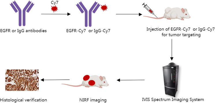
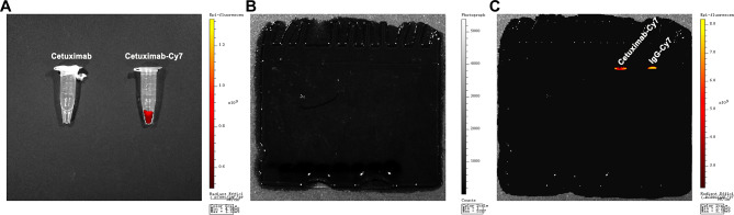
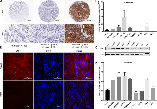
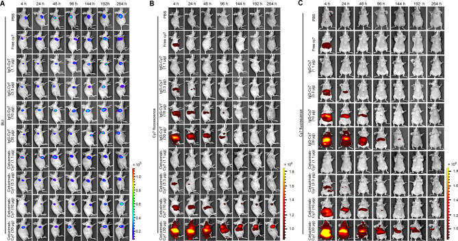
Methods: We determined the expression of EGFR in EOC. NIR fluorescent molecular probe with cetuximab (cetuximab-Cy7) was chemically engineered and identified. The subcutaneous xenografted tumor model of EOC was induced using SKOV3-Luc cell line with positive expression of EGFR. Cetuximab-Cy7 was used for in vivo fluorescence imaging, and phosphate-buffered saline, free Cy7 dye and mouse isotype immunoglobulin G-Cy7 were used as controls. NIRF imaging system was performed to study the distribution and targeting of the probes. Tumors were imaged in situ and ex vivo, and fluorescent intensity was quantified. Resected specimens were analyzed to confirm diagnosis, and immunohistochemical (IHC) staining was used to identify EGFR expression.

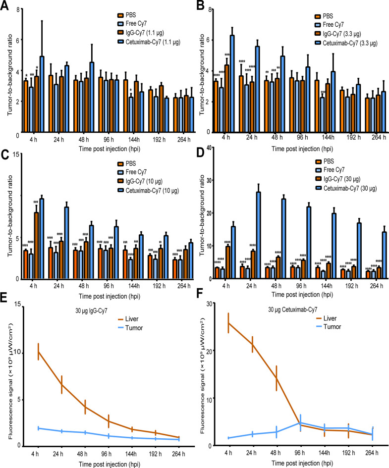
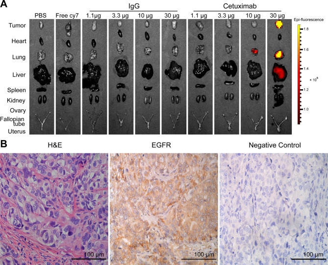
Results: EGFR expression was increased in EOC tissues than fallopian tube tissues. The high expression of EGFR was significantly correlated with well-differentiation, residual lesions ≤ 1 cm, no recurrence and increased survival. NIRF imaging showed that the cetuximab-Cy7 enabled detection of tumor lesions in EOC-bearing mice with the optimal dose of 30 µg. The suitable imaging time window may be 24-96 h post-injection. Ex vivo fluorescence imaging indicated that fluorescent signal was mainly detected in the tumor and the lung. IHC results confirmed that xenografts were EGFR positive.

Conclusion: Cetuximab-Cy7 can specifically target the tumors of EOC xenografted nude mice. This research lays the foundation for future studies on EOC surgery navigation.

Keywords: Animal model; Cetuximab; Cy7; Epithelial ovarian cancer; Near-infrared fluorescence imaging.


Conflict of interest statement

Declarations Ethics approval and consent to participate This experiment was approved by the Medical Ethics Committee of Peking University People’s Hospital (No.2016PHC078), and all patients signed informed consent. Consent for publication Not required. Competing interests The authors declare no competing interests.

Figures

Fig. 1
Fig. 1
Scheme for using cetuximab-Cy7 probe in NIRF guided surgery in OC. EGFR-Cy7: epidermal growth factor receptor-cyanine7 (cetuximab-cyanine7); NIRF: near-infrared fluorescence; OC: ovarian cancer; IgG-Cy7: immunoglobulin G-cyanine7
Fig. 2
Fig. 2
Characterization of cetuximab-Cy7 probe (A) The Cy7-labeled EGFR antibody solution was screened using NIRF imaging. SDS-PAGE of Cy7-labeled antibodies under (B) bright-field condition and (C) NIRF imaging. NIRF: near-infrared fluorescence; SDS-PAGE: sodium dodecyl sulfate - polyacrylamide gel electrophoresis; Cetuximab-Cy7: cetuximab-cyanine7; IgG-Cy7: immunoglobulin G-cyanine7
Fig. 3
Fig. 3
EGFR expression in OC tissues and cell lines. (A) IHC staining of EGFR protein in TMAs. FT tissue with a low H-score (4.13); a serous OC tumor (grade 3) with a low H-score (3.20); and a serous OC tumor (grade 3) with a high H-score (217.50). Black scale bar, 200 µm; yellow scale bar, 50 µm. (B) EGFR mRNA expression in OC cell lines was detected by qPCR. (C) WB of EGFR protein in OC cell lines and (D) quantification analyses. (E) IF staining of EGFR in OC SKOV3 cells (positive) and A2780 cells (negative). EGFR, red; DAPI, blue. Magnification 400×; white scale bar, 100 µm. OC: ovarian cancer; IHC: immunohistochemical; TMAs: tissue microarrays; FT: fallopian tube; qPCR: quantitative real-time PCR; WB: western blotting; IF: immunofluorescence; DAPI: 4’,6-diamidino-2-phenylindole
Fig. 4
Fig. 4
In vivo imaging of subcutaneous OC tumors and biodistribution of cetuximab-Cy7 probe. (A) BLI imaging was used to detect SKOV3-Luc OC tumor cells at 4, 24, 48, 96, 144, 192, and 264 h post-injection of cetuximab-Cy7. In vivo NIRF imaging of mice post-injection of cetuximab-Cy7 in (B) the lateral position and (C) the ventral position at the same timepoints. The control groups including PBS, free Cy7 dye and mouse isotype IgG-Cy7 were also used in our previous work [22]. The corresponding images in the control groups in Fig. 4A and B were reprinted with permission from reference 22. OC: ovarian cancer; BLI: bioluminescent; Cetuximab-Cy7: cetuximab-cyanine7; IgG-Cy7: immunoglobulin G-cyanine7; NIRF: near-infrared fluorescence; PBS: phosphate-buffered saline
Fig. 5
Fig. 5
Quantification of the fluorescence intensity of in vivo NIRF imaging using cetuximab-Cy7 probe in OC tumor models. The T/B ratios for (A) 1.1 µg cetuximab-Cy7, (B) 3.3 µg cetuximab-Cy7, (C) 10 µg cetuximab-Cy7 and (D) 30 µg cetuximab-Cy7 groups at all time points. Quantification of fluorescence intensity of (E) 30 µg mouse isotype IgG-Cy7 and (F) 30 µg cetuximab-Cy7 groups were shown for tumor and liver (n = 3). The mice were divided into 10 groups with 3 mice in each group, respectively intravenously injected with PBS, free Cy7, 1.1 µg, 3.3 µg, 10 µg, 30 µg dose of cetuximab-Cy7 or IgG-Cy7. *, p < 0.05; **, p < 0.01; ***, p < 0.001; ****, p < 0.0001. The control groups including PBS, free Cy7 dye and mouse isotype IgG-Cy7 were also used in our previous work [22]. The corresponding images in the control groups in Fig. 5A -E were reprinted with permission from reference 22. NIRF: near-infrared fluorescence; Cetuximab-Cy7: cetuximab-cyanine7; OC: ovarian cancer; T/B: tumor-to-background; IgG-Cy7: immunoglobulin G-cyanine7; PBS: phosphate-buffered saline; hpi: time post injection
Fig. 6
Fig. 6
Ex vivo imaging and IHC staining of EGFR in OC tumors. (A) Ex vivo NIRF imaging of tumor and normal organs in different groups. (B) IHC staining of EGFR protein in OC tumor tissue. Magnification 400×; scale bar, 100 μm. The control groups including PBS, free Cy7 dye and mouse isotype IgG-Cy7 were also used in our previous work [22]. The corresponding images in the control groups in Fig. 6A were reprinted with permission from reference 22. IHC: immunohistochemical; OC: ovarian cancer; NIRF: near-infrared fluorescence; IgG: immunoglobulin G; PBS: phosphate-buffered saline; H&E, hematoxylin-eosin

References

    1. Siegel RL, Giaquinto AN, Jemal A. Cancer statistics, 2024. CA Cancer J Clin. 2024;74:12–49. - PubMed
    1. du Bois A, Reuss A, Pujade-Lauraine E, Harter P, Ray-Coquard I, Pfisterer J. Role of surgical outcome as prognostic factor in advanced epithelial ovarian cancer: a combined exploratory analysis of 3 prospectively randomized phase 3 multicenter trials: by the Arbeitsgemeinschaft Gynaekologische Onkologie Studiengruppe Ovarialkarzinom (AGO-OVAR) and the Groupe d’Investigateurs Nationaux Pour Les Etudes des Cancers de l’Ovaire (GINECO). Cancer. 2009;115:1234–44. - PubMed
    1. Eisenkop SM, Friedman RL, Wang HJ. Complete cytoreductive surgery is feasible and maximizes survival in patients with advanced epithelial ovarian cancer: a prospective study. Gynecol Oncol. 1998;69:103–8. - PubMed
    1. Lauwerends LJ, van Driel P, Baatenburg de Jong RJ, Hardillo JAU, Koljenovic S, Puppels G, et al. Real-time fluorescence imaging in intraoperative decision making for cancer surgery. Lancet Oncol. 2021;22:e186–95. - PubMed
    1. Sun C, Huang Y, Jiang C, Li Z. Updates on fluorescent probes and open-field imaging methods for fluorescence-guided cytoreductive surgery for epithelial ovarian cancer: a review. BJOG. 2022;129(Suppl 2):50–9. - PMC - PubMed

MeSH terms