

Hepatobiliary-Phase Hypointense Nodules Without Arterial-Phase Hyperenhancement: Developing a Risk Stratification for Hypervascular Transformation Based on a Real-World Observational Cohort Study
- PMID: 39544080
- PMCID: PMC11565688
- DOI: 10.1177/15330338241299003
Hepatobiliary-Phase Hypointense Nodules Without Arterial-Phase Hyperenhancement: Developing a Risk Stratification for Hypervascular Transformation Based on a Real-World Observational Cohort Study
Abstract
Purpose: To develop a risk stratification based on MRI features to predict hypervascular transformation for hepatobiliary-phase (HBP) hypointense nodules without arterial-phase hyperenhancement (APHE).
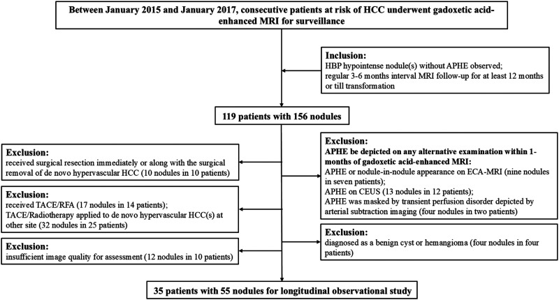
Materials and methods: This retrospective observational cohort study included 55 HBP hypointense nodules without APHE in 35 patients with chronic liver disease, cirrhosis, or current hepatocellular carcinoma (HCC) who underwent gadoxetic acid-enhanced MRI. The hypervascular transformation during the follow-up MRI(s) was the primary endpoint analyzed for the nodules. Univariable and multivariable Cox proportional hazard regression analyses were performed to identify risk features predicting transformation and assess their predictive value.
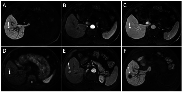
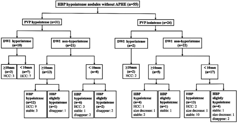
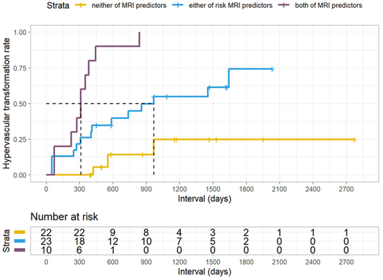
Results: Among the 55 nodules, 27 developed hypervascular transformation, while 28 did not. Diffusion-weighted imaging (DWI) hyperintensity (hazard ratio [HR], 4.98; 95% confidence interval [CI]: 1.60, 15.54; p = 0.006) and portal venous phase (PVP) hypointensity (HR, 4.08; 95% CI: 1.43, 11.64; p = 0.009) were associated with hypervascular transformation. DWI hyperintensity and PVP hypointensity had 44.4% (95% CI: 26.0%, 64.4%) and 81.9% (95% CI: 61.3%, 93.0%) sensitivity, while their specificity was 78.2% (95% CI: 64.6%, 87.8%) and 67.9 (95% CI: 47.6%, 83.4%), respectively. The specificity of the combination of two features was 100% (95% CI: 85.0%, 100%). The hypervascular transformation rates for nodules with both, either and neither of the risk MRI findings were 100% (10/10), 60.9% (14/23), and 13.6% (3/22), respectively; the median intervals for transformation were 312 (range: 73-838), 409 (range: 50-1643) and 555 (range: 423-968) days, respectively.
Conclusion: The combination of DWI hyperintensity and PVP hypointensity may be used as a high-risk indicator for the hypervascular transformation of HBP hypointense nodules without APHE; nodules without either feature may be treated as low-risk nodules and could adopt an extended interval follow-up schedule.
Keywords: cancer risk; cirrhosis; gadolinium ethoxybenzyl DTPA; hepatocellular carcinoma; magnetic resonance imaging.
Conflict of interest statement
Declaration of Conflicting InterestsThe authors declared no potential conflicts of interest with respect to the research, authorship, and/or publication of this article.
Figures
Similar articles
-
Hypervascular transformation of hepatobiliary phase hypointense nodules without arterial phase hyperenhancement on gadoxetic acid-enhanced MRI: long-term follow-up in a surveillance cohort.Eur Radiol. 2022 Aug;32(8):5064-5074. doi: 10.1007/s00330-022-08623-8. Epub 2022 Feb 28. Eur Radiol. 2022. PMID: 35229195
-
Hypervascular Transformation of Hypovascular Hypointense Nodules in the Hepatobiliary Phase of Gadoxetic Acid-Enhanced MRI: A Systematic Review and Meta-Analysis.AJR Am J Roentgenol. 2017 Oct;209(4):781-789. doi: 10.2214/AJR.16.17711. Epub 2017 Jul 25. AJR Am J Roentgenol. 2017. PMID: 28742376
-
Diagnostic performances of adding transition and hepatobiliary phase to washout in gadoxetic acid-enhanced MRI for subcentimeter hepatocellular carcinoma.BMC Gastroenterol. 2025 Jul 1;25(1):483. doi: 10.1186/s12876-025-04071-y. BMC Gastroenterol. 2025. PMID: 40596938 Free PMC article.
-
Non-hypervascular Hypointense Nodules on Hepatocyte Phase Gadoxetic Acid-Enhanced MR Images: Transformation of MR Hepatobiliary Hypointense Nodules into Hypervascular Hepatocellular Carcinomas.Gut Liver. 2018 Jan 15;12(1):79-85. doi: 10.5009/gnl17046. Gut Liver. 2018. PMID: 28798287 Free PMC article.
-
Contrast-enhanced ultrasound for the diagnosis of hepatocellular carcinoma in adults with chronic liver disease.Cochrane Database Syst Rev. 2022 Sep 2;9(9):CD013483. doi: 10.1002/14651858.CD013483.pub2. Cochrane Database Syst Rev. 2022. PMID: 36053210 Free PMC article.
Cited by
-
Long-term outcomes of indeterminate focal hepatic observations less than 20 mm followed up with gadoxetic acid-enhanced magnetic resonance imaging (Gd-EOB-DTPA-MRI).J Gastrointest Oncol. 2025 Jun 30;16(3):1078-1091. doi: 10.21037/jgo-2025-302. Epub 2025 Jun 27. J Gastrointest Oncol. 2025. PMID: 40672063 Free PMC article.
References
Publication types
MeSH terms
Substances
LinkOut - more resources
Full Text Sources
Medical
Miscellaneous
