Review
. 2024 Nov 1;14(11):7937-7957.
doi: 10.21037/qims-24-365. Epub 2024 Oct 8.

Myosteatosis: diagnostic significance and assessment by imaging approaches

Affiliations
Review

Myosteatosis: diagnostic significance and assessment by imaging approaches

Ana Isabel Garcia-Diez et al. Quant Imaging Med Surg. .

Abstract

Myosteatosis has emerged as an important concept in muscle health as it is associated with an increased risk of adverse health outcomes, a higher rate of complications, and increased mortality associated with ageing, chronic systemic and neuromuscular diseases, cancer, metabolic syndromes, degenerative events, and trauma. Myosteatosis involves ectopic infiltration of fat into skeletal muscle, and it exhibits a negative correlation with muscle mass, strength, and mobility representing a contributing factor to decreased muscle quality. While myosteatosis serves as an additional biomarker for sarcopenia, cachexia, and metabolic syndromes, it is not synonymous with sarcopenia. Myosteatosis induces proinflammatory changes that contribute to decreased muscle function, compromise mitochondrial function, and increase inflammatory response in muscles. Imaging techniques such as computed tomography (CT), particularly opportunistic abdominal CT scans, and magnetic resonance imaging (MRI) or magnetic resonance spectroscopy (MRS), have been used in both clinical practice and research. And in recent years, ultrasound has emerged as a promising bedside tool for measuring changes in muscle tissue. Various techniques, including CT-based muscle attenuation (MA) and intermuscular adipose tissue (IMAT) quantification, MRI-based proton density fat fraction (PDFF) and T1-T2 mapping, and musculoskeletal ultrasound (MSUS)-based echo intensity (EI) and shear wave elastography (SWE), are accessible in clinical practice and can be used as adjunct biomarkers of myosteatosis to assess various debilitating muscle health conditions. However, a stan¬dard definition of myosteatosis with a thorough understanding of the pathophysiological mechanisms, and a consensus in assessment methods and clinical outcomes has not yet been established. Recent developments in image acquisition and quantification have attempted to develop an appropriate muscle quality index for the assessment of myosteatosis. Additionally, emerging studies on artificial intelligence (AI) may provide further insights into quantification and automated assessment, including MRS analysis. In this review, we discuss the pathophysiological aspects of myosteatosis, all the current imaging techniques and recent advances in imaging assessment as potential biomarkers of myosteatosis, and the most common clinical conditions involved.

Keywords: Myosteatosis; computed tomography (CT); imaging biomarkers; magnetic resonance (MR); ultrasound (US).


Conflict of interest statement

Conflicts of Interest: All authors have completed the ICMJE uniform disclosure form (available at https://qims.amegroups.com/article/view/10.21037/qims-24-365/coif). The special issue “Advances in Diagnostic Musculoskeletal Imaging and Image-guided Therapy” was commissioned by the editorial office without any funding or sponsorship. X.T. served as the unpaid Guest Editor of the issue. The authors have no other conflicts of interest to declare.

Figures

Figure 1
Figure 1
Axial CT image at the level of L3 in a 66-year-old male patient with lung and gastrointestinal cancer and a BMI of 36.9 kg/m2 (classified as obese with BMI >30 kg/m2) (A). CT-based quantification with thresholds for MA of the (B) SMA, (C) NAMA, (D) LAMA, (E) IMAT, and (F) SAT were obtained using Syngo.via software (Siemens, Erlangen, Germany). CT-based parameters indicate myosteatosis. The MA was 31 HU (myosteatosis criteria for males is <33 HU for BMI ≥25.0 kg/m2). The NAMA/TAMA index was 47% [myosteatosis criteria for males is <66.4% (T-score <2.0)]. The SAT was −94 HU and the MA/SAT index was −0.32 HU (myosteatosis criteria for males is >−0.44 HU). CT, computed tomography; BMI, body mass index; MA, muscle attenuation; SMA, skeletal muscle area; NAMA, normal attenuation muscle area; LAMA, low attenuation muscle area; IMAT, intermuscular adipose tissue; SAT, subcutaneous tissue attenuation; TAMA, total abdominal muscle area.
Figure 2
Figure 2
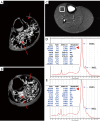
MRI-based quantification on T1-weighted images of the calf in a male patient with HIV-lipodystrophy and metabolic syndrome (A) and a healthy control (B), using Analyze 10.0 software with thresholds for IMAT (red long arrows) and MV (red short arrows). The patient (A) exhibited lower IMAT and higher MV than the healthy control (B). Localized 1-dimensional 1H-MRS using point-resolve spectroscopy data were collected from a voxel within the tibialis anterior muscle on axial T1-weighted image (C). LCModel software was employed to obtain spectra results, revealing that the patient (D) had higher IMCL and EMCL than the healthy control (E) (red arrowheads). EMCL, extramyocellular lipids; IMCL, intramyocellular lipids; SD, standard deviation; MRI, magnetic resonance imaging; IMAT, intermuscular adipose tissue; MV, muscular volume; 1H-MRS, proton magnetic resonance spectroscopy.
Figure 3
Figure 3
Whole-body muscle MRI in a patient with congenital muscular dystrophy (GNE or Nonaka myopathy). Axial (A) and coronal (B) T1-weighted images of various regions reveal a nearly symmetrical bilateral pattern of muscle fatty infiltration grades 3 (30–60%) and 4 (>60%), as assessed by the 5-point modified Mercuri scale, in several muscles of the different levels. MRI, magnetic resonance imaging.
Figure 4
Figure 4
Axial Dixon FF images of a healthy 26-year-old male with a BMI of 23 kg/m2 (within the healthy weight range of BMI 18.5 to <25 kg/m2), showing non-segmented (A) and segmented (B) images; and a healthy 31-year-old male with a BMI of 27 kg/m2 (within the overweight range of BMI 25.0 to <30 kg/m2), displaying non-segmented (C) and segmented (D) images. The male with overweight (C,D) exhibited myosteatosis with higher values of IMAT and FF in both flexor (red) and extensor (blue) compartments compared to the healthy weight male (A,B). FF, fat fraction; BMI, body mass index; IMAT, intermuscular adipose tissue.
Figure 5
Figure 5
Localized 2D-COSY spectrum recorded of the vastus lateralis muscle (top left) in a healthy 46-year-old female subject. Post-processing was conducted using FELIX NMR software (Accelrys, San Diego, CA, USA), and a total of 22 peaks were measured. Monounsaturated lipid pools can be distinguished by the cross peaks C1 (5.3/2.1 and 2.1/5.3 ppm), and PUFAs by the cross peaks C2 (5.3–5.5/2.75–2.9 and 2.75–2.9/5.3–5.5 ppm). Ratios of these cross-peak volumes were used to define the degree of unsaturation. 2D-COSY, 2-dimensional correlation spectroscopy; NMR, nuclear magnetic resonance; PUFAs, polyunsaturated lipid pools.
Figure 6
Figure 6
B-mode ultrasound transverse scan images of the rectus femoris muscle in a healthy 17-year-old female with preserved muscle architecture and echogenicity (A); a healthy 83-year-old female (B), and a healthy 68-year-old female (C), both exhibiting a moderate grade 2 of muscle degeneration (moderate/partial loss of muscle architecture and increased echogenicity). Muscle EI in grayscale and histographic analysis was performed using ImageJ software. The young healthy subject (A) demonstrated low EI. The middle-aged subject (C) with moderate muscle degeneration exhibited higher EI compared to the older subject (B). EI, echo intensity.

References

    1. Cruz-Jentoft AJ, Sayer AA. Sarcopenia. Lancet 2019;393:2636-46. 10.1016/S0140-6736(19)31138-9 - DOI - PubMed
    1. Cruz-Jentoft AJ, Bahat G, Bauer J, Boirie Y, Bruyère O, Cederholm T, Cooper C, Landi F, Rolland Y, Sayer AA, Schneider SM, Sieber CC, Topinkova E, Vandewoude M, Visser M, Zamboni M; Writing Group for the European Working Group on Sarcopenia in Older People 2 (EWGSOP2), and the Extended Group for EWGSOP2. Sarcopenia: revised European consensus on definition and diagnosis. Age Ageing 2019;48:601. 10.1093/ageing/afz046 - DOI - PMC - PubMed
    1. Xia L, Zhao R, Wan Q, Wu Y, Zhou Y, Wang Y, Cui Y, Shen X, Wu X. Sarcopenia and adverse health-related outcomes: An umbrella review of meta-analyses of observational studies. Cancer Med 2020;9:7964-78. 10.1002/cam4.3428 - DOI - PMC - PubMed
    1. Prado CM, Ford KL, Gonzalez MC, Murnane LC, Gillis C, Wischmeyer PE, Morrison CA, Lobo DN. Nascent to novel methods to evaluate malnutrition and frailty in the surgical patient. JPEN J Parenter Enteral Nutr 2023;47 Suppl 1:S54-68. 10.1002/jpen.2420 - DOI - PMC - PubMed
    1. Correa-de-Araujo R, Addison O, Miljkovic I, Goodpaster BH, Bergman BC, Clark RV, Elena JW, Esser KA, Ferrucci L, Harris-Love MO, Kritchevsky SB, Lorbergs A, Shepherd JA, Shulman GI, Rosen CJ. Myosteatosis in the Context of Skeletal Muscle Function Deficit: An Interdisciplinary Workshop at the National Institute on Aging. Front Physiol 2020;11:963. 10.3389/fphys.2020.00963 - DOI - PMC - PubMed

LinkOut - more resources