Incarcerated Incisional Hernia: An Unusual Presentation of Metastatic Endometrial Carcinoma
- PMID: 39544575
- PMCID: PMC11561335
- DOI: 10.7759/cureus.71497
Incarcerated Incisional Hernia: An Unusual Presentation of Metastatic Endometrial Carcinoma
Abstract
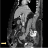
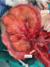
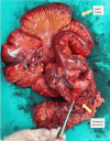
Abdominal wall hernia is a common condition seen in the clinical practice of surgery. However, malignant tumors in the hernia sac are rare and there are limited studies on this subject. We report a case of a 77-year-old female who presented with generalized abdominal pain and vomiting. She was treated for an incarcerated incisional hernia and underwent an exploratory laparotomy, which showed a multiseptated incisional hernia sac. Histopathological examination revealed a metastatic endometrial serous carcinoma (ESC). ESC is an aggressive variant associated with poor prognosis, characterized by metastasis and extrauterine spread. Its treatment mainly involves a multidisciplinary approach, including surgical treatment and chemoradiotherapy. This report highlights the importance of considering malignant tumors in the differential diagnosis of hernia sac contents. Raising awareness among healthcare professionals and the general public can aid in the prompt diagnosis, appropriate treatment, and improved outcomes for individuals with such rare presentations.
Keywords: endometrial serous carcinoma; gynaecological malignancy; incarcerated incisional hernia; incisional ventral hernia; strangulated ventral hernia.
Copyright © 2024, Ong et al.
Conflict of interest statement
Human subjects: Consent was obtained or waived by all participants in this study. Conflicts of interest: In compliance with the ICMJE uniform disclosure form, all authors declare the following: Payment/services info: All authors have declared that no financial support was received from any organization for the submitted work. Financial relationships: All authors have declared that they have no financial relationships at present or within the previous three years with any organizations that might have an interest in the submitted work. Other relationships: All authors have declared that there are no other relationships or activities that could appear to have influenced the submitted work.
Figures


References
-
- SSAT patient care guidelines. Surgical repair of incisional hernias. J Gastrointest Surg. 2007;11:1231–1232. - PubMed
-
- An evidence map and synthesis review with meta-analysis on the risk of incisional hernia in colorectal surgery with standard closure. Stabilini C, Garcia-Urena MA, Berrevoet F, et al. Hernia. 2022;26:411–436. - PubMed
-
- Malignant epithelial tumors observed in hernia sacs. Val-Bernal JF, Mayorga M, Fernández FA, Val D, Sánchez R. Hernia. 2014;18:831–835. - PubMed
-
- Malignant tumors in hernia sac: clinicopathological and immunohistochemical studies of 21 cases at a single institution. Zhang D, Yang Q, Katerji R, Drage MG, Huber A, Liao X. Pathol Int. 2020;70:975–983. - PubMed
Publication types
LinkOut - more resources
Full Text Sources
