

Antivascular Endothelial Growth Factor Injections for the Chronic Treatment of Macula-off, Fovea-on Diabetic Tractional Retinal Detachment With Vitreous Hemorrhage
- PMID: 39544710
- PMCID: PMC11558646
- DOI: 10.1177/24741264241297684
Antivascular Endothelial Growth Factor Injections for the Chronic Treatment of Macula-off, Fovea-on Diabetic Tractional Retinal Detachment With Vitreous Hemorrhage
Abstract
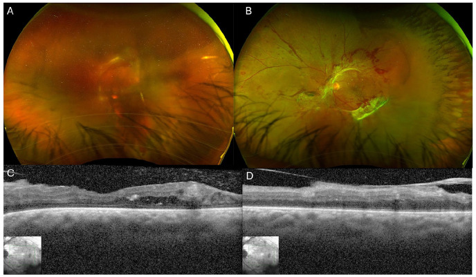
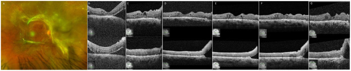
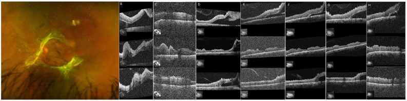
Purpose: To evaluate the use of antivascular endothelial growth factor (anti-VEGF) as treatment for tractional retinal detachments (TRDs) involving the macula. Methods: A case report was evaluated. Results: A 40-year-old man with a medical history notable for congenital heart disease and proliferative diabetic retinopathy presented with decreased vision. Surgery was not feasible for medical reasons, and panretinal photocoagulation was limited by retinal traction and hemorrhage. Despite initial progression of bilateral macula-off, foveal-on TRDs, the patient received intravitreal (IVT) bevacizumab regularly every 8 to 12 weeks. After initial improvement, both eyes remained anatomically stable with attached foveas over more than 1.5 years of follow-up. Conclusions: In cases in which surgery is not possible, macula-involving TRDs with neovascularization can be managed successfully with repeated IVT anti-VEGF injection monotherapy.
Keywords: antivascular endothelial growth factor; surgery; tractional retinal detachment.
© The Author(s) 2024.
Conflict of interest statement
Although the authors declared no potential conflicts of interest with respect to the research, authorship, and/or publication of this article, the following declared financial disclosures: Dr. Patel is a consultant to Regeneron, Dutch Ophthalmic, Genentech, EyePoint Pharmaceuticals, and Alcon Vision. Dr. Kim is a consultant to Ingenia Therapeutics and CureVac AG, receives research support from CureVac AG and Valo Health, and receives grant support from the National Eye Institute (R01EY027739) and US Department of Defense (VR220059). None of the other authors declared potential conflicts of interest with respect to the research, authorship, and/or publication of the article.
Figures
Similar articles
-
Current management of diabetic tractional retinal detachments.Indian J Ophthalmol. 2018 Dec;66(12):1751-1762. doi: 10.4103/ijo.IJO_1217_18. Indian J Ophthalmol. 2018. PMID: 30451175 Free PMC article. Review.
-
Progression to macula-off tractional retinal detachment after a contralateral intraoperative intravitreal bevacizumab injection for proliferative diabetic retinopathy.Clin Ophthalmol. 2015 Jan 6;9:69-71. doi: 10.2147/OPTH.S69466. eCollection 2015. Clin Ophthalmol. 2015. PMID: 25609907 Free PMC article.
-
Intravitreal bevacizumab (Avastin) and panretinal photocoagulation in the treatment of high-risk proliferative diabetic retinopathy.J Ocul Pharmacol Ther. 2013 Jul-Aug;29(6):550-5. doi: 10.1089/jop.2012.0202. Epub 2013 Mar 15. J Ocul Pharmacol Ther. 2013. PMID: 23495932 Free PMC article. Clinical Trial.
-
Anti-VEGF crunch syndrome in proliferative diabetic retinopathy: A review.Surv Ophthalmol. 2021 Nov-Dec;66(6):926-932. doi: 10.1016/j.survophthal.2021.03.001. Epub 2021 Mar 8. Surv Ophthalmol. 2021. PMID: 33705807 Review.
-
Management of proliferative diabetic retinopathy.Compr Ophthalmol Update. 2007 Sep-Oct;8(5):245-56. Compr Ophthalmol Update. 2007. PMID: 18201511 Review.
Cited by
-
Issues in Medicine: Equity and Site Neutrality.J Vitreoretin Dis. 2025 Mar 16;9(2):125-130. doi: 10.1177/24741264251322832. eCollection 2025 Mar-Apr. J Vitreoretin Dis. 2025. PMID: 40103668 No abstract available.
References
Publication types
LinkOut - more resources
Full Text Sources
