. 2024 Oct 31:15:1412699.
doi: 10.3389/fimmu.2024.1412699. eCollection 2024.

The microglial innate immune receptor TREM2 participates in fear memory formation through excessive prelimbic cortical synaptic pruning

Affiliations

The microglial innate immune receptor TREM2 participates in fear memory formation through excessive prelimbic cortical synaptic pruning

Le-le Zhang et al. Front Immunol. .

Abstract

Introduction: Fear memory formation has been implicated in fear- and stress-related psychiatric disorders, including post-traumatic stress disorder (PTSD) and phobias. Synapse deficiency and microglial activation are common among patients with PTSD, and induced in animal models of fear conditioning. Increasing studies now focus on explaining the specific mechanisms between microglia and synapse deficiency. Though newly-identified microglia regulator triggering receptor expressed on myeloid cells 2 (TREM2) plays a role in microglial phagocytic activity, its role in fear-formation remains unknown.

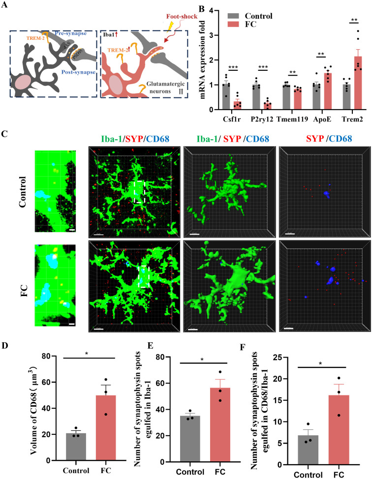
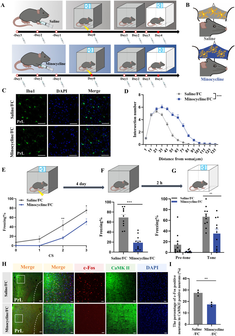
Methods: We successfully constructed a fear- formation model by foot-shock. Four days after foot-shock, microglial capacity of synaptic pruning was investigated via western blotting, immunofluorescence and Golgi-Cox staining. Prelimbic chemical deletion or microglia inhibition was performed to detect the role of microglia in synaptic loss and neuron activity. Finally, Trem2 knockout mice or wild-type mice with Trem2 siRNA injection were exposed to foot-shock to identify the involvement of TREM2 in fear memory formation.

Results: The results herein indicate that the foot-shock protocol in male mice resulted in a fear formation model. Mechanistically, fear conditioning enhanced the microglial capacity for engulfing synapse materials, and led to glutamatergic neuron activation in the prelimbic cortex. Prelimbic chemical deletion or microglia inhibition improved fear memory formation. Further investigation demonstrated that TREM2 regulates microglial phagocytosis, enhancing synaptic pruning. Trem2 knockout mice showed remarkable reductions in prelimbic synaptic pruning and reduced neuron activation, with decreased fear memory formation.

Discussion: Our cumulative results suggest that prelimbic TREM2-mediated excessive microglial synaptic pruning is involved in the fear memory formation process, leading to development of abnormal stress-related behavior.

Keywords: TREM2; fear memory formation; microglia; prelimbic; synaptic pruning.


Conflict of interest statement

The authors declare that the research was conducted in the absence of any commercial or financial relationships that could be construed as a potential conflict of interest.

Figures

Figure 1
Figure 1
Fear conditioning-induced microglial activation and fear memory formation. (A) Experimental diagram of control and fear condition. (B) Freezing level during three CS–US pairings or control CS exposures (n = 9 mice, ***P < 0.001). (C, D) Freezing level during contextual (C) and cued (D) fear memory test compared with control mice (n = 9 mice, ***P < 0.001).
Figure 2
Figure 2
Activation of prelimbic glutamatergic neurons during fear conditioning. (A) Representative image of prelimbic c-Fos (red) and CaMKII (green) (scale bars = 200 μm [first and third rows]; 20 μm [second and fourth rows]). (B) Quantification of c-Fos+ proportion among CaMKII+ neurons (n = 3 mice, 2 slices per mouse, **P < 0.01). (C) Quantification of CaMKII+ proportion among c-Fos+ neurons (n = 3 mice). (D) Representative image illustrating prelimbic AAV expression (scale bars = 100 μm). (E) Percentage of freezing in mice with AAV-CaMKIIα-hM4D(Gi)-mCherry injection during fear conditioning compared with control mice (n = 8 mice, ***P < 0.001). (F, G) Percentage of freezing during contextual (F) and cued (G) fear memory test (n = 8 mice, ***P < 0.001). (H) Representative image illustrating prelimbic AAV expression (scale bars = 100 μm). (I) Percentage of freezing in mice with AAV-CaMKIIα-hM3D(Gq)-mCherry injection during fear conditioning compared with control mice (n = 8 mice). (J, K) Percentage of freezing during contextual (J) and cued (K) fear memory test (n = 8 mice).
Figure 3
Figure 3
Fear memory formation drives increased microglial phagocytosis. (A) Schematic diagram showing fear conditioning-induced microglial activation and enhanced microglial capacity for engulfing synapse materials. (B) Relative mRNA expression in the prelimbic by qRT-PCR (n = 6 mice, **P < 0.01, ***P < 0.001). (C) Detailed figure (first row) and representative confocal (second row) and Imaris (middle and bottom rows) images of Iba1 (green), CD68 (blue), and SYP (red) from control and fear conditioned mice. Third row: reconstruction of Iba1 and CD68 staining. Fourth row: reconstruction of CD68 and SYP staining inside microglia. (D) CD68 volume inside microglia. (E) SYP puncta inside microglia. (F) SYP puncta inside CD68 phagosomes (n = 3 mice, 10 cells per mouse, *P < 0.05, scale bars = 5 μm and scale bars = 1 μm in detailed figure of the top row).
Figure 4
Figure 4
Fear memory formation drives increased microglial phagocytosis. (A) Representative confocal (top row) and Imaris (middle and bottom rows) images of Iba1 (green), CD68 (blue), and PSD95 (red) from control and fear conditioned mice. Middle row: reconstruction of Iba1 and CD68 staining. Bottom row: reconstruction of CD68 and PSD95 staining inside microglia. (B) PSD95 puncta inside microglia. (C) and PSD95 puncta inside CD68 phagosomes (n = 3 mice, 10 cells per mouse. *P < 0.05, **P < 0.01, scale bars = 5 μm and scale bars = 1 μm in detailed figure). (D, E) Protein level of prelimbic SYP and PSD95 detected by WB, and quantitative results of WB analyses (n = 6 mice, **P < 0.01). (F) Representative dendritic spine density of prelimbic neurons, and dendritic spine density quantification (n = 6 mice, 3 dendritic segments per mouse, **P < 0.01, Scale bars = 10 μm).
Figure 5
Figure 5
Ablation of microglia with PLX5622 protects against increased microglial phagocytosis and formation of fear memory. (A) Time course for PLX5622 and AIN-76A administrations and behavior tests. (B) Schematic diagram showing PLX5622 efficacy. (C) Representative confocal images of Iba1 immunostaining (green) of prelimbic microglia of mice with PLX5622 and AIN-76A feeding (scale bars = 100 μm). (D, E) Quantification of Iba1+ cells and Iba1+ area (n = 3 mice, 2 slices per mouse, ***P < 0.001). (F) Percentage of freezing in mice with control or PLX5622 feeding during fear conditioning (n =9 mice, *P < 0.05). (G, H) Percentage of freezing in control or PLX5622 feeding mice during contextual (G) and cued (H) fear memory test (n =9 mice, *P < 0.05, **P < 0.01). (I) Representative images of prelimbic c-Fos (red) and CaMKII (green) in mice with PLX5622 and AIN-76A feeding (scale bars = 100 μm [first row]; 20 μm [the other rows]). (J) Number of prelimbic CaMKII and c-Fos double-labeled neurons (n = 3 mice, 2 slices per mouse, *P < 0.05). (K, L) Protein level of prelimbic SYP and PSD95 detected by WB, and quantitative results for mice treated with or without PLX5622 feeding (n = 6 mice, ***P < 0.001). (M) Representative prelimbic neuron dendritic spine density, and dendritic spine density quantification for mice treated with or without PLX5622 feeding (n = 6 mice, 3 dendritic segments per mouse, **P < 0.01, scale bars = 10 μm).
Figure 6
Figure 6
Microglia inhibition with minocycline protects against increased neuronal activity and fear memory formation. (A) Time course for i.p. minocycline or saline injections and behavior tests. (B) Schematic diagram showing minocycline efficacy. (C) Representative confocal images of Iba1 immunostaining (green) of microglia in the prelimbic of mice with minocycline injection (scale bars = 50 μm). (D) Sholl analysis of microglial morphology of mice with or without minocycline (n = 3 mice, 10 cells per mouse, ***P < 0.001). (E) Percentage of freezing in control or minocycline-treated mice during fear conditioning (n = 11 mice, *P < 0.05, **P < 0.01). (F, G) Percentage of freezing in control or minocycline-treated mice during contextual (F) and cued (G) fear memory test (n =11 mice, **P < 0.01, ***P < 0.001). (H) Representative image of prelimbic c-Fos (red) and CaMKII (green) of mice with minocycline microinjection (scale bars = 100 μm [first row]; 20 μm [the other rows]). (I) Number of prelimbic CaMKII and c-Fos double-labeled neurons (n = 3 mice, 2 slices per mouse, **P < 0.01).
Figure 7
Figure 7
Inhibition of microglia with minocycline protects against increased microglial phagocytosis. (A) Representative confocal (top row) and Imaris (middle and bottom rows) images of Iba1 (green), CD68 (blue), and SYP (red) from saline- and minocycline-treated mice. Middle row: reconstruction of Iba1 and CD68 staining. Bottom row: reconstruction of CD68 and SYP staining inside microglia. (B) CD68 volume inside microglia. (C) SYP puncta inside microglia. (D) SYP puncta inside CD68 phagosomes (n = 3 mice, 10 cells per mouse, *P < 0.05, scale bars = 5 μm and scale bars = 1 μm in detailed figure). (E) Representative confocal (top row) and Imaris (middle and bottom rows) images of Iba1 (green), CD68 (blue), and PSD95 (red) from saline- and minocycline-treated mice. Middle row: reconstruction of Iba1 and CD68 staining. Bottom row: reconstruction of CD68 and PSD95 staining inside microglia. (F) PSD95 puncta inside microglia. (G) PSD95 puncta inside CD68 phagosomes (n = 3 mice, 10 cells per mouse, *P < 0.05, scale bars = 5 μm and scale bars = 1 μm in detailed figure). (H, I) Protein levels of prelimbic SYP and PSD95 detected by WB, and WB quantification (n = 6 mice, **P < 0.01). (J) Representative prelimbic neuron dendritic spine density, and dendritic spine density quantification (n = 6 mice, 3 dendritic segments per mouse, ***P < 0.001, scale bars = 10 μm).
Figure 8
Figure 8
Prelimbic TREM2 expression is upregulated after foot-shock exposure. (A) Representative plots of CD11b+CD45int iLy6Gmicroglia, CD11b+CD45hiLy6G macrophages, and CD11b+CD45hiLy6Ghi PMNs populations (3000 cells in total) after foot-shock exposure. (B) Representative histograms of microglial TREM2 expression. (C) Prelimbic microglial TREM2 MFI (n = 5 mice, **P < 0.01). (D) Prelimbic TREM2 protein level detected by WB, and WB quantification (n = 6 mice, ***P < 0.001).
Figure 9
Figure 9
TREM2 inhibition affects neuronal activity and fear memory formation. (A) Representative confocal images of prelimbic microglia Iba1 immunostaining (green) in Trem2 KO mice (scale bars = 50 μm). (B) Sholl analysis of microglial morphology (n = 3 mice, 10 cells per mouse, ***P < 0.001). (C) Percentage of freezing in Trem2 KO mice during fear conditioning (n = 8 mice, ***P < 0.001). (D, E) Percentage of freezing during contextual (D) and cued (E) fear memory test (n = 8 mice, *P < 0.05, **P < 0.01). (F) Representative image of prelimbic c-Fos (red) and CaMKII (green) in Trem2 KO mice (scale bars = 100 μm [first row]; 20 μm [the other rows]). (G) Schematic diagram showing that TREM2 inhibition protects against prelimbic glutamatergic neuron activation after exposed to foot-shock. (H) Number of prelimbic CaMKIIα and c-Fos double-labeled neurons (n = 3 mice, 2 slices per mouse, **P < 0.01).
Figure 10
Figure 10
TREM2 inhibition protects against increased microglial phagocytosis. (A) Representative confocal (top row) and Imaris (middle and bottom rows) images of Iba1 (green), CD68 (blue), and SYP (red) from WT and Trem2 KO control mice and WT and Trem2 KO mice with three CS–US pairings exposure. Middle row: reconstruction of Iba1 and CD68 staining. Bottom row: reconstruction of CD68 and SYP staining inside microglia. (B) CD68 volume inside microglia. (C) SYP puncta inside microglia. (D) SYP puncta inside CD68 phagosomes (n = 3 mice, 10 cells per mouse, *P < 0.05, **P < 0.01, scale bars = 5 μm and scale bars = 1 μm in detailed figure). (E) Representative confocal (top row) and Imaris (middle and bottom rows) images of Iba1 (green), CD68 (blue), and PSD95 (red). Middle row: reconstruction of Iba1 and CD68 staining. Bottom row: reconstruction of CD68 and PSD95 staining inside microglia. (F) PSD95 puncta inside microglia. (G) PSD95 puncta inside CD68 phagosomes (n = 3 mice, 10 cells per mouse, *P < 0.05, ***P < 0.001, scale bars = 5 μm and scale bars = 1 μm in detailed figure). (H, I) Protein level of prelimbic SYP and PSD95 detected by WB, and quantitative WB results (n = 6 mice, *P < 0.05, **P < 0.01). (J) Representative prelimbic neuron dendritic spine density, and dendritic spine density quantification (n = 6 mice, 3 dendritic segments per mouse, **P < 0.01, scale bars = 10 μm).
Figure 11
Figure 11
Microglial TREM2 participates in fear memory formation through excessive prelimbic cortex synaptic pruning. Fear conditioning induced microglial activation and enhanced microglial capacity for engulfing synapse materials, which are mainly on glutamatergic neurons. TREM2 also served as a key microglial regulator, leading to increasing synaptic pruning.

Similar articles

Cited by

References

    1. Bocchio M, Nabavi S, Capogna M. Synaptic plasticity, engrams, and network oscillations in amygdala circuits for storage and retrieval of emotional memories. Neuron. (2017) 94:731–43. doi: 10.1016/j.neuron.2017.03.022 - DOI - PubMed
    1. Careaga MBL, Girardi CEN, Suchecki D. Understanding posttraumatic stress disorder through fear conditioning, extinction and reconsolidation. Neurosci Biobehav Rev. (2016) 71:48–57. doi: 10.1016/j.neubiorev.2016.08.023 - DOI - PubMed
    1. Ilovich O, Dines M, Paul BK, Barkai E, Lamprecht R. Nck1 activity in lateral amygdala regulates long-term fear memory formation. Trans Psychiatry. (2022) 12:475. doi: 10.1038/s41398-022-02244-x - DOI - PMC - PubMed
    1. Enomoto S, Kato TA. Involvement of microglia in disturbed fear memory regulation: possible microglial contribution to the pathophysiology of posttraumatic stress disorder. Neurochem Int. (2021) 142:104921. doi: 10.1016/j.neuint.2020.104921 - DOI - PubMed
    1. Chen MB, Jiang X, Quake SR, Sudhof TC. Persistent transcriptional programmes are associated with remote memory. Nature. (2020) 587:437–42. doi: 10.1038/s41586-020-2905-5 - DOI - PMC - PubMed

LinkOut - more resources