. 2024 Oct 31:15:1461841.
doi: 10.3389/fimmu.2024.1461841. eCollection 2024.

Dexamethasone and IFN-γ primed mesenchymal stem cells conditioned media immunomodulates aberrant NETosis in SLE via PGE2 and IDO

Affiliations

Dexamethasone and IFN-γ primed mesenchymal stem cells conditioned media immunomodulates aberrant NETosis in SLE via PGE2 and IDO

Khushbu Priya et al. Front Immunol. .

Abstract

Background: Systemic Lupus Erythematosus (SLE) is characterized by dysregulated immune responses, with neutrophil extracellular traps (NETs) playing a significant role. NETs are recognized by autoantibodies in SLE patients, exacerbating pathology. Both excessive NET formation and impaired degradation contribute to SLE pathophysiology.

Objective: To investigate the immunomodulatory effects of Dexamethasone-primed Wharton's jelly (WJ) derived MSCs CM (DW) and IFN-γ-primed WJ-MSCs-CM (IW) on NETosis and associated protein markers in SLE patients' LPS or ribonucleoprotein immune complexes (RNP ICs) induced neutrophils and in pristane induced lupus (PIL) model. And to elucidate the mechanism involved therein.

Methods: We investigated the immunomodulatory effects of DW and IW on NETosis in SLE. Utilizing ex vivo and in vivo models, we assessed the impact of preconditioned media on NET formation and associated protein markers neutrophil elastase (NE), citrullinated histone (citH3), myeloperoxidase (MPO), cytoplasmic and mitochondrial ROS production. We also examined the involvement of key immunomodulatory factors present in DW and IW, including prostaglandin E2 (PGE2), indoleamine 2,3-dioxygenase (IDO), and transforming growth factor-beta (TGF-β).

Results: Preconditioned media effectively suppressed NETosis and reduced ROS generation in SLE neutrophils, indicating their immunomodulatory potential. Inhibition studies implicated IDO and PGE2 in mediating this effect. Combined treatment with DW or IW together with hydroxychloroquine (HCQ) demonstrated superior efficacy over HCQ alone, a standard SLE medication. In PIL mouse model, DW and IW treatments reduced NETosis, ROS generation, as evidenced by decreased NET-associated protein expression in vital organs.

Conclusion: Our study highlights the multifaceted impact of IW and DW on NETosis, ROS dynamics, and lupus severity in SLE. These findings underscore the potential of preconditioned media for the development of targeted, personalized approaches for SLE treatment.

Keywords: NETosis; hydroxychloroquine; lupus model; neutrophil extracellular trap formation; reactive oxygen species; ribonucleoprotein immune complexes; systemic lupus erythematosus.


Conflict of interest statement

The authors declare that the research was conducted in the absence of any commercial or financial relationships that could be construed as a potential conflict of interest. The author(s) declared that they were an editorial board member of Frontiers, at the time of submission. This had no impact on the peer review process and the final decision.

Figures

Figure 1
Figure 1
Immunomodulatory effect of DW and IW on NET formation, neutrophil viability and ROS generation in LPS induced SLE patients’ neutrophils ex vivo. The image reveals (A) NET formation in healthy adults and SLE patients’ neutrophils (B) decreased NET formation and (C) low expression of NET associated markers in DW and IW treated cells. NET formation was detected by confocal microscopy using two binding dyes; Sytox orange (red) and syto13 (green). Netting neutrophils were confirmed (as NE/CitH3/DNA overlap) by immunofluorescence staining with anti-NE (red), CitH3 (green) and DNA (blue). (At 40X, Scale Bar -100 um). The data in this figure are from 3 independent experiments. DW and IW treatments restored the compromised (D) cell viability (n=9) and (E) NETosis (n=20) incurred by LPS induction. DW and IW treatment suppress (F) mitochondrial as well as (G) cytoplasmic ROS generation in LPS induced SLE patients’ neutrophils. The line graphs illustrate mean data from a sample OF 6 SLE patient. Error bars show mean ± SEM. p values indicate significant changes as follows: non-significant (ns) p > 0.05, *p < 0.05, **p < 0.01, ***p < 0.001 and ****p < 0.0001; One-way ANOVA.
Figure 2
Figure 2
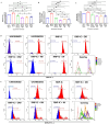
Immunomodulatory effect of DW and IW on NETosis and ROS generation in non-induced SLE patients’ neutrophils and healthy adult. DW and IW treatment suppress (A) extracellular DNA release (B) mitochondrial as well as (C) cytoplasmic ROS generation in uninduced SLE patients’ (n=3) neutrophils and (D) LPS induced NETosis in healthy adults (n=3). The line graphs illustrate mean data from a sample of 3 SLE patient and healthy adult. Error bars show mean ± SEM. p values indicate significant changes as follows: non-significant (ns) p > 0.05, *p < 0.05, **p < 0.01, ***p < 0.001 and ****p < 0.0001; One-way ANOVA.
Figure 3
Figure 3
Immunomodulatory effect of DW and IW on NETosis and ROS generation in RNP-IC induced SLE patients’ neutrophils. DW and IW treatment suppress (A) extracellular DNA release and (B) mitochondrial as well as (C) cytoplasmic ROS generation in RNP-IC induced SLE patients’ neutrophils. (D) Mitochondrial and (E) cytoplasmic ROS was also determined by flow cytometry. The line graphs illustrate mean data and histogram represent individual data from a sample of 3 SLE patient. Error bars show mean ± SEM. p values indicate significant changes as follows: non-significant (ns) p > 0.05, *p < 0.05, **p < 0.01 and ****p < 0.0001; One-way ANOVA.
Figure 4
Figure 4
Immunomodulatory effect of DW/IW and HCQ combination on NETosis and ROS generation in RNP-IC positive serum induced SLE patients’ neutrophils. DW/IW and HCQ treatment suppress (A) extracellular DNA release and (B) mitochondrial as well as (C) cytoplasmic ROS generation in RNP-IC induced SLE patients’ neutrophils. The line graphs illustrate mean data from a sample of 3 SLE patient. Error bars show mean ± SEM. p values indicate significant changes as follows: non-significant (ns) p > 0.05, *p < 0.05, **p < 0.01, ***p < 0.001 and ****p < 0.0001; One-way ANOVA.
Figure 5
Figure 5
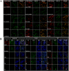
DW and IW immunomodulates NETosis and ROS generation through IDO and PGE2. The image reveals (A) NET formation (B) NET associated markers in SLE patients’ neutrophils upon inhibition by using specific inhibitors for TGF-β (SB-431542), PGE2 (NS398) and IDO (1MT). NET formation was detected by confocal microscopy using two binding dyes; Sytox orange (red) and syto13 (green). Netting neutrophils were confirmed (as NE/CitH3/DNA overlap) by immunofluorescence staining with anti-NE (red), CitH3 (green) and DNA (blue). (At 63X objective, Scale Bar -100 um). The data in this figure are from 3 independent experiments.
Figure 6
Figure 6
Role of TGF-β and IDO, TGF-β and PGE2, IDO and PGE2 together in DW and IW mediated suppression of NETosis and ROS generation. The data reveals changes in NETosis, as well as mitochondrial and cytoplasmic ROS generation upon inhibition of (A) TGF-β and IDO, TGF-β and PGE2 and IDO and PGE2 in DW (A–C) and IW treated neutrophils (D–F). The violin plot illustrate mean data from a sample of 3 SLE patient. Error bars show mean ± SEM. p values indicate significant changes as follows: non-significant (ns) p > 0.05, *p < 0.05, **p < 0.01, ***p < 0.001 and ****p < 0.0001; One-way ANOVA.
Figure 7
Figure 7
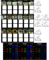
Immunomodulatory effect of DW and IW on NET formation and ROS generation in PIL mice. (A) The image reveals decreased NET formation in DW and IW treated PIL mice. NET formation was detected by confocal microscopy using two binding dyes; Sytox orange (red) and syto13 (green). DW and IW treatment suppress (B) extracellular DNA release (C) mitochondrial as well as (D) cytoplasmic ROS generation in PIL mice. The line graphs illustrate mean data from 5 mice. (E) The image reveals low expression of NET associated markers (CitH3 and NE) in DW and IW treated PIL mice. Netting neutrophils were confirmed (as NE/CitH3/DNA overlap) by immunofluorescence staining with anti-NE (red), CitH3 (green) and DNA (blue). (At 63X, Scale Bar - 20 µm). The data in this figure are from 3 independent experiments. Error bars show mean ± SEM. p values indicate significant changes as follows: non-significant (ns) p > 0.05, *p < 0.05, **p < 0.01, ***p < 0.001 and ****p < 0.0001; One-way ANOVA. ##P < 0.01, ###P < 0.001, ####P < 0.0001 and ns (non-significant). * - compared to Healthy (Normal Saline) and # - compared to PIL (Normal Saline).
Figure 8
Figure 8
Immunomodulatory effect of DW and IW on extracellular DNA release and MPO in different organs of PIL mice. (A) Both DW and IW efficiently reduced extracellular DNA release and MPO in (A–C) liver, (D–H) kidneys and (I–K) heart of PIL mice in vivo as observed in PAI system. (B, C) Data are presented as mean ± standard deviation from n=6 mice/group. Error bars show mean ± SEM. p values indicate significant changes as follows: non-significant (ns) p > 0.05, *p < 0.05, **p < 0.01, ***p < 0.001 and ****p < 0.0001; One-way ANOVA. The color green denotes MPO and red denotes Sytox orange. (L) DW and IW reduced NET associated markers, NE and CitH3 in lungs, heart and kidney of PIL mice. The image reveals low expression of NET associated markers (CitH3 and NE) in different organs upon DW and IW treatment in PIL mice. NET formation was detected by confocal microscopy. Netting neutrophils were confirmed (as NE/CitH3/DNA overlap) by immunofluorescence staining with anti-NE (red), CitH3 (green) and DNA (blue). (At 63X, Scale Bar - 1000 µm). The data in this figure are from 3 independent experiments.

References

    1. Pan L, Lu MP, Wang JH, Xu M, Yang SR. Immunological pathogenesis and treatment of systemic lupus erythematosus. World J Pediatr. (2020) 16:19–30. doi: 10.1007/s12519-019-00229-3 - DOI - PMC - PubMed
    1. Fatemi A, Alipour R, Khanahmad H, Alsahebfosul F, Andalib A, Pourazar A. The impact of neutrophil extracellular trap from patients with systemic lupus erythematosus on the viability, CD11b expression and oxidative burst of healthy neutrophils. BMC Immunol. (2021) 22:12. doi: 10.1186/s12865-021-00402-2 - DOI - PMC - PubMed
    1. Kaplan MJ, Radic M. Neutrophil extracellular traps: double-edged swords of innate immunity. J Immunol. (2012) 189:2689–95. doi: 10.4049/jimmunol.1201719 - DOI - PMC - PubMed
    1. Brinkmann V, Reichard U, Goosmann C, Fauler B, Uhlemann Y, Weiss DS, et al. . Neutrophil extracellular traps kill bacteria. Science. (2004) 303:1532–5. doi: 10.1126/science.1092385 - DOI - PubMed
    1. Hakkim A, Fürnrohr BG, Amann K, Laube B, Abed UA, Brinkmann V, et al. . Impairment of neutrophil extracellular trap degradation is associated with lupus nephritis. Proc Natl Acad Sci USA. (2010) 107:9813–8. doi: 10.1073/pnas.0909927107 - DOI - PMC - PubMed

MeSH terms