

Solitary pituitary metastasis from breast cancer masquerading as a pituitary macroadenoma: a report of 2 rare cases
- PMID: 39544980
- PMCID: PMC11558294
- DOI: 10.21037/gs-24-284
Solitary pituitary metastasis from breast cancer masquerading as a pituitary macroadenoma: a report of 2 rare cases
Abstract
Background: Breast cancer is one of the most common malignant tumors, occurring in the mammary glands, which often metastasizes to bones, lungs, and liver. However, pituitary metastasis (PM) originating from breast cancer is a rare phenomenon that can easily be mistaken for benign pituitary macroadenoma.
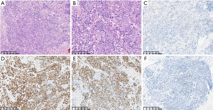
Case description: This report details two cases of middle-aged and elderly Chinese women who presented with serious neurological symptoms, each with a history of breast malignancy. Both patients underwent magnetic resonance imaging (MRI), which showed a lesion in the sellar region, suggesting a pituitary adenoma. In one case, intraoperative frozen biopsy samples initially suggested a benign pituitary macroadenoma. However, post-surgical resection and permanent pathology combined with immunohistochemical stains confirmed both cases as symptomatic PM from breast cancer. Following surgery, one patient had a favorable postoperative prognosis, while the other unfortunately succumbed to systemic disease progression 5 months later.
Conclusions: A history of a malignancy should raise the suspicion for metastatic disease in patients presenting with a lesion in the sellar region accompanied by symptoms. Due to its low incidence, diagnosing PM preoperatively is challenging. This case report aims to raise awareness among healthcare providers that this condition is crucial for timely and accurate diagnosis.
Keywords: Breast cancer; case report; pituitary adenoma; pituitary metastasis (PM).
2024 AME Publishing Company. All rights reserved.
Conflict of interest statement
Conflicts of Interest: Both authors have completed the ICMJE uniform disclosure form (available at https://gs.amegroups.com/article/view/10.21037/gs-24-284/coif). The authors have no conflicts of interest to declare.
Figures


Similar articles
-
Melanoma metastasis to a nonfunctioning pituitary macroadenoma: illustrative case.J Neurosurg Case Lessons. 2021 Jun 7;1(23):CASE2167. doi: 10.3171/CASE2167. eCollection 2021 Jun 7. J Neurosurg Case Lessons. 2021. PMID: 36046510 Free PMC article.
-
Early and isolated breast cancer metastasis to the pituitary: A case report and systematic review.Surg Neurol Int. 2022 Oct 14;13:462. doi: 10.25259/SNI_1053_2021. eCollection 2022. Surg Neurol Int. 2022. PMID: 36324911 Free PMC article.
-
Pituitary Metastasis Presenting With Central Diabetes Insipidus and Panhypopituitarism.AACE Clin Case Rep. 2021 Jun 16;8(1):15-18. doi: 10.1016/j.aace.2021.06.006. eCollection 2022 Jan-Feb. AACE Clin Case Rep. 2021. PMID: 35097195 Free PMC article.
-
Primary sellar melanocytic tumor mimicking hemorrhagic pituitary macroadenoma: Case report and literature review.Br J Neurosurg. 2015 Apr;29(2):298-302. doi: 10.3109/02688697.2014.967752. Epub 2015 Jan 23. Br J Neurosurg. 2015. PMID: 25613300 Review.
-
Diabetes insipidus and panhypopituitarism as a first presentation of silent adenocarcinoma of lung: a case report and literature review.BMC Endocr Disord. 2019 Oct 29;19(1):114. doi: 10.1186/s12902-019-0445-5. BMC Endocr Disord. 2019. PMID: 31664980 Free PMC article. Review.
References
Publication types
LinkOut - more resources
Full Text Sources