. 2024 Oct 1;134(22):e179527.
doi: 10.1172/JCI179527.

Inducible CCR2+ nonclassical monocytes mediate the regression of cancer metastasis

Affiliations

Inducible CCR2+ nonclassical monocytes mediate the regression of cancer metastasis

Xianpeng Liu et al. J Clin Invest. .

Abstract

A major limitation of immunotherapy is the development of resistance resulting from cancer-mediated inhibition of host lymphocytes. Cancer cells release CCL2 to recruit classical monocytes expressing its receptor CCR2 for the promotion of metastasis and resistance to immunosurveillance. In the circulation, some CCR2-expressing classical monocytes lose CCR2 and differentiate into intravascular nonclassical monocytes that have anticancer properties but are unable to access extravascular tumor sites. We found that in mice and humans, an ontogenetically distinct subset of naturally underrepresented CCR2-expressing nonclassical monocytes was expanded during inflammatory states such as organ transplant and COVID-19 infection. These cells could be induced during health by treatment of classical monocytes with small-molecule activators of NOD2. The presence of CCR2 enabled these inducible nonclassical monocytes to infiltrate both intra- and extravascular metastatic sites of melanoma, lung, breast, and colon cancer in murine models, and they reversed the increased susceptibility of Nod2-/- mutant mice to cancer metastasis. Within the tumor colonies, CCR2+ nonclassical monocytes secreted CCL6 to recruit NK cells that mediated tumor regression, independent of T and B lymphocytes. Hence, pharmacological induction of CCR2+ nonclassical monocytes might be useful for immunotherapy-resistant cancers.

Keywords: Cancer; Immunology; Lung cancer.


Conflict of interest statement

Conflict of interest: The authors have declared that no conflict of interest exists.

Figures

Figure 1
Figure 1. Ontogenetic dichotomy of I-NCMs and N-NCMs.
(AC) NOD2 activation in vivo by MDP but not isomer control induced conversion of blood CMs in WT and Nr4a1–/–, but not Nod2–/– or double-mutant Nr4a1–/– Nod2–/–, mice to I-NCMs (A), in a dose- (B) and time-dependent (C) manner. (D and E) Inhibition of the NOTCH2 signaling pathway by either gliotoxin (10 mg/kg) or DAPT (25 mg/kg) did not impair the development of I-NCMs in Nr4a1–/– mice but reduced N-NCMs in Nod2–/– mice. (F and G) Development of I-NCMs was not altered by conditional genetic depletion of TLR7 in CCR2+ CM or TLR7/8 activation by R848 treatment (50 μg/mouse). (HL) Four weeks after splenectomy or sham surgery, mice were treated with MDP or control for 36 hours, and the monocyte subsets were analyzed by flow cytometry. (HJ) The increase in I-NCMs in the bone marrow (H), blood (I), and lung (J) was not impaired by splenectomy (Splx) in Nr4a1–/– mice. Interstitial macrophages (IMs); monocyte-derived dendritic cells (MoDCs). (K) The ratio of LY6Clo NCMs in blood monocytes was stabilized 2 weeks after splenectomy in the blood of WT B6 mice. n = 5. (L) Ratio of monocyte subsets in total monocytes in BM, blood, and lung in Nod2–/– mice (n = 3–4). Unless otherwise mentioned, MDP treatment indicates 1–2 doses of 10 mg/kg MDP via retro-orbital injection for 36 hours; n = 3–12 mice in each group; data are presented as mean ± SEM. *P < 0.05; **P < 0.01, ***P < 0.001; 2-tailed t test was used for A, F, and G; Kruskal-Wallis (nonparametric) test for C: IntM and NCM, J: NCM, and K: IntM; 1-way ANOVA for the rest of the panels.
Figure 2
Figure 2. I-NCMs and N-NCMs are phenotypically and transcriptionally distinct.
(AC) Ultrastructural examination of monocyte subsets by TEM. (A and B) Experimental design and representative TEM images. Blood LY6Chi or LY6Clo monocyte subsets were sorted from untreated Nod2–/– or MDP-treated Nr4a1–/– mice and embedded with agarose for TEM. Scale bars, left columns: 1 μm; right column: 500 μm; magnification, left columns ×6,800; right columns, ×13,000. (C) Statistical analysis of cell size, number of pseudopods (P), mitochondria (M), and liposomes (L) and ratio of euchromatin (Eu) in the 4 monocyte subsets shown in A and B based on the TEM data. Approximately 7–28 cells were counted or measured in each group; data are presented as mean ± SEM; *P < 0.05, **P < 0.01, ***P < 0.001; Kruskal-Wallis test or Brown-Forsythe and Welch’s ANOVA. (DH) Transcriptomic profiling of I-NCMs and N-NCMs. Blood samples were taken from the facial vein of MDP-treated Nr4a1–/– (I-NCMs) or Nod2–/– (N-NCMs) mice and used for RNA extraction and subsequent bulk RNA-Seq. (D) Experimental design schematic showing sample preparation. (E) Volcano plot demonstrating significantly (FDR <0.05) differentially expressed genes (DEGs) in I-NCMs and N-NCMs. (F) Heatmap analysis (left) demonstrated differential gene expression and pathway enrichment analysis (right) in I-NCMs and N-NCMs. (G and H) Pairwise comparison of gene expression of the selected gene signatures in blood monocyte subsets from MDP-treated Nr4a1–/– or Nod2–/– mice. TFs, transcription factors. In G and H, RNA-Seq CPM data are presented as mean ± SEM; n = 3-4 in each group; *P < 0.05, **P < 0.01, ***P < 0.001; 1-way ANOVA.
Figure 3
Figure 3. I-NCMs mediate the regression of melanoma metastasis.
(A) Representative 2-photon images showing accumulation and colonization of B16F10-GFP in the lung at the indicated time points after retro-orbital injection into Nod2–/– mice. Scale bar: 100 μm. Dx, dextran (B) Accumulation of B16F10-LUC2 cells was detected and determined by luciferase activity using LAGO on day 3 after retro-orbital injection (d.p.i.). (C) Detection of melanoma cells in lungs in WT B6, Nr4a1–/–, and Nod2–/– mice by LAGO at the indicated dose and time points. The lung was isolated and photographed to visualize the accumulation of black dots in the metastatic B16F10-LUC2 cells after LAGO detection. (D) Metastatic B16F10 colonization in lung and other tissues was analyzed in monocyte-depleted mice. (E and F) Adoptive transfer of I-NCMs reduced metastatic colonization (E) and attenuated established metastatic B16F10 melanoma colonies (F). Approximately 1 × 10⁵ I-NCMs from MDP-treated Nr4a1–/– mice were retro-orbitally injected into Nod2–/– mice prior to or after the injection of 3 × 10⁵ B16F10-LUC2 cells. Colonization of B16 in the lung was determined by LAGO detection of luciferase activity. In E and F, the experimental design is shown in the upper panel; a representative image and quantification of LAGO detection of luciferase activity in B16F10-LUC2 cells in the lung are shown in the lower-left panel and lower-right panel, respectively. The data are presented as mean ± SEM; n = 3–13 in each group; *P < 0.05, **P < 0.01, ***P < 0.001; 2-tailed t test in B, E, and F; 1-way ANOVA in C.
Figure 4
Figure 4. Pharmacological induction of I-NCMs promotes regression of hematogenous metastasis from multiple cancers.
MDP or L-MTP-PE attenuates metastatic colonization of multiple cancers. (A and B) MDP (A) or the liposome-conjugated MDP analog MTP (L-MTP-PE, or mifamurtide) (B) reduced the number of established B16F10 clusters in Nr4a1–/– mice. (C) MDP treatment attenuated the accumulation of established LL/2-LUC lung cancer cells in Nr4a1–/– mice. (DF) MDP treatment attenuated accumulation of the established breast cancer 4T1-LUC-GFP (D) or colon cancer CT26-LUC-GFP (E), or melanoma B16F10-LUC2 (F) in WT BALB/c mouse lung. Data are presented as mean ± SEM; n = 5–10 mice in each group; *P < 0.05, **P < 0.01, ***P < 0.001; 2-tailed t test in AF.
Figure 5
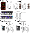
Figure 5. Retention of CCR2 on I-NCMs is crucial for their migration to tumor metastasis.
(A and B) Intravascular and extravascular distribution of established B16F10-GFP clusters in mouse lung. (A) Representative confocal images show immunostaining of CD31 (endothelial cell marker, red) and B16-GFP (green) in Nr4a1–/– mice. B16F10-GFP clusters on the lung surface are indicated by dashed white lines. White and yellow arrows indicate the areas of extravascular and intravascular B16F10-GFP cells, respectively. White and yellow scale bars: 100 and 20 μm, respectively. (B) GFP intensity in 10–22 areas from multiple slides from the deeper regions in lung from WT B6, Nod2–/–, and Nr4a1–/– mice (n = 3–4) was determined by use of ImageJ and quantified. Data are presented as mean ± SEM; **P < 0.01; 1-way ANOVA. (C and D) Experimental design (C) and representative 2-photon images (D) showing colocalization of I-NCMs with the established B16F10-GFP metastatic clusters in mouse lung. The I-NCMs were sorted, stained with Hoechst 33342, and adoptively transferred into Nod2–/– mice bearing B10F10-GFP cells in the lung as described in C. Intravital 2-photon imaging was used for tracing I-NCMs. Blue: Hoechst 33342–stained I-NCMs; green: B16F10-GFP; red: Dx-rhodamine–labeled blood vessel. Colocalization of blue and green signals is indicated in the area circled with a dashed white line in D. Scale bars: 50 μm. (E and F) RFP-labeled I-NCMs (3.5 × 10⁵ cells) were sorted and adoptively transferred into Nr4a1–/– Nod2–/– double-mutant mice bearing B10F10-GFP cells in the lung as described for E. Accumulation and distribution of the adoptively transferred RFP–I-NCMs were detected and determined by confocal imaging of lung sections immunostained with CD31 (endothelial cell marker; white), GFP (B16F10-GFP), and RFP (RFP-I-NCMs) in F. Yellow and blue arrows indicate the typical RFP-labeled I-NCMs outside and inside the blood vessel within the B16F10-GFP clusters, respectively. White and yellow scale bars: 50 and 20 μm, respectively. (G and H) Expression of CCL2 in metastatic B16F10 clusters and adjacent areas in mouse lungs. Expression of CCL2 was detected by IHC in the lungs (G) of untreated WT B6 control mice (n = 5, left) and Nr4a1–/– mice bearing B16F10-GFP melanoma cells (n = 4, right). White and yellow scale bars: 100 and 10 μm, respectively. RFP intensity in 34–96 areas from multiple slides was measured by use of ImageJ and quantified (H). FI, fluorescence intensity. The FI data are presented as mean ± SEM; n = 4–5 mice in each group; *P < 0.05; Forsythe’s and Welch’s ANOVA. (I and J) CCR2 deficiency impaired the accumulation and extravasation of MDP-triggering CCR2+ monocytes from blood vessels at the B16F10 metastasis site. Experimental design (I) and representative confocal images (J) showing typical extravasated (J, top row, indicated with white arrows) or intravascular (J, bottom row, indicated with yellow arrows) MDP-triggering CCR2+ monocytes in Nr4a1–/–Nod2–/– mouse lungs. Scale bars: 20 μm.
Figure 6
Figure 6. I-NCMs recruit NK cells to promote tumor regression.
(AE) Detection of the uptake of B10F10-GFP by I-NCMs in vitro (AC) and in vivo (D and E). Experimental design (A) and uptake of B16F10-GFP by RFP-labeled splenic (B) or blood (C) I-NCMs at 36 hours following coculture. The GFP signal in I-NCMs was detected by flow cytometry (B) or ICC (C). Monocytes and B16 cells were cocultured at a ratio of 5:1. White and yellow scale bars: 100 and 10 μm, respectively, in C. Experimental design (D) and FACS detection of uptake of B16F10-GFP materials by monocyte subsets in blood (E, left) or lung (right) in Nr4a1–/– mice after retro-orbital injection of MDP and B16F10-GFP injection. (FK) I-NCMs recruit NK cells through CCL6 release to sites of B16F10 melanoma metastasis. (F and G) Adoptive transfer of I-NCMs (F) and MDP treatment (G) increased NK cells in the lungs of B16F10-bearing Nod2–/– and Nr4a1–/– mice, respectively. (H) MDP-triggered attenuation of B16F10 colonization in Nr4a1–/– mice was inhibited by depletion of NK cells using NK1.1 antibody. (I) Bulk RNA-Seq data showing higher expression of Ccl6 and Ccl9, but not other detected chemokine genes, in I-NCMs. (J) Anti-CCL6 antibody reduced NK cells and (K) suppressed the MDP-mediated attenuation of B16F10 colonization in Nr4a1–/– mice. Data are presented as mean ± SEM; n = 4–10 in each group; *P < 0.05, **P < 0.01, ***P < 0.001; 2-tailed t test in F and J, Kruskal-Wallis test in H, and 1-way ANOVA in G, I, and K.
Figure 7
Figure 7. Regression of metastasis by I-NCMs is independent of T and B lymphocytes.
(A and B) MDP treatment reduced the established B16F10 clusters in the lung in T cell–deficient Rag1–/– mice but not in T cell–deficient and NK-impaired NOD/SCID mice. (C and D) MDP treatment suppressed the established B16F10 clusters in Nr4a1–/– lungs (C) in the absence of CD4+ and CD8+ T cells (D). Data are presented as mean ± SEM; n = 3–10 in each group; **P < 0.01, ***P < 0.001; 2-tailed t test in A and B (for 2-column comparison), 1-way ANOVA in B (for multiple-column comparison) and C and D.

References

    1. Xu M, et al. Role of the CCL2-CCR2 signalling axis in cancer: mechanisms and therapeutic targeting. Cell Prolif. 2021;54(10):e13115. doi: 10.1111/cpr.13115. - DOI - PMC - PubMed
    1. Li C, et al. Tumor-associated macrophages: potential therapeutic strategies and future prospects in cancer. J Immunother Cancer. 2021;9(1):e001341. doi: 10.1136/jitc-2020-001341. - DOI - PMC - PubMed
    1. Chen C, et al. LNMAT1 promotes lymphatic metastasis of bladder cancer via CCL2 dependent macrophage recruitment. Nat Commun. 2018;9(1):3826. doi: 10.1038/s41467-018-06152-x. - DOI - PMC - PubMed
    1. Cassetta L, Pollard JW. Targeting macrophages: therapeutic approaches in cancer. Nat Rev Drug Discov. 2018;17(12):887–904. doi: 10.1038/nrd.2018.169. - DOI - PubMed
    1. Nywening TM, et al. Targeting tumour-associated macrophages with CCR2 inhibition in combination with FOLFIRINOX in patients with borderline resectable and locally advanced pancreatic cancer: a single-centre, open-label, dose-finding, non-randomised, phase 1b trial. Lancet Oncol. 2016;17(5):651–662. doi: 10.1016/S1470-2045(16)00078-4. - DOI - PMC - PubMed

LinkOut - more resources