. 2025 Jan;24(1):e16638.
doi: 10.1111/jocd.16638. Epub 2024 Nov 15.

Microfocused Ultrasound With Visualization Induces Remodeling of Collagen and Elastin Within the Skin

Affiliations

Microfocused Ultrasound With Visualization Induces Remodeling of Collagen and Elastin Within the Skin

Kay Marquardt et al. J Cosmet Dermatol. 2025 Jan.

Abstract

Purpose: Microfocused ultrasound with real-time visualization (MFU-V) is often used for noninvasive skin lifting, by precisely targeting dermal and subcutaneous tissues to create thermal coagulation points (TCPs). These TCPs denature collagen and initiate a transient inflammatory response, ultimately attracting dermal fibroblasts and inducing efficient neocollagenesis and extracellular matrix (ECM) remodeling, yielding to MFU-V's desired skin-lifting effects. The current study investigates MFU-V's underlying mode of action based on the histological progression of TCPs in the skin, providing new insight into the technology's regenerative effect.

Methods: Following standard triple-depth MFU-V treatment, in vivo skin samples were assessed using histology and immunohistochemistry to evaluate TCPs, heat shock protein (HSP47), and elastin expression in fibroblasts.

Results: MFU-V treatment induced elongated, flame-like TCPs with denatured collagen at focal depths of 1.5, 3.0, and 4.5 mm within the skin-each corresponding to its respective transducer depth. Time-dependent progression of TCPs showed significantly increased scores of fibroblasts and mature collagen along with recruitment of HSP47-positive fibroblasts to TCP areas on Day 90. Collagen formation and later maturation were visualized. Newly synthesized elastin significantly increased in the TCP area on Day 90 compared to Day 14.

Conclusion: This work provides histological evidence of stimulation and regeneration of newly synthesized elastin fibers after TCP induction. MFU-V-generated TCPs triggered the body's own healing cascade of collagen denaturation, transient inflammation, proliferation, and tissue remodeling, resulting in attraction of HSP47-positive fibroblasts to the TCP sites, and new collagen and elastin fiber regeneration by fibroblasts. Besides the well-described neocollagenesis, this study demonstrates that MFU-V treatment induces elastin neogenesis that may result not only in skin lifting but also in improved skin elasticity, providing an overall regenerative effect.

Keywords: energy‐based device (EBD); extracellular matrix (ECM); healing process; microfocused ultrasound; regeneration; thermal coagulation point (TCP).


Conflict of interest statement

K. Marquardt, C. Hartmann, F. Wegener, and T. Hengl were employees of Merz Aesthetics GmbH. D. Halbert and S. Hsu were employees of Merz North America, Inc. J.‐Y. Park has no financial interest to disclose in relation to the content of the article. All authors contributed to the development and review of this work, agreed with the content, and report no conflicts of interest in this work.

Figures

FIGURE 1
FIGURE 1
MFU‐V induced TCPs in the dermal layer of skin: Skin sections displaying small, inverted cone‐like multifoci with collagen denaturation in the dermis at regular intervals (A). Detailed TCPs showing focal collagen denaturation, characterized by loss of fine fibrillar structure at the different depths of 1.5 mm (*) (B) and 3.0 mm (**) (C). Images represent sections at 14 days posttreatment. Scale bars indicate 500 μm.
FIGURE 2
FIGURE 2
Time‐dependent histological progression of TCPs after MFU‐V treatment. Fibroblast consistently started to grow around Day 14 through Day 90 along with collagen maturity. Transient inflammation on Day 14 together with increased macrophage levels is present, but strongly decreases till Day 90.
FIGURE 3
FIGURE 3
Visualization of HSP47‐positive fibroblasts within and in proximity to TCPs on Day 14 and Day 90 post–MFU‐V treatment. Overview image with asterisk indicating TCPs at the depth of 1.5 mm (*) and 3.0 mm (**) on Days 14 (A) and 90 (D). Red encircled area indicates the direct TCP area, green encircled area indicates the 300 μm2 expansion area around the TCP on Days 14 (B) and 90 (E). HSP47‐positive fibroblasts were analyzed within TCPs (red circle) and in an expansion area of 300 μm2 surrounding the TCPs (green circle) on Days 14 (C) and 90 (F). Scale bars indicate 500 μm.
FIGURE 4
FIGURE 4
Quantification of HSP47‐positive fibroblasts in TCPs and in the respective expansion area after Day 14 and Day 90 post–MFU‐V treatment. Total cell number per mm2 increased in TCPs after Day 90 compared to cell number on Day 14 and compared to the pooled expansion area on Day 14 and Day 90 (A, ANOVA Šídák's multiple comparison ****p < 0.0001). Number of HSP47‐positive fibroblasts is reduced in the TCP area compared to the expansion area on Day 14 but increased significantly on Day 90 compared to its respective expansion area (B, ANOVA Šídák's multiple comparison ****p < 0.0001). Bars represent mean, and error bars represent standard error of the mean.
FIGURE 5
FIGURE 5
Visualization of elastic fibers and elastin in proximity and within the TCPs on Day 14 and Day 90 post–MFU‐V treatment. Overview image with asterisk indicating TCPs at a depth of 1.5 mm on Days 14 (A) and 90 (C) and encircling of the direct TCP area (indicated by the red circle) and the expansion area (indicated by the green circle) on Days 14 (B) and 90 (D). Arrowheads show newly formed elastin fibers. Scale bars indicate 500 μm.
FIGURE 6
FIGURE 6
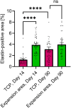
Semiquantification of elastin‐positive area in proximity and within the TCPs on Day 14 and Day 90 post–MFU‐V treatment. The elastin‐positive area within the TCP is significantly increased on Day 90 compared to Day 14 (****p < 0.0001) but remains similar to its respective expansion area (ANOVA Šídák's multiple comparison). Bars represent mean, and error bars represent standard error of the mean.
FIGURE 7
FIGURE 7
Proposed stages of histological response to MFU‐V based on the current results of this study.

References

    1. Fabi S. G., “Noninvasive Skin Tightening: Focus on New Ultrasound Techniques,” Clinical, Cosmetic and Investigational Dermatology 8 (2015): 47–52, 10.2147/CCID.S69118. - DOI - PMC - PubMed
    1. Gliklich R. E., White W. M., Slayton M. H., Barthe P. G., and Makin I. R. S., “Clinical Pilot Study of Intense Ultrasound Therapy to Deep Dermal Facial Skin and Subcutaneous Tissues,” Archives of Facial Plastic Surgery 9, no. 2 (2007): 88–95, 10.1001/archfaci.9.2.88. - DOI - PubMed
    1. Alam M., White L. E., Martin N., Witherspoon J., Yoo S., and West D. P., “Ultrasound Tightening of Facial and Neck Skin: A Rater‐Blinded Prospective Cohort Study,” Journal of the American Academy of Dermatology 62, no. 2 (2010): 262–269, 10.1016/j.jaad.2009.06.0394. - DOI - PubMed
    1. Lee H. S., Jang W. S., Cha Y. J., et al., “Multiple Pass Ultrasound Tightening of Skin Laxity of the Lower Face and Neck,” Dermatologic Surgery 38, no. 1 (2012): 20–27, 10.1111/j.1524-4725.2011.02158.x. - DOI - PubMed
    1. Contini M., Hollander M. H. J., Vissink A., Schepers R. H., Jansma J., and Schortinghuis J., “A Systematic Review of the Efficacy of Microfocused Ultrasound for Facial Skin Tightening,” International Journal of Environmental Research and Public Health 20, no. 2 (2023): 1522, 10.3390/ijerph20021522. - DOI - PMC - PubMed