. 2024 Nov 15;148(1):65.
doi: 10.1007/s00401-024-02820-z.

Microglia contribute to the production of the amyloidogenic ABri peptide in familial British dementia

Affiliations

Microglia contribute to the production of the amyloidogenic ABri peptide in familial British dementia

Charles Arber et al. Acta Neuropathol. .

Abstract

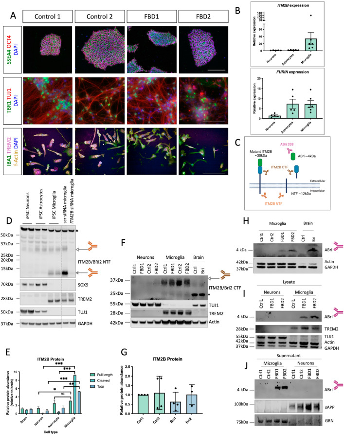
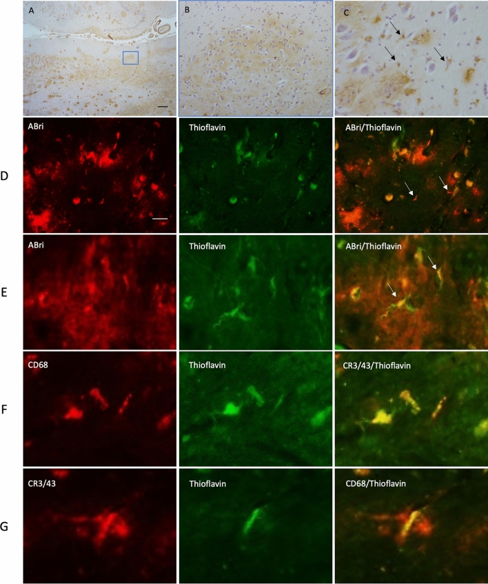
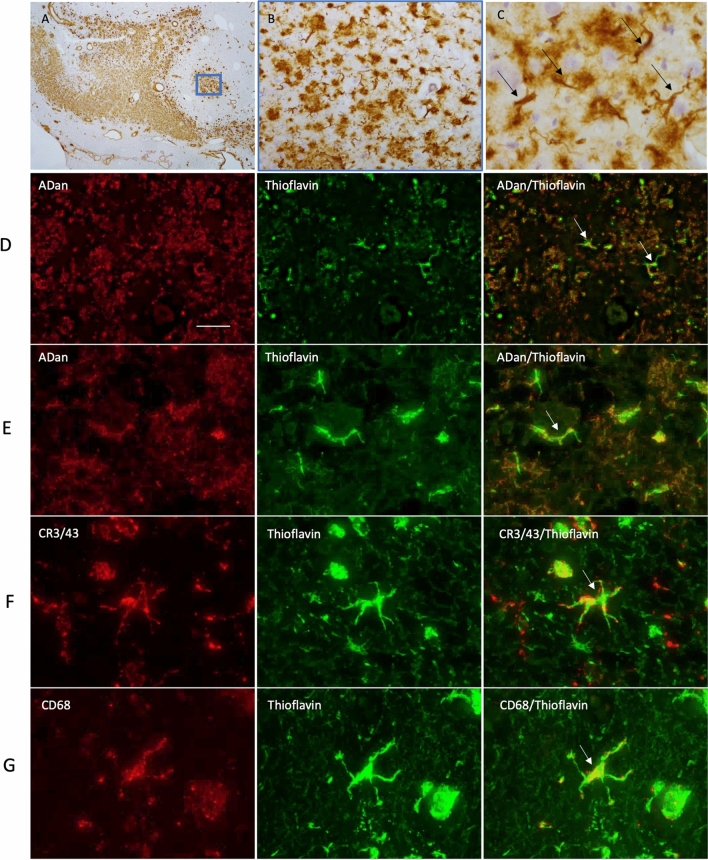
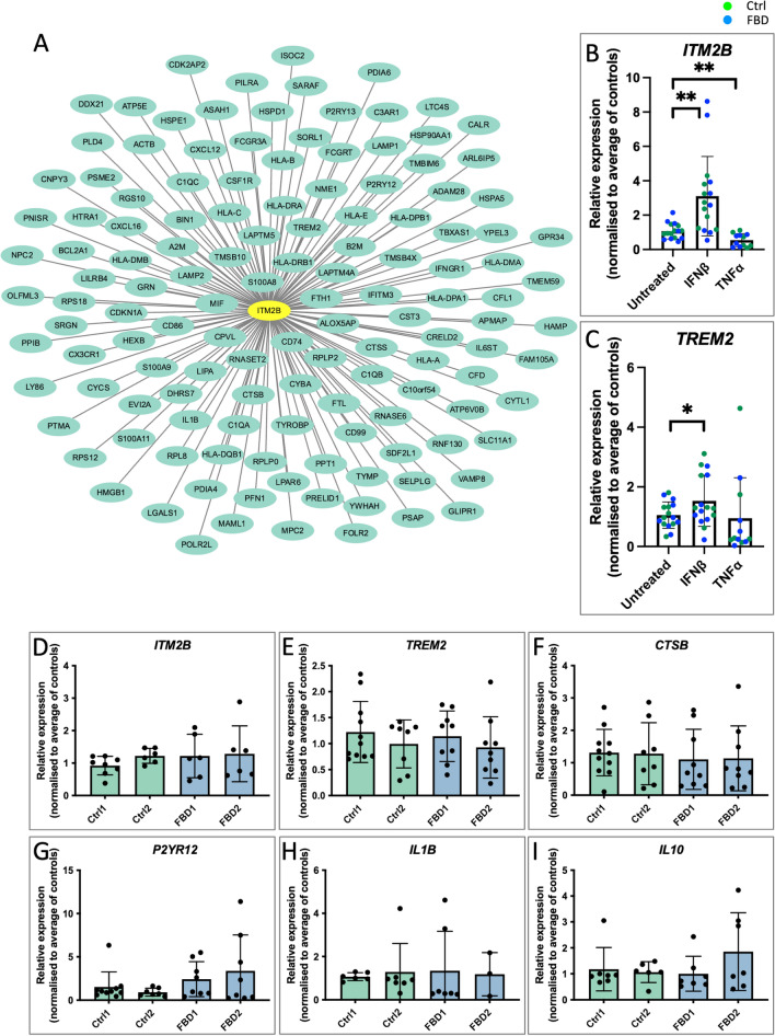
Mutations in ITM2B cause familial British, Danish, Chinese, and Korean dementias. In familial British dementia (FBD), a mutation in the stop codon of the ITM2B gene (also known as BRI2) causes a C-terminal cleavage fragment of the ITM2B/BRI2 protein to be extended by 11 amino acids. This fragment, termed amyloid-Bri (ABri), is highly insoluble and forms extracellular plaques in the brain. ABri plaques are accompanied by tau pathology, neuronal cell death and progressive dementia, with striking parallels to the aetiology and pathogenesis of Alzheimer's disease. The molecular mechanisms underpinning FBD are ill-defined. Using patient-derived induced pluripotent stem cells, we show that expression of ITM2B/BRI2 is 34-fold higher in microglia than neurons and 15-fold higher in microglia compared with astrocytes. This cell-specific enrichment is supported by expression data from both mouse and human brain tissue. ITM2B/BRI2 protein levels are higher in iPSC-microglia compared with neurons and astrocytes. The ABri peptide was detected in patient iPSC-derived microglial lysates and conditioned media but was undetectable in patient-derived neurons and control microglia. The pathological examination of post-mortem tissue supports the presence of ABri in microglia that are in proximity to pre-amyloid deposits. Finally, gene co-expression analysis supports a role for ITM2B/BRI2 in disease-associated microglial responses. These data demonstrate that microglia are major contributors to the production of amyloid forming peptides in FBD, potentially acting as instigators of neurodegeneration. Additionally, these data also suggest ITM2B/BRI2 may be part of a microglial response to disease, motivating further investigations of its role in microglial activation. These data have implications for our understanding of the role of microglia and the innate immune response in the pathogenesis of FBD and other neurodegenerative dementias including Alzheimer's disease.

Keywords: Alzheimer’s disease; Amyloid; Dementia; Familial British dementia; Microglia; iPSC.


Conflict of interest statement

Declarations Conflict of interest The authors declare no competing interests.

Figures

Fig. 1
Fig. 1
ABri is produced by iPSC-derived microglia. A Immunocytochemistry of iPSCs (upper panels), iPSC-derived neurons (middle panels) and iPSC-derived microglia (lower panels). SSEA4 and OCT4 are pluripotency markers, scale bar 200 μm. TUJ1 is a pan-neuronal marker and TBR1 labels deep layer cortical neurons, scale bar 200 μm. IBA1 labels microglia-like cells, scale bar 50 μm. B qPCR analysis of ITM2B/BRI2 and FURIN expression in control iPSC-derived cortical neurons, astrocytes and microglia. Neuronal cDNA represents 5 independent inductions with 2 independent control iPSC lines, astrocytic cDNA was generated from 5 independent inductions of two independent control iPSC lines and microglial cDNA was generated from 6 harvests from 4 inductions and represents two independent control iPSC lines. Significant differences are abolished by the outlier at two standard deviations above the mean. C Representation of antibodies used in this figure (produced in biorender). D Western blotting of iPSC-derived neurons, iPSC-derived astrocytes and iPSC-derived microglia. ITM2B/BRI2 knockdown via siRNA depicts antibody specificity for bands at around 30 kDa and 12 kDa. TUJ1, SOX9 and TREM2 are markers for neurons, astrocytes and microglia respectively. Samples represent two independent control lines for each cell type. Microglial samples from independent batches are separated by a dotted line. E Quantification of specific bands (30 kDa and 12 kDa) from 3 independent neuronal, astrocyte and microglia inductions of at least two control lines in each cell type. F Western blotting of iPSC neurons, iPSC microglia and post-mortem brain tissue for ITM2B/BRI2 as well as neuronal TUJ1, microglial TREM2 and loading control (Actin). G Quantification of Western blotting of ITM2B/BRI2 in control and patient-derived microglia from four harvests from three independent batches of microglia. HJ) Western blotting for ABri in iPSC-derived microglia lysates and brain tissue showed a band of 4 kDa. J) Western blotting for ABri in iPSC-derived microglial conditioned media, with secreted APP (sAPP) and GRN as neuronal and microglial loading controls respectively. White arrowheads show full length ITM2B/BRI2, grey arrowheads show cleaved fragments of ITM2B/BRI2 and asterisks show potential unspecific bands. Comparisons represent two tailed t-tests where * = p < 0.05, ** = p < 0.01, *** = p < 0.001
Fig. 2
Fig. 2
Immunohistochemical staining in FBD for ABri, Thioflavin and microglial markers. ABri pathology is observed in the hippocampus (A) in the form of extracellular amyloid and preamyloid deposits. ABri is also found within parenchymal and leptomeningeal blood vessels as cerebral amyloid angiopathy. ABri pre-amyloid plaques are shown at higher magnification in (B). The preamyloid deposits contained cells resembling microglia morphology (C, arrows). The bar represents 500 µm in (A) and 50 µm in (B) and 25 µm in (C). ABri immunohistochemistry (red, row D and E) combined with Thioflavin staining highlights the presence of ABri in cells resembling microglia. Microglial antibodies were used to determine the Thioflavin positive structures identified in the cells (rows F and G). The bar represents 100 µm in row (D) and 20 µm in row (E) and 10 µm rows in (F) and (G). Data are from one donor
Fig. 3
Fig. 3
Immunohistochemical staining in FDD for ADan, thioflavin and microglial markers. ADan pathology was observed in the hippocampus (A) in the form of extracellular pre-amyloid deposits and cerebral amyloid angiopathy. At higher magnification we observed the preamyloid deposits (B). Structures resembling microglia are also found to be highlighted with the ADan immunohistochemical preparations in the pre-amyloid deposits (C, arrows). The bar represents 500 µm in (A) and 50 µm in (B) and 25 µm in (C). ADan immunohistochemistry (red, row D and E) combined with Thioflavin staining (green) highlights the presence of ADan in cells resembling microglia. Microglial antibodies were used to determine the Thioflavin positive structures identified in the cells (rows F and G, arrows). The bar represents 100 µm in row (D) and 20 µm in row (E) and 10 µm rows in (F) and (G). Data is from one donor
Fig. 4
Fig. 4
ITM2B is coexpressed with ARM network genes and responds to inflammatory cues in a similar manner to TREM2. A Genetic network plot of a module containing ITM2B detected in microglial cells isolated from human Alzheimer’s disease patients and individuals with MCI analysed by scRNA-seq [44] demonstrating ITM2B is coexpressed with ARM network genes in microglia isolated from human individuals showing neurodegeneration (and collectively varying between AD and MCI donors). Genes varying in response to AD with the highest connectivity to ITM2B from the co-expression network were plotted based on ranking the connectivity matrix of the expression data. This module contains genes associated with the DAM/ARM state (the full network and the strength of each interaction is given in Supplementary Table 1). Genes most strongly co-expressed with ITM2B include genes known to be associated with neurodegeneration including LAPTM5, HLA genes, CTSB, CTSS, GRN, TREM2 and TYROBP. ITM2B is highlighted with a yellow oval. B, C qPCR expression analyses of ITM2B and TREM2 in response to a 24 h treatment with IFNβ and TNFα. Data represent n = 4 for untreated and IFNβ and n = 3 for TNFα, each from 2 independent batches of microglia and for two control iPSC lines and two FBD lines. Data separated by genotype is presented in Fig S8B. Paired t-tests were performed for each treatment relative to untreated samples, * = p < 0.05, ** = p < 0.01. DI qPCR analyses of genes associated with microglial state changes under basal conditions. Data represent 6 ≤ n ≤ 11 from 3 independent batches of microglia

Update of

References

    1. Akiyama H, Kondo H, Arai T, Ikeda K, Kato M, Iseki E et al (2004) Expression of BRI, the normal precursor of the amyloid protein of familial British dementia, in human brain. Acta Neuropathol 107:53–58. 10.1007/s00401-003-0783-1 - PubMed
    1. Arber C, Belder CRS, Tomczuk F, Gabriele R, Buhidma Y, Farrell C et al (2024) The presenilin 1 mutation P436S causes familial Alzheimer’s disease with elevated Aβ43 and atypical clinical manifestations. Alzheimer’s Dement. 10.1002/alz.13904 - PMC - PubMed
    1. Arber C, Lovejoy C, Harris L, Willumsen N, Alatza A, Casey JM et al (2021) Familial Alzheimer’s disease mutations in PSEN1 lead to premature human stem cell neurogenesis. Cell Rep 34:108615. 10.1016/j.celrep.2020.108615 - PMC - PubMed
    1. Arber C, Toombs J, Lovejoy C, Ryan NS, Paterson RW, Willumsen N et al (2020) Familial Alzheimer’s disease patient-derived neurons reveal distinct mutation-specific effects on amyloid beta. Mol Psychiatry 25:2919–2931. 10.1038/s41380-019-0410-8 - PMC - PubMed
    1. Audo I, Bujakowska K, Orhan E, El Shamieh S, Sennlaub F, Guillonneau X et al (2014) The familial dementia gene revisited: a missense mutation revealed by whole-exome sequencing identifies ITM2B as a candidate gene underlying a novel autosomal dominant retinal dystrophy in a large family. Hum Mol Genet 23:491–501. 10.1093/hmg/ddt439 - PubMed

Publication types

Substances

LinkOut - more resources