. 2024 Nov 15;26(1):11.
doi: 10.1007/s10522-024-10154-2.

Whole-body vibration elicits 40 Hz cortical gamma oscillations and ameliorates age-related cognitive impairment through hippocampal astrocyte synapses in male rats

Affiliations

Whole-body vibration elicits 40 Hz cortical gamma oscillations and ameliorates age-related cognitive impairment through hippocampal astrocyte synapses in male rats

Mingsong Liu et al. Biogerontology. .

Abstract

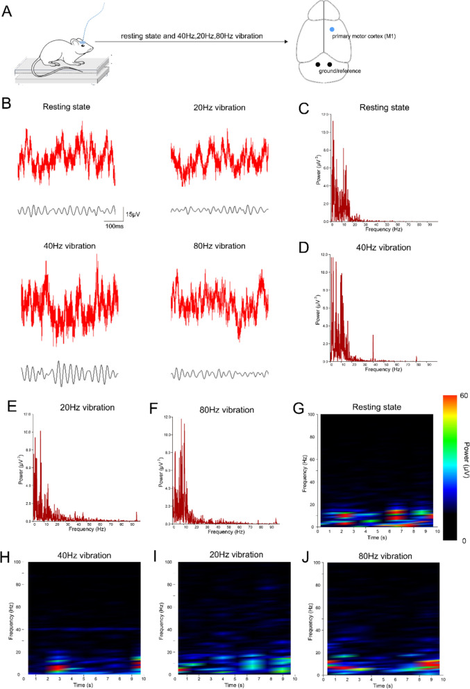
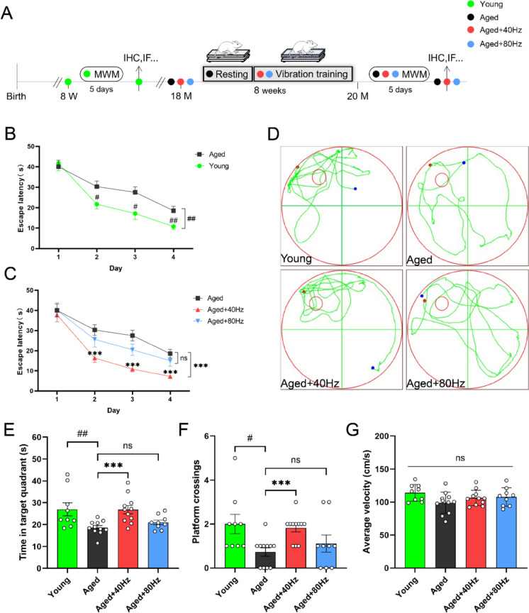
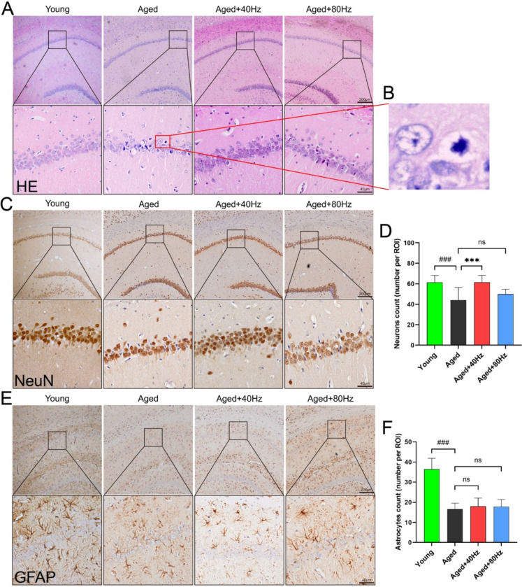
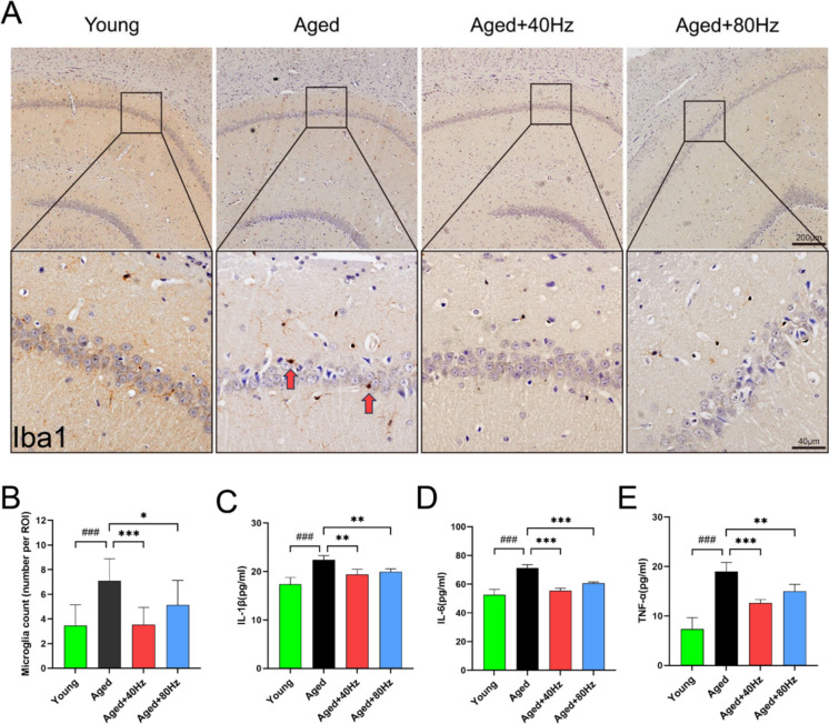
Age-related cognitive impairment is a prevalent issue in developed societies. Gamma oscil2lations at 40 Hz have been identified as a potential therapeutic approach for age-related cognitive decline and can be induced through various modalities, including auditory, visual, electrical, and magnetic stimulation. In this study, we investigated a novel modality of stimulation: whole-body vibration at 40 Hz. We examined the effects of 40 Hz vibration on cognitive performance and associated neuronal activity in the brains of aged male rats. Our findings revealed that only vibration at 40 Hz, rather than 20 Hz or 80 Hz, elicited cortical gamma oscillations in aged male rats. Additionally, following 8 weeks of prolonged treatment, the implementation of 40 Hz whole-body vibration significantly augmented the cognitive function of aged male rats as evidenced by behavioral assessments. Mechanistic studies demonstrated that these beneficial effects were attributed to the reduction of neuronal apoptosis in hippocampal CA1 through regulation of synaptic connections between astrocytes and neurons via 40 Hz gamma oscillations. Collectively, this suggests a promising intervention for age-related cognitive decline and identifies neuron-astrocyte synapses as potential therapeutic targets.

Keywords: Cognitive dysfunction; Gamma oscillations; Neuron-astrocyte synapses; Vibration.


Conflict of interest statement

Declarations Conflict of interest The authors declare no competing interests. Ethical approval All experimental procedures received approval from Guilin Medical College's Animal Ethics Committee (Approval Number: GLMC202105081) and adhered to guidelines outlined by European Union Council Directive (86/609/European Economic Community).

Figures

Fig. 1
Fig. 1
Whole-body vibration selectively and persistently enhances gamma oscillations in the M1 region of aged rats. A Experimental protocol and target regions for analyses are indicated. B Representative raw M1 ECoG recordings of a correct trial (top traces) and bandpass-filtered ECoG for 40 Hz gamma oscillations (bottom traces) from aged rats during resting state and vibrations at 40, 20, and 80 Hz. C-F Representative M1 ECoG power values from aged rats during resting state during resting state and vibrations at various frequencies. G-J The spectra of M1 ECoG recorded from aged rats during resting state and vibrations at various frequencies
Fig. 2
Fig. 2
Effect of 40 Hz WBV on spatial learning and memory in rats. A Experimental design. The 18-month-old rats underwent 8 weeks of whole-body vibration intervention. After 8 weeks, MWM was performed to assess memory functions. B Latency for finding a platform during training in young (n = 9) and aged rats (n = 11). C Latency of plateau finding during training in aged rats exposed to 40 Hz vibration (n = 11), 80 Hz vibration (n = 9) and no intervention (n = 11). D Representative trails from MWM probe trial. E Time spent in the target quadrant during probe test. F Number of platform crossings in the probe test. G Swimming velocity during MWM. # *P < 0.05, ## **P < 0.01, ### ***P < 0.001, ns indicates not significant, data are shown as the mean ± SEM
Fig. 3
Fig. 3
Immunohistochemical staining was performed on the CA1 region of the rat hippocampus. A HE staining images of the hippocampal CA1 region from each rat group, picture above scale bar: 200 μm, picture below scale bar: 40 μm. B Left: HE staining of normal neuronal nuclei, right: HE staining of apoptotic neuronal nuclei. C NeuN immunohistochemical staining images of the hippocampal CA1 region were obtained for each group of rats., picture above scale bar: 200 μm, picture below scale bar: 40 μm. D Neuronal counts within the visual field of the CA1 region in the hippocampus were assessed for each group of rats, utilizing a 40X magnification. E GFAP immunohistochemical staining images of the hippocampal CA1 region were obtained for each group of rats., picture above scale bar: 200 μm, picture below scale bar: 40 μm. F Astrocyte counts within the visual field of the CA1 region in the hippocampus were assessed for each group of rats, utilizing a 40× magnification. n = 4 rat per group # *P < 0.05, ## **P < 0.01, ### ***P < 0.001, ns indicates not significant, data are shown as the mean ± SD
Fig. 4
Fig. 4
Immunofluorescence staining and western blot analysis were performed on the rat hippocampus. A Immunofluorescence staining images of the CA1 region were obtained from each group of rats. Scale bar: 40 μm. B Extensive co-staining of PSD95 and GFAP was observed surrounding apoptotic neurons in the CA1 region of aged rats. C The positive area of PSD95 and GFAP co-staining in the CA1 region of rats was normalized to that in young rats. D The mean fluorescence intensity of PSD95 and GFAP co-staining in the CA1 region was measured for each group of rats. E The positive area of GFAP expression in the CA1 region was normalized to that in young rats. F The mean fluorescence intensity of GFAP expression in the CA1 region was quantified. G The positive area of PSD95 expression in the CA1 region was normalized to that in young rats. H The mean fluorescence intensity of PSD95 expression in the CA1 region was determined. I A representative western blot showed levels of GFAP and GAPDH proteins in the rat hippocampus. J The relative fold change of GFAP, normalized to the young group. K A representative western blot demonstrated levels of PSD95 and GAPDH proteins in the rat hippocampus. L The relative fold change of GFAP, normalized to the young group. n = 4 rat per group. # *P < 0.05, ## **P < 0.01, ### ***P < 0.001, ns indicates not significant, data are shown as the mean ± SD
Fig. 5
Fig. 5
The detection of inflammatory markers in the hippocampus of rats A Iba1 immunohistochemical staining images of the hippocampal CA1 region were obtained for each group of rats. Red arrow: microglia. Picture above, scale bar: 200 μm, picture below scale bar: 40 μm. B Microglia counts within the visual field of the CA1 region in the hippocampus were assessed for each group of rats, utilizing a 40× magnification. CE Relevant levels of pro-inflammatory factors in the hippocampus of rats were measured

References

    1. Adaikkan C, Middleton SJ, Marco A et al (2019) Gamma entrainment binds higher-order brain regions and offers neuroprotection. Neuron 102:929-943.e8. 10.1016/j.neuron.2019.04.011 - PMC - PubMed
    1. Adamsky A, Kol A, Kreisel T et al (2018) Astrocytic activation generates de novo neuronal potentiation and memory enhancement. Cell 174:59-71.e14. 10.1016/j.cell.2018.05.002 - PubMed
    1. Anderson ND (2019) State of the science on mild cognitive impairment (MCI). CNS Spectr 24:78–87. 10.1017/S1092852918001347 - PubMed
    1. Asahina Y, Sakaguchi Y, Kajimoto S et al (2023) A randomized controlled trial of whole-body vibration on gait ability and balance among older hemodialysis patients. Clin J Am Soc Nephrol 18:84–90. 10.2215/CJN.0000000000000018 - PMC - PubMed
    1. Başar E, Başar-Eroğlu C, Güntekin B, Yener GG (2013) Brain’s alpha, beta, gamma, delta, and theta oscillations in neuropsychiatric diseases: proposal for biomarker strategies. Suppl Clin Neurophysiol 62:19–54. 10.1016/b978-0-7020-5307-8.00002-8 - PubMed

LinkOut - more resources