

Peri-implant bone regeneration in pigs
- PMID: 39546101
- PMCID: PMC11568104
- DOI: 10.1186/s40729-024-00572-9
Peri-implant bone regeneration in pigs
Abstract
Purpose: To review the current literature to answer the focused question: in the experimental pig model (population), which types of peri-implant bone defects (exposure) have been used evaluate different modes of therapy and what is their capacity for spontaneous healing and regeneration (outcome)?
Methods: Following PRISMA guidelines, electronic databases were searched for studies reporting peri-implant bone defects in the maxillae or mandibles of pigs. Those studies which reported a control group of untreated defects with assessment of spontaneous regeneration [new bone area (BA)] and/or re-osseointegration [new bone-to-implant contact (BIC)] via quantitative radiography or histomorphometry were included in a random effects meta-analysis for the outcomes BA and BIC.
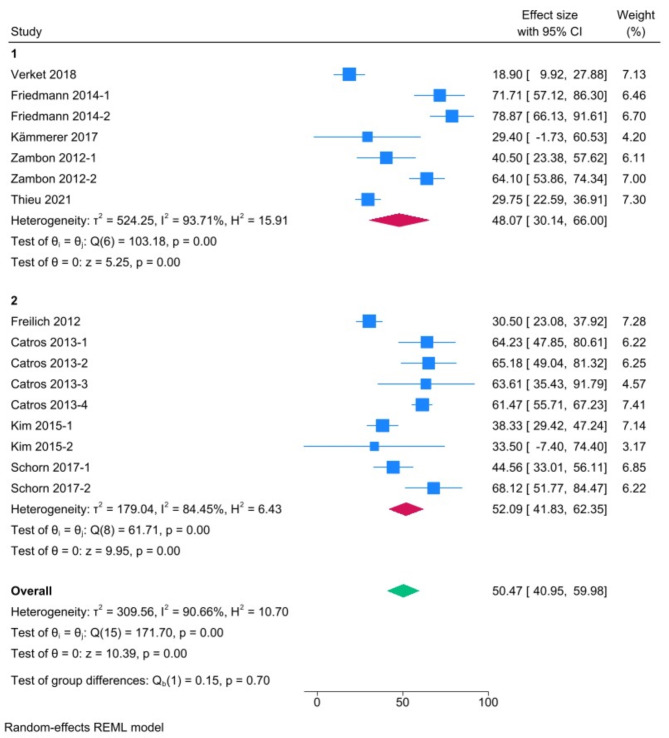
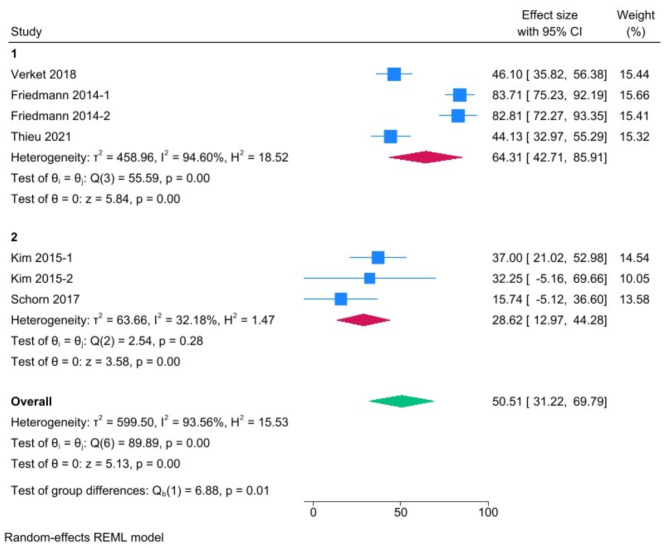
Results: Overall, 21 studies, mostly performed in the mandibles of minipigs, were included. Most studies reported 'acute' intrabony (circumferential and/or dehiscence; n = 12) or supra-alveolar defects (horizontal; n = 4). Five studies attempted to induce 'chronic' peri-implantitis lesions using ligatures with conflicting results. Meta-analyses revealed pooled estimates (with 95% confidence intervals) of 48.07% BIC (30.14-66%) and 64.31% BA (42.71-85.91%) in intrabony defects, and 52.09% BIC (41.83-62.35%) and 28.62% BA (12.97-44.28%) in supra-alveolar defects. Heterogeneity in the meta-analysis was high (I2 > 90%).
Conclusion: Current evidence for peri-implant bone regeneration in pigs is mainly based on acute intrabony defects, which demonstrate a high capacity for spontaneous regeneration and re-osseointegration. The evidence for chronic peri-implantitis is limited and does not clearly indicate a spontaneous progression of the disease in this animal model.
Keywords: Animal models; Bone regeneration; Peri-implantitis; Systematic reviews.
© 2024. The Author(s).
Conflict of interest statement
Figures
References
-
- Chiapasco M, Zaniboni M. Clinical outcomes of GBR procedures to correct peri-implant dehiscences and fenestrations: a systematic review. Clin Oral Implants Res. 2009;20(Suppl 4):113–23. - PubMed
-
- Renvert S, Persson GR. Periodontitis as a potential risk factor for peri-implantitis. J Clin Periodontol. 2009;36(Suppl 10):9–14. - PubMed
-
- Herrera D, Berglundh T, Schwarz F, Chapple I, Jepsen S, Sculean A, et al. Prevention and treatment of peri-implant diseases-the EFP S3 level clinical practice guideline. J Clin Periodontol. 2023;50(Suppl 26):4–76. - PubMed
-
- Monje A, Pons R, Insua A, Nart J, Wang HL, Schwarz F. Morphology and severity of peri-implantitis bone defects. Clin Implant Dent Relat Res. 2019;21(4):635–43. - PubMed
Publication types
MeSH terms
Substances
LinkOut - more resources
Full Text Sources
