. 2024 Nov 19;121(47):e2408086121.
doi: 10.1073/pnas.2408086121. Epub 2024 Nov 15.

S-adenosyl-L-methionine is the unexpected methyl donor for the methylation of mercury by the membrane-associated HgcAB complex

Affiliations

S-adenosyl-L-methionine is the unexpected methyl donor for the methylation of mercury by the membrane-associated HgcAB complex

Kaiyuan Zheng et al. Proc Natl Acad Sci U S A. .

Abstract

Mercury (Hg) is a heavy metal that exhibits high biological toxicity. Monomethylmercury and dimethylmercury are neurotoxins and a significant environmental concern as they bioaccumulate and biomagnify within the aquatic food web. Microbial Hg methylation involves two proteins, HgcA and HgcB. Here, we show that HgcA and HgcB can be heterologously coexpressed, and the HgcAB complex can be purified. We demonstrated that HgcA is a membrane-associated cobalamin-dependent methyltransferase and HgcB is a ferredoxin-like protein containing two [4Fe-4S] clusters. Further, spectroscopic and kinetic results demonstrate that S-adenosyl-L-methionine (SAM) donates the methyl group to Hg in a two-step reaction involving a methylcob(III)alamin intermediate including Co-thiolate ligation from a conserved Cys residue. Our findings uncover a biological role for SAM in microbial Hg methylation.

Keywords: S-adenosyl-L-methionine; cobalamin; iron-sulfur; methyl mercury; methyltransferase.


Conflict of interest statement

Competing interests statement:The authors declare no competing interest.

Figures

Fig. 1.
Fig. 1.
Hg methylation assays using P. mercurii ND132 cell lysate with different methyl donors and using E. coli BL21 cell lysates containing HgcAB. (A) Mercury methylation assays for P. mercurii ND132 cell lysate with various methyl donors. (B) Mercury methylation results for P. mercurii ND132 cell lysate with increasing concentrations of SAM. (C) The Left panel (rows 1 to 6) presents results of Hg methylation assays using E. coli BL21 cell lysates containing HgcAB or N-terminal His6-tagged HgcAB incubated with SAM or MeTHF. Row 1: negative control for Hg methylation assays using only ND132(∆hgcAB) cell lysate. Row 2: positive control for Hg methylation assays using E. coli BL21 cell lysates containing HgcAB. Rows 3, 5: E. coli BL21 cell lysates containing HgcAB or N-terminal His6-tagged HgcAB incubated with Me-THF. Rows 4, 6: Hg methylation assays performed as in rows 3, 5 except using SAM as methyl donor. The Middle panel presents results of Hg methylation assays using E. coli BL21 cell lysates containing HgcAB or N-terminal His6-tagged HgcAB + P. mercurii ND132 (∆hgcAB) cell lysates incubated with SAM or MeTHF. Rows 7 to 10: Hg methylation assays performed as in rows 3 to 6 but included P. mercurii ND132 (∆hgcAB) cell lysates. Row 11 to 12: Assays as in rows 9 and 10, respectively, except using E. coli BL21(DE3) membrane debris separate from E. coli BL21-expressed N-terminal His6-tagged HgcAB. The Right panel presents results of Hg methylation assays using purified N-terminal His6-tagged HgcAB and reference controls, including E. coli BL21 cell lysates + P. mercurii ND132 (∆hgcAB) cell lysates containing N-terminal His6-tagged HgcAB incubated ±SAM or ±Sarkosyl. Rows 13 to 14: Hg methylation assays with purified HgcAB and ND132(∆hgcAB) cell lysate with SAM/MeTHF. Row 15: SAM methylated purified HgcAB were incubated with ND132(∆hgcAB) cell lysate, and mercury methylation assays were performed. Row 16: E. coli BL21 cell lysates containing N-terminal His6-tag HgcAB were incubated with Sarkosyl and P. mercurii ND132 (∆hgcAB) cell lysates, and mercury methylation assays were performed. Row 17: Same as row 16, except with Sarkosyl. We conducted the assays described in rows 16 and 17 because we use Sarkosyl in our purification procedure, and these assays revealed that Sarkosyl disrupts the second half-reaction. Row 18: Same as Row 10. All P values were obtained through a t test using GraphPad Prism 9. Error bars represent SD. N = 3 for each bar. ns: P > 0.05; *P ≤ 0.05; **P ≤ 0.01; ***P ≤ 0.001; ****P ≤ 0.0001. Details of assay conditions are described in SI Appendix, Table S2. Experimental details are provided in the Materials and Methods and SI Appendix.
Fig. 2.
Fig. 2.
UV-visible spectroscopic and LC–MS characterization of HgcAB methylation. (AD) UV-vis spectra of (A) 10 µM Cob(III)-HgcAB before and after addition of 100 µM Ti(III) citrate reductant to obtain Cob(I)-HgcAB. (B) 10 µM Cob(III)-HgcAB and after reduction to Cob(I)-HgcAB as in A; then, after addition of 50 µM Me-THF at 0, 5, and 20 min. (C) 10 µM Cob(I)-HgcAB formed as in A and at 0, 5, and 20 min after addition of 50 µM of SAM. (D) 5 µM Cob(I)-HgcAB after reaction with 50 µM of SAM for 0, 0.1, 0.5, 1, 5, 10, 100, and 600 s. Insets in each figure show the UV-vis spectral differences between reduced HgcAB or methylated HgcAB and initial HgcAB spectra [Cob(III)-HgcAB or Cob(I)-HgcAB]. (E) Triple quadruple time of flight liquid chromatography/mass spectrometric (Q-TOF LC/MS) identification of SAH as product of the SAM methylation reaction, as in “d”. (a) 50 µM Cob(I)-HgcAB, (b) quenched “a” + 200 µM SAM, (c) “a”+ 150 µM SAM, and (d) “a” + 400 µM SAM, under photolysis conditions to enable multiple turnovers as described in the scheme above the data and the main text. The Y axis represents SAM or SAH (ppm peak height from the LC–MS signal, and µM SAM remaining or SAH generated). Error bars represent the SD (N = 3). Other details are provided in the Materials and Methods and SI Appendix.
Fig. 3.
Fig. 3.
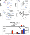
EPR spectroscopic characterization of the HgcAB methylation reaction. (A) EPR spectra at 60 K (Left) and 10 K (Right) of Cob(III)-HgcAB, Cob(II)-HgcAB, Cob(I)-HgcAB, and SAM-generated Me-Cob(III)-HgcAB. The middle figures show the UV-vis spectra associated with the corresponding EPR spectra on the Left and Right. (B) Detailed view of Cob(II)-HgcAB EPR spectrum, which indicates its base-on, Cys-off coordination mode. For further details, see the Materials and Methods.
Fig. 4.
Fig. 4.
Co K-edge XAS of HgcAB. (A) Co K-edge XAS of methylated (red) and as-isolated (black) HgcAB. (Inset) Expanded Co K-pre-edge region. (B) Non-phase-shift-corrected Fourier transform and (Inset) corresponding EXAFS for methylated-HgcAB. Data (black), fit (red). For further details, see the Materials and Methods and SI Appendix.
Fig. 5.
Fig. 5.
Proposed mechanism for enzymatic mercury methylation catalyzed by HgcAB. For the as-isolated Co(III) state, the upper axial ligand is likely to be water or OH. Two-electron reduction by the two ferredoxin-like [Fe4S4]1+ clusters of HgcB yields the nucleophilic Co(I) state, which attacks the electrophilic methyl group of SAM to generate methyl-Co(III)-thiolato cobalamin, which likely transfers its methyl group as a methyl radical (or methyl carbanion) to Hg(II) to generate MeHg.

References

    1. Clarkson T. W., Human toxicology of Mercury. J. Trace Elem. Exp. Med. 11, 303–317 (1998).
    1. Driscoll C. T., Mason R. P., Chan H. M., Jacob D. J., Pirrone N., Mercury as a global pollutant: Sources, pathways, and effects. Environ. Sci. Technol. 47, 4967–4983 (2013). - PMC - PubMed
    1. Parks J. M., et al. , The genetic basis for bacterial mercury methylation. Science 339, 1332–1335 (2013). - PubMed
    1. Gilmour C. C., et al. , Mercury methylation by novel microorganisms from new environments. Environ. Sci. Technol. 47, 11810–11820 (2013). - PubMed
    1. Gilmour C. C., Bullock A. L., McBurney A., Podar M., Elias D. A., Robust mercury methylation across diverse methanogenic archaea. mBio 9, e02403-17 (2018). - PMC - PubMed

LinkOut - more resources