. 2024 Nov 15;10(46):eadf9758.
doi: 10.1126/sciadv.adf9758. Epub 2024 Nov 15.

Cell response to extracellular matrix viscous energy dissipation outweighs high-rigidity sensing

Affiliations

Cell response to extracellular matrix viscous energy dissipation outweighs high-rigidity sensing

Carla Huerta-López et al. Sci Adv. .

Abstract

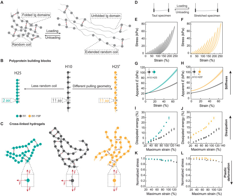
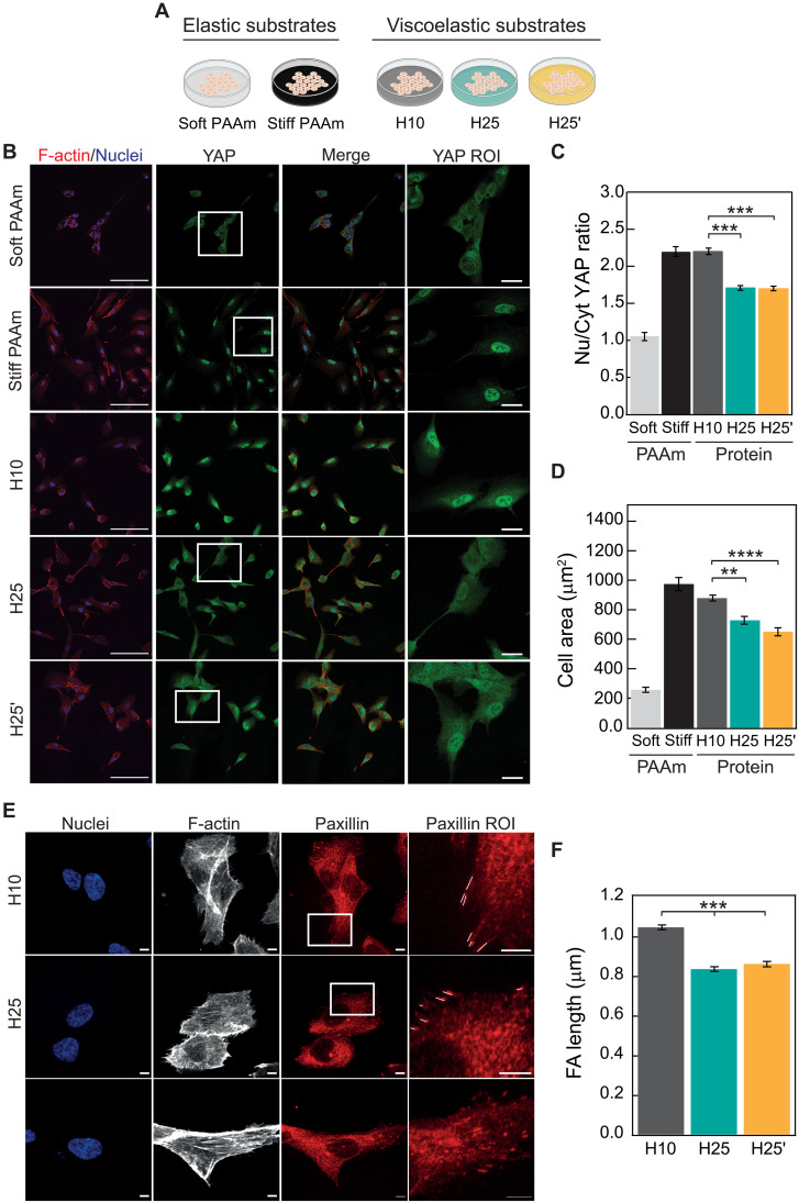
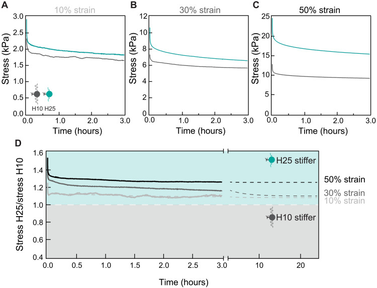
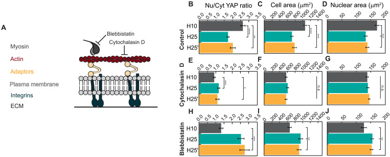
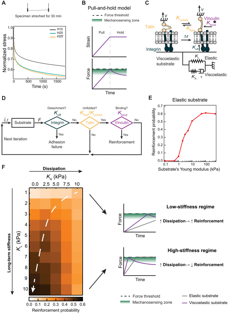
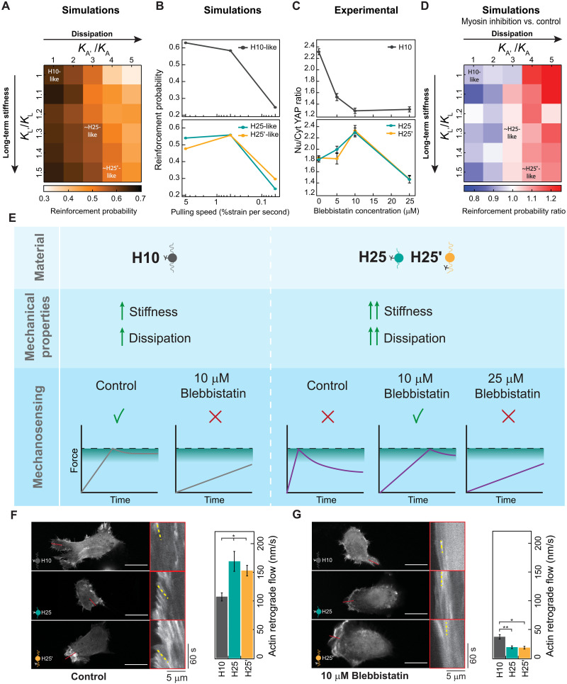
The mechanics of the extracellular matrix (ECM) determine cell activity and fate through mechanoresponsive proteins including Yes-associated protein 1 (YAP). Rigidity and viscous relaxation have emerged as the main mechanical properties of the ECM steering cell behavior. However, how cells integrate coexisting ECM rigidity and viscosity cues remains poorly understood, particularly in the high-stiffness regime. Here, we have exploited engineered stiff viscoelastic protein hydrogels to show that, contrary to current models of cell-ECM interaction, substrate viscous energy dissipation attenuates mechanosensing even when cells are exposed to higher effective rigidity. This unexpected behavior is however readily captured by a pull-and-hold model of molecular clutch-based cell mechanosensing, which also recapitulates opposite cellular response at low rigidities. Consistent with predictions of the pull-and-hold model, we find that myosin inhibition can boost mechanosensing on cells cultured on dissipative matrices. Together, our work provides general mechanistic understanding on how cells respond to the viscoelastic properties of the ECM.


Figures

Fig. 1.
Fig. 1.. Bottom-up engineering of protein hydrogels with different viscoelastic properties.
(A) Representation of a protein hydrogel covalently cross-linked through tyrosine residues. Building blocks include immunoglobulin (Ig)–like domains and random coil regions. Under uniaxial traction, random coil regions extend elastically, and Ig domains unfold, dissipating energy. (B) Schematics of the polyprotein building blocks used in this study. The I91 Ig domain of titin was engineered into octameric repeats separated by 11 amino acid (aa)–long random coil linkers (H10, dark gray). Two more constructs were designed starting from H10. First, H25 (teal) was produced by shortening the linkers between Ig domains to only two amino acids. To build H25′ (mustard), tyrosines were mutated out from Ig domains and included as one of the 11 amino acids in the random coil linkers. (C) Schematic representation of the network structure of I91 matrices. Cross-linking takes place at tyrosine positions, leading to different mechanical force distributions (dashed insets). (D) Loading and unloading stress-strain tests used to determine apparent elastic moduli, dissipated energy, and plasticity of hydrogels. (E and F) Stress-strain curves of H10 (E) and H25′ (F) hydrogels pulled at 5 mm/s. For clarity, stress-strain curves are offset in the x axis. Actual stress-strain curves can be found in fig. S5. Curves were measured immediately one after another from low to high strains. (G and H) Strain dependency of apparent elastic moduli obtained in stress-strain experiments to 75% maximum strain. Lines represent average values, and shaded areas are SEM. (I and J) Dissipated energy is measured in stress-strain experiments to different maximum strains. (K and L) Normalized stress at 15% strain for consecutive stress-strain curves to different maximum strains. Data are obtained from four specimens of H10 and H25 and six specimens of H25′ produced in independent cross-linking reactions from two purification batches.
Fig. 2.
Fig. 2.. Blunted mechanosensing in cells grown on rigid viscoelastic substrates.
(A) Outline of experimental conditions. (B) Confocal immunofluorescence images of YAP localization in RPE-1 cells grown on elastic stiff or soft PAAm or on H10, H25, or H25′ protein matrices. F-actin was stained with Alexa Fluor 647-conjugated phalloidin (red; first column), and nuclei were stained with 4′,6-diamidino-2-phenylindole (DAPI; blue in merged images; first and third columns). YAP was labeled with Alexa-Fluor-488-conjugated antibody in green (second column). The fourth column shows zoomed views of the YAP region of interest (ROI) (boxed in white in the YAP column). Scale bars, 100 μm (first to third columns) and 20 μm (fourth column). (C) Quantification of nuclear versus cytoplasmic YAP distribution. (D) Cell spreading quantified as cell area. A minimum of n = 30 cells per condition were quantified in a total of four independent experiments. (E) Confocal immunofluorescence images of focal adhesions in RPE-1 cells grown on H10, H25, or H25′ matrices. Nuclei were stained with DAPI (first column), and F-actin was stained with Alexa-Fluor-647-conjugated phalloidin (second column). Paxillin in focal adhesions was labeled with Alexa-Fluor-568-conjugated antibody in red (third column). The fourth column shows zoomed views of the paxillin ROI (boxed in white in the paxillin column; focal adhesions are marked with white lines). Scale bars, 5 μm. (F) Focal adhesion (FA) length quantification. A minimum of n = 30 cells per condition were quantified in a total of three independent experiments. PAAm groups are not considered for statistical analysis in (C) and (D). Data are presented as mean ± SEM, ordinary one-way analysis of variance (ANOVA); **P < 0.01, ***P < 0.001, and ****P < 0.0001.
Fig. 3.
Fig. 3.. H25 hydrogels are effectively stiffer than H10 counterparts.
Stress relaxation of H10 (dark gray) and H25 (teal) hydrogels for 3 hours at a (A) 10%, (B) 30%, and (C) 50% strain. (D) Ratio of the stress generated by H25 and H10 hydrogels in the relaxation experiments. Ratios above 1 indicate that H25 hydrogels are stiffer than H10 counterparts. Gray dashed lines are double-exponential fits to the experimental data. Results were obtained with three H10 and three H25 specimens coming from three cross-linking reactions and two different protein purifications.
Fig. 4.
Fig. 4.. Actomyosin contractility mediates cell response to viscous energy dissipation.
(A) Representation of mechanical communication between the ECM and the cytoskeleton through molecular clutches of integrins and adaptor proteins like talin. Blebbistatin and cytochalasin D treatments inhibit myosin activity and actin polymerization, respectively. (B to D) Quantification of nuclear versus cytoplasmic YAP distribution, cell spreading, and nuclear area of cells seeded on protein matrices in control conditions. (E to G) Quantification of YAP translocation to the nucleus, cell spreading, and nuclear area of cells treated with 1 μM cytochalasin D. (H to J) Quantification of YAP translocation to the nucleus, cell spreading, and nuclear area of cells treated with 10 μM blebbistatin. A minimum of 40 cells coming from three independent experiments were analyzed for quantifications in (B) to (J). Data are presented as mean ± SEM, ordinary one-way ANOVA. n.s., not significant; *P < 0.05, **P < 0.01, ***P < 0.001, and ****P < 0.0001.
Fig. 5.
Fig. 5.. Pull-and-hold model of ECM-triggered mechanosensing.
(A) Stress relaxation at 50 kPa of H10, H25, and H25′ materials. Data obtained from three specimens of each hydrogel from independent cross-linking reactions and two purification batches. (B) Pull-and-hold substrate straining model. Initially, cells strain the substrate monotonically leading to an increase in force (pull phase). When a force threshold is reached (black dashed line), cells enter length-clamp mode (hold phase). During hold phase, the force generated by a viscoelastic substrate drops due to energy dissipation. Green indicates the region where talin unfolding, and therefore mechanosensing, is most probable. (C) Single-chain molecular clutch. A single integrin binds to a two-element generalized Maxwell-Wiechert substrate. Integrin detachment occurs according to a Koff rate. Inside the cell, talin attached to integrins can be in a folded or unfolded state according to Kunfold and Kfold rates. Vinculin binds unfolded talin given a Kbind rate. (D) Iterative Monte Carlo algorithm to estimate adhesion reinforcement. Substrate generates force according to a given strain rate (ε·). Simulations resulting in integrin detachment are considered adhesion failures. If no detachment occurs, and if vinculin binds to talin, then the run is considered a successful reinforcement event. If there is no detachment nor vinculin binding, then a new Monte Carlo iteration starts with updated strain values. This cycle is repeated until adhesion failure or reinforcement occurs. (E) Simulated reinforcement probability for elastic substrates increases with substrate stiffness. (F) Left: Heatmap of simulated reinforcement probability according to elastic long-term stiffness (KL) and additional viscous stiffness (KA). The white dashed arrow highlights regions of maximum reinforcement probability for substrates with equal KL. Right: In the low long-term stiffness regime, KA increases overall stiffness ensuring reinforcement. In the high long-term stiffness region, viscosity leads to stress relaxation after reaching the force threshold, blunting mechanosensing.
Fig. 6.
Fig. 6.. The pull-and-hold molecular clutch model reproduces cell mechanosensing of protein viscoelastic matrices.
(A) Simulated reinforcement probability in different stiff viscoelastic substrates considering H10-like parameters as reference. Position of parameters similar to H25/H25′ materials is indicated. (B) Dependency of simulated reinforcement probability with cell pulling speed for H10-like and H25-like substrates. (C) Nuclear versus cytoplasmic YAP in cells seeded on H10, H25, or H25′ substrates and treated with increasing concentrations of blebbistatin. Data are obtained from three specimens of H10, H25, and H25′ hydrogels produced in independent cross-linking reactions from two protein purification batches. (D) Reinforcement probability ratio obtained by comparing simulations at pulling speeds of 0.5 (representing myosin inhibition) and 5 (representing control conditions) %strain per second. H10-like parameters are used as a reference and position of parameters similar to H25/H25′ materials are indicated. (E) Interpretation of cell mechanosensing in different viscoelastic substrates and under different degrees of myosin inhibition according to the pull-and-hold model. (F and G) Actin retrograde flow of cells seeded on H10, H25, or H25′ matrices in control (F) and in the presence of 10 μM blebbistatin (G). Representative static images are shown on the left of the panels, and kymographs on red boxes show actin movement toward the cell center measured in regions of interest indicated by red lines. Representative actin trajectories are highlighted in yellow. At least 30 measurements per condition were used to quantify actin retrograde flow (right). Scale bars, 20 μm. Data are presented as mean ± SEM, ordinary one-way ANOVA; *P < 0.05 and **P < 0.01.

References

    1. Bonnans C., Chou J., Werb Z., Remodelling the extracellular matrix in development and disease. Nat. Rev. Mol. Cell. Biol. 15, 786–801 (2014). - PMC - PubMed
    1. Humphrey J. D., Dufresne E. R., Schwartz M. A., Mechanotransduction and extracellular matrix homeostasis. Nat. Rev. Mol. Cell. Biol. 15, 802–812 (2014). - PMC - PubMed
    1. Engler A. J., Sen S., Sweeney H. L., Discher D. E., Matrix elasticity directs stem cell lineage specification. Cell 126, 677–689 (2006). - PubMed
    1. Elosegui-Artola A., Trepat X., Roca-Cusachs P., Control of mechanotransduction by molecular clutch dynamics. Trends Cell Biol. 28, 356–367 (2018). - PubMed
    1. Segel M., Neumann B., Hill M. F. E., Weber I. P., Viscomi C., Zhao C., Young A., Agley C. C., Thompson A. J., Gonzalez G. A., Sharma A., Holmqvist S., Rowitch D. H., Franze K., Franklin R. J. M., Chalut K. J., Niche stiffness underlies the ageing of central nervous system progenitor cells. Nature 573, 130–134 (2019). - PMC - PubMed

Publication types