. 2024 Nov 15;8(2):e202402583.
doi: 10.26508/lsa.202402583. Print 2025 Feb.

Vinculin-Arp2/3 interaction inhibits branched actin assembly to control migration and proliferation

Affiliations

Vinculin-Arp2/3 interaction inhibits branched actin assembly to control migration and proliferation

John James et al. Life Sci Alliance. .

Abstract

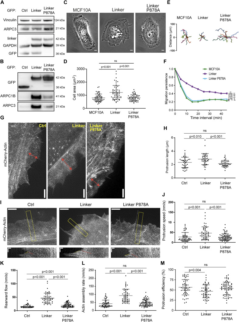
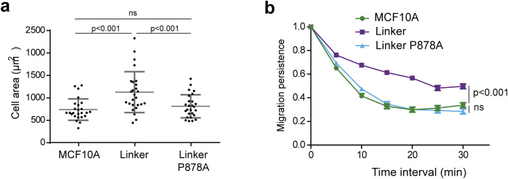
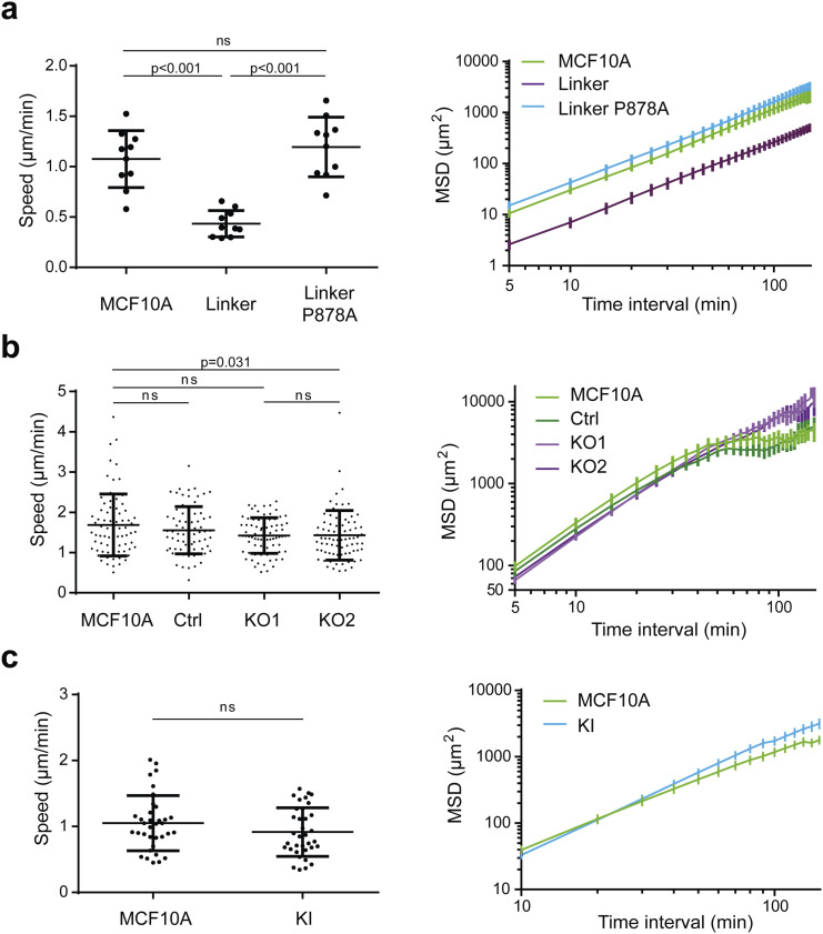
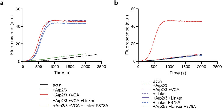
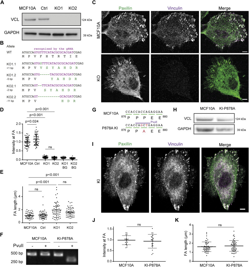
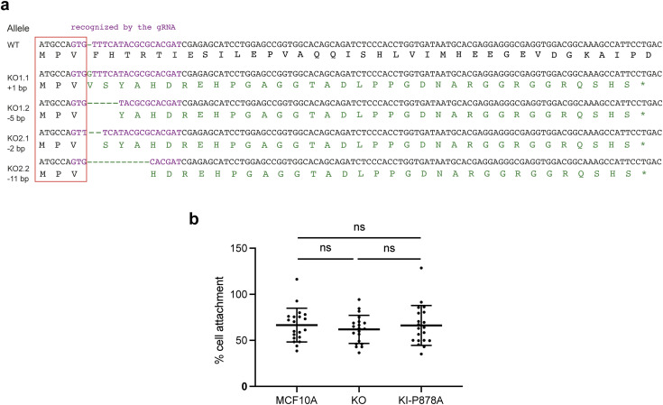
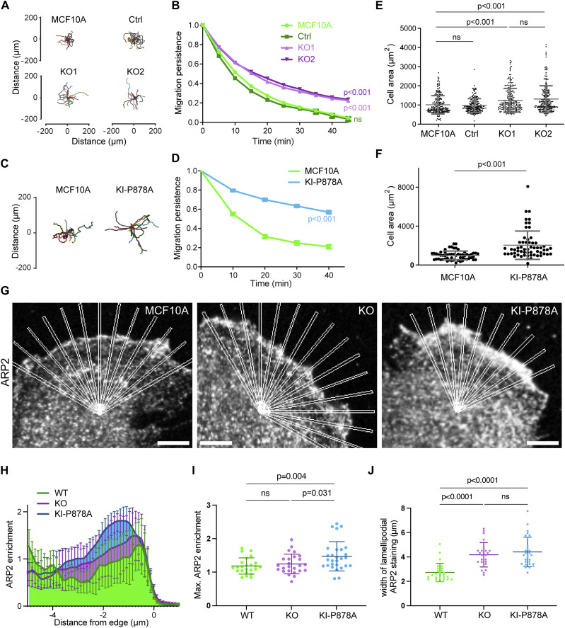
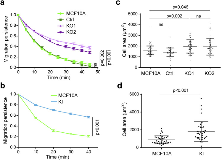
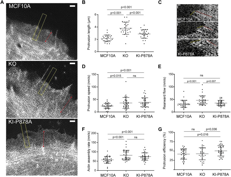
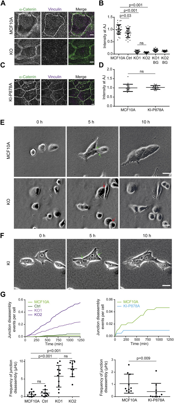
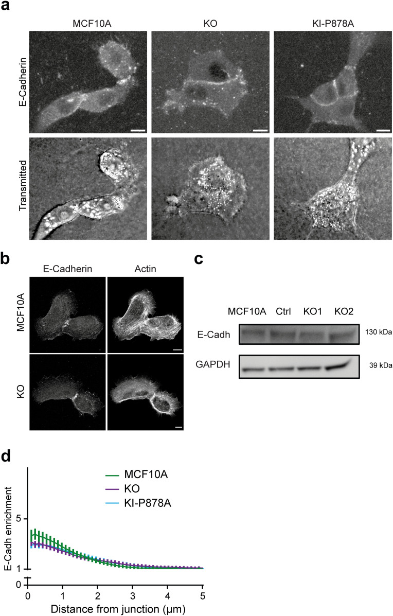
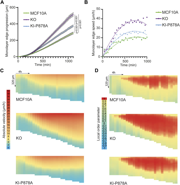
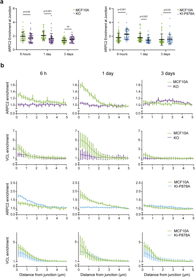
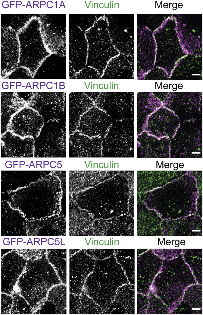
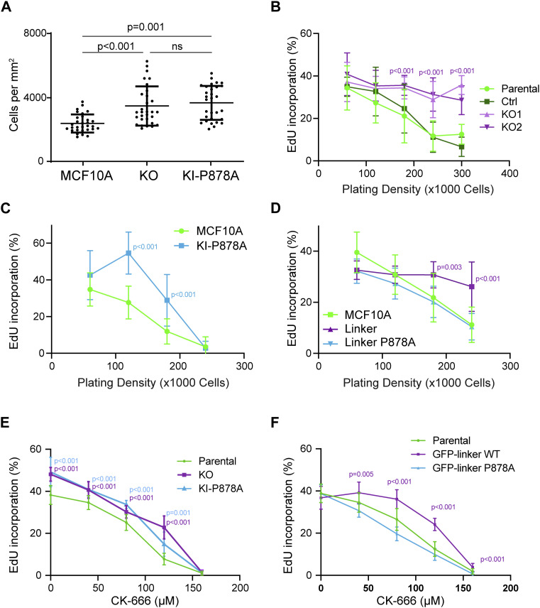
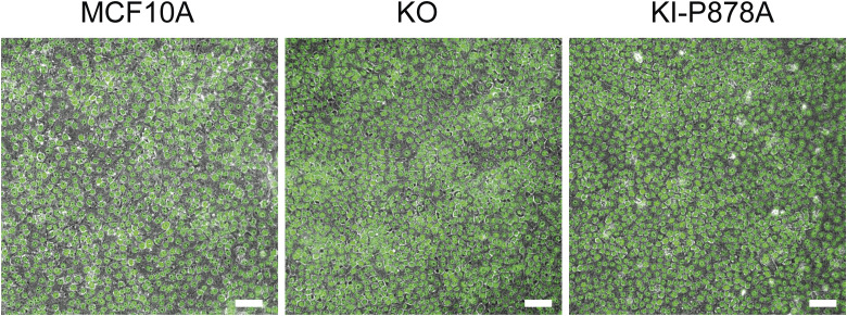
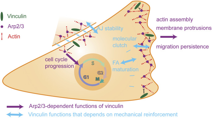
Vinculin is a mechanotransducer that reinforces links between cell adhesions and linear arrays of actin filaments upon myosin-mediated contractility. Both adhesions to the substratum and neighboring cells, however, are initiated within membrane protrusions that originate from Arp2/3-nucleated branched actin networks. Vinculin has been reported to interact with the Arp2/3 complex, but the role of this interaction remains poorly understood. Here, we compared the phenotypes of vinculin knock-out (KO) cells with those of knock-in (KI-P878A) cells, where the point mutation P878A that impairs the Arp2/3 interaction is introduced in the two vinculin alleles of MCF10A mammary epithelial cells. The interaction of vinculin with Arp2/3 inhibits actin polymerization at membrane protrusions and decreases migration persistence of single cells. In cell monolayers, vinculin recruits Arp2/3 and the vinculin-Arp2/3 interaction participates in cell-cell junction plasticity. Through this interaction, vinculin controls the decision to enter a new cell cycle as a function of cell density.


Conflict of interest statement

The authors declare that they have no conflict of interest.

Figures

Figure 1.
Figure 1.. Expression of the vinculin linker that binds to the Arp2/3 complex increases actin polymerization and membrane protrusion.
(A, B) Arp2/3 complex co-immunoprecipitates with the vinculin linker (amino acids 811-881). (A, B) MCF10A cells stably expressing GFP, the GFP-tagged linker in a WT or P878A form, were lysed and subjected either to Western blot analysis (A), or to GFP immunoprecipitation and Western blot analysis (B). N = 3; 1 representative experiment shown. (C) Phase-contrast images of the same cell lines. Scale bar: 5 μm. (D) Cell area of the cell lines stained for actin. Mean ± SD, n = 56, t test. N = 3; pooled measurements from the three independent repeats are plotted. (E, F) Single-cell migration. (E, F) Cell trajectories (E) and migration persistence (F). Mean ± SD, n = 35, linear mixed-effect model. N = 3; pooled measurements from the three independent repeats are plotted. (G, H) Membrane protrusions of the stable MCF10A cell lines expressing the vinculin linker transiently transfected with mCherry–actin. (G, H) TIRF-SIM images of mCherry–actin (G) and quantification of protrusion length (H). Double-headed arrows in red indicate the length of lamellipodia. Scale bar: 2 μm. Mean ± SD, n = 40, t test. N = 3; pooled measurements from the three independent repeats are plotted. (I) Kymographs (bottom panels, scale bars: 0.4 μm horizontal, 40 s vertical) were generated along a line centered in the region boxed in yellow in the TIRF-SIM video (scale bar: 2 μm). (J, K) Dashed yellow and red lines indicate protrusion speed (J) and rearward flow (K), respectively. (L) Actin assembly rate (L) is the sum of protrusion speed and rearward flow. (M) Protrusion efficiency (M) is the ratio of protrusion speed to actin assembly rate. Mean ± SD, n = 40, t test. N = 3 with similar results; pooled measurements from the three independent repeats are plotted.
Figure S1.
Figure S1.. (Extended data of Fig 1) One representative experiment of migration persistence and cell area assays showing the same tendency as the three pooled experiments shown in Fig 1.
(A) Cell area of the cell lines stained for actin. Mean ± SD, n = 25, t test; 1 representative experiment shown. (B) Migration persistence. Mean ± SD, n = 10, linear mixed-effect model; 1 representative experiment shown.
Figure S2.
Figure S2.. Speed and mean square displacement (MSD).
(A) MCF10A cells expressing the vinculin linker (n = 10). (B) KO cells (n = 74). (C) KI cells (n = 35). Mean ± SD, t test, N = 3; 1 representative experiment shown.
Figure S3.
Figure S3.. Pyrene–actin polymerization assays.
(A) Vinculin linker does not regulate Arp2/3 activity in vitro. (B) Vinculin linker does not affect actin polymerization in vitro. Conditions: 1.5 μM of 10% pyrenyl-labeled actin, 20 nM Arp2/3, 250 nM VCA, and 1 μM vinculin linker or P878A linker as indicated. All these curves were acquired the same day, and the Arp2/3 + VCA is replotted in the two panels for comparison.
Figure 2.
Figure 2.. Characterization of VCL knock-out and knock-in cell lines.
(A) Parental MCF10A, and isolated clones transfected with a VCL-targeting gRNA or a non-targeting gRNA were analyzed by Western blot. (B) Sequences of the two alleles in each KO cell line. All mutations induce a frameshift and thus a premature stop codon in the ORF. (C) Staining of vinculin and paxillin in parental and KO cells. Scale bar: 5 μm. (D) Quantification of vinculin staining in focal adhesions (FAs) and normalization by the intensity of parental cells. BG refers to the background in the non-FA cytoplasm. Mean ± SD, n = 45, t test. N = 3 with similar results; pooled measurements from the three independent repeats are plotted. (E) Quantification of the length of FAs. Mean ± SD, n = 45, t test. N = 3 with similar results; pooled measurements from the three independent repeats are plotted. (F) Genome analysis of the KI. Part of the VCL ORF containing the P878A mutation was amplified by PCR and digested with the PvuII restriction enzyme. Agarose gel electrophoresis of digested or undigested PCR fragment. (G) Sequencing of the genome-amplified PCR fragment confirmed the presence of the P878A mutation on the two alleles and the introduction of the PvuII restriction site in the genome of the KI-P878 line. (H) Cell lysates of parental MCF10A, and the isolated clone of the KI-P878A were analyzed by Western blot using anti-vinculin and anti-GAPDH antibodies. (I) Staining of vinculin and paxillin in the KI-P878A cells. Scale bar: 5 μm. (J) Quantification of vinculin staining in focal adhesions (FAs) and normalization by the intensity of parental cells. Mean ± SD, n = 19, t test. N = 3 with similar results; pooled measurements from the three independent repeats are plotted. (K) Quantification of the length of FAs. Mean ± SD, n = 50, t test. N = 3 with similar results; pooled measurements from the three independent repeats are plotted. Ctrl refers to the MCF10A cell line that has been genome-edited using a Ctrl gRNA.
Figure S4.
Figure S4.. Characterization of VCL knock-out and knock-in cell lines.
(A) Full nucleotide sequences of the two alleles for each KO cell line, and translated peptides in the ORF. All mutations induce a frameshift after the third codon, leading to translation by each allele of a similar peptide of 3.4–3.6 kD, which is unrelated to vinculin. (B) Quantification of the cell adhesion to fibronectin-coated plates. Mean ± SD, t test; five replicates of the three independent repeats are plotted.
Figure 3.
Figure 3.. Vinculin decreases migration persistence and Arp2/3 recruitment in membrane protrusions through its interaction with the Arp2/3 complex.
(A, B, C, D) Single-cell migration of KO and KI-P878A cells. Cell trajectories (A, C) and migration persistence (B, D). (B, D) Mean ± SD, n = 75 in (B) and n = 35 in (D), linear mixed-effect model. N = 3; pooled measurements from the three independent repeats are plotted. (E, F) Cell area of KO and KI-P878A lines. (E, F) Mean ± SD, n = 228 in (E) and n = 59 in (F), t test. N = 3; pooled measurements from the three independent repeats are plotted. (G) Staining of ARP2 in the lamellipodium of parental, KO, and KI-P878A cells on single-plane confocal microscopy images. Scale bar: 5 μm. (H) Recruitment of ARP2 assessed by multiple radial line scans. Average profiles of ARP2 enrichment in the lamellipodium of parental, KO, and KI-P878A cells upon registering line scans to the cell edge. (I, J) Maximum intensity (I) and average width (J) of ARP2 staining at the edge of the lamellipodium. Mean ± SD, n = 29, t test. N = 3 with similar results; pooled measurements from the three independent repeats are plotted.
Figure S5.
Figure S5.. (Extended data of Fig 3) One representative experiment of migration persistence and cell area assays showing the same tendency as the three pooled experiments.
(A, B) Migration persistence of KO (A) and KI cells (B). (B, D) Mean ± SD, n = 34 in (B) and n = 15 in (D), linear mixed-effect model; 1 representative experiment shown. (C, D) Cell area of KO and KI lines. (C, D) Mean ± SD, n = 50 in (C) and n = 39 in (D), t test; 1 representative experiment shown.
Figure 4.
Figure 4.. Vinculin–Arp2/3 interaction decreases actin dynamics in membrane protrusions.
(A, B) Membrane protrusions of the parental MCF10A, KO, and KI-P878A cell lines transiently transfected with mCherry–actin. (A, B) TIRF-SIM images of mCherry–actin (A) and quantification of the protrusion length (B). Double-headed arrows in red indicate the length of lamellipodia, and the dashed yellow box indicates the position of kymograph analysis. (A) Double-headed red arrows measure lamellipodium length (A). Scale bar: 1 μm. Mean ± SD, n = 25, t test. N = 3 with similar results; pooled measurements from the three independent repeats are plotted. (A, C, D, E, F, G) Kymograph analysis of KO and KI-P878A lines drawn along a line centered in the region boxed in yellow depicted in panel (A). (C) Scale bars: 0.25 μm horizontal, 10 s vertical (C). (D, E) Protrusion speed (D) is measured from yellow dashed lines, and rearward flow (E), from red dashed lines. (F) Actin assembly rate (F) is the sum of protrusion speed and rearward flow. (G) Protrusion efficiency (G) is the ratio of protrusion speed to actin assembly rate. Mean ± SD, n = 29, t test. N = 3 with similar results; pooled measurements from the three independent repeats are plotted. Ctrl refers to the MCF10A cell line that has been genome-edited using a Ctrl gRNA.
Figure 5.
Figure 5.. Vinculin stabilizes cell–cell junctions.
(A, B, C, D) Presence of vinculin at cell–cell junctions in KO and KI-P878A cells. (A, B, C, D) Staining of vinculin and α-catenin (A, C) and quantification of intensity at cell–cell junctions (B, D). Max z-projection from confocal microscopy. Scale bar: 5 μm. (B, D) Mean ± SD, n = 37 for (B) and n = 20 for (D), t test. N = 3 with similar results; pooled measurements from three independent repeats are plotted. (E, F) Time-lapse imaging of KO (E) and KI-P878A (F) cells in 3D collagen gels by phase contrast. Green arrows point at cell–cell junctions that were present at 5 h and that did not disassemble at 10 h, and red arrows point at cell–cell junctions that were present at 5 h and that were disassembled at 10 h. (A, B) Scale bars: 50 μm in (A); 25 μm in (B). (G) Quantification of cell–cell junction disassembly events per cell and frequency of cell–cell junction disassembly. Mean ± SD, n = 10, t test. N = 3 with similar results; pooled measurements from three independent repeats are plotted. Ctrl refers to the MCF10A cell line that has been genome-edited using a Ctrl gRNA.
Figure S6.
Figure S6.. E-cadherin in KO and KI clones.
(A, B) Staining of E-cadherin, and transmitted light or F-actin using phalloidin as indicated in parental KO and KI cells, 1 d after plating on 3D collagen gels (A) or glass coverslips (B). Scale bars: 10 μm. (C) Western blot analysis of KO clones. (D) Enrichment of E-cadherin at cell–cell junctions of parental, KO, and KI cell monolayers, 6 h after plating. Mean ± SD at each distance are plotted. N = 3; 1 representative experiment shown.
Figure 6.
Figure 6.. Vinculin controls collective migration upon wound healing.
Collective migration of MCF10A, KO, and KI-P878A cells over the wound was imaged by phase contrast over time and analyzed by particle image velocimetry. (A) Quantification of monolayer edge progression over time. (B) Quantification of monolayer edge speed over time. (C, D) Heat map representation of velocity ((C), length of displacement vectors) and local order parameter ((D), cosine of angles between adjacent displacement vectors). The vertical axis corresponds to coordinates along the perpendicular axis relative to the monolayer edge (edge position kept constant on the top of heat maps), whereas the horizontal axis corresponds to time course (from left to right). N = 3 with similar results; 1 representative experiment shown.
Figure 7.
Figure 7.. Vinculin retains Arp2/3 at cell–cell junctions.
(A) Staining of vinculin, ARPC2, and E-cadherin 6 h, 1 d, or 3 d after plating. Scale bar: 5 μm. Max z-projection from confocal microscopy. (B, C) Quantification of ARPC2 enrichment at adherens junctions in KO (B) and KI-P878A (C) cells. Mean ± SD, n = 10, t test. N = 3; 1 representative experiment shown.
Figure S7.
Figure S7.. Enrichment of ARPC2 and vinculin at cell–cell junctions of KO and KI cells.
(A) Pooled measurements of three independent quantifications of ARPC2 enrichment at adherens junctions in KO and KI cells. Mean ± SD, n = 50, t test. N = 3. (B) Normalized intensity of staining plotted against distance from junctions, 6 h, 1 d, and 3 d after plating. Mean ± SD at each distance are plotted. n = 10, N = 3; 1 representative experiment shown.
Figure S8.
Figure S8.. Localization of various Arp2/3 subunits at cell–cell junctions.
Stable MCF10A clones expressing the indicated GFP fusion proteins were fixed 1 d after plating and stained with vinculin antibodies. Max z-projection from confocal microscopy. Scale bars: 5 μm.
Figure 8.
Figure 8.. Vinculin controls cell cycle progression through its interaction with the Arp2/3 complex.
(A) Saturation density of KO and KI-P878A cells. Mean ± SD, n = 30, t test. N = 3 with similar results; pooled measurements from three independent repeats are plotted. (B, C, D) Cell cycle progression of KO- (B), KI-P878A– (C), and linker-expressing MCF10A cells (D). The percentage of cells incorporating EdU is represented as a function of cell density. N = 3 with similar results; pooled measurements from the three independent repeats are plotted. (E, F) Cell cycle progression of KO and KI-P878A cells (E), or linker-expressing MCF10A cells (F) plated at a density of 5 × 10⁴ cells and treated with increasing doses of the Arp2/3 inhibitory compound CK-666. Mean ± SD, n = 8 fields of views per repeat with more than 15,000 cells in total, t test. N = 3 with similar results; pooled measurements from the three independent repeats are plotted. (B, C, D, E, F) P-values are shown only when both KOs and KI-P878A cells are different from both controls (B, C, E) and when the linker is significantly different from parental MCF10A (D, F).
Figure S9.
Figure S9.. Saturation density of KO and KI cells.
DAPI staining (green) was overlaid onto phase-contrast images. Scale bar: 50 μm.
Figure 9.
Figure 9.. Vinculin controls cell migration and cell cycle progression through its ability to interact with the Arp2/3 complex.
The vinculin–Arp2/3 interaction antagonizes branched actin polymerization in membrane protrusion and inhibits migration persistence of single cells. The actin reinforcement provided by vinculin stabilizes cell–cell junctions. Subsequent vinculin-dependent recruitment of Arp2/3 at cell–cell junctions is likely to contribute to the Arp2/3-dependent role of vinculin in collective migration and density-dependent inhibition of cell cycle progression.

Similar articles

References

    1. Abella JVG, Galloni C, Pernier J, Barry DJ, Kjær S, Carlier M-F, Way M (2016) Isoform diversity in the Arp2/3 complex determines actin filament dynamics. Nat Cell Biol 18: 76–86. 10.1038/ncb3286 - DOI - PubMed
    1. Agudelo D, Duringer A, Bozoyan L, Huard CC, Carter S, Loehr J, Synodinou D, Drouin M, Salsman J, Dellaire G, et al. (2017) Marker-free coselection for CRISPR-driven genome editing in human cells. Nat Methods 14: 615–620. 10.1038/nmeth.4265 - DOI - PubMed
    1. Alexandrova AY, Arnold K, Schaub S, Vasiliev JM, Meister J-J, Bershadsky AD, Verkhovsky AB (2008) Comparative dynamics of retrograde actin flow and focal adhesions: Formation of nascent adhesions triggers transition from fast to slow flow. PLoS One 3: e3234. 10.1371/journal.pone.0003234 - DOI - PMC - PubMed
    1. Artym VV, Matsumoto K (2010) Imaging cells in three-dimensional collagen matrix. Curr Protoc Cell Biol Chapter 10: 10.18.20. 10.1002/0471143030.cb1018s48 - DOI - PMC - PubMed
    1. Atherton P, Stutchbury B, Jethwa D, Ballestrem C (2016) Mechanosensitive components of integrin adhesions: Role of vinculin. Exp Cell Res 343: 21–27. 10.1016/j.yexcr.2015.11.017 - DOI - PMC - PubMed

LinkOut - more resources