. 2024 Nov 15;14(1):140.
doi: 10.1186/s13578-024-01320-7.

CXCL11 reprograms M2-biased macrophage polarization to alleviate pulmonary fibrosis in mice

Affiliations

CXCL11 reprograms M2-biased macrophage polarization to alleviate pulmonary fibrosis in mice

Ji-Young Kim et al. Cell Biosci. .

Abstract

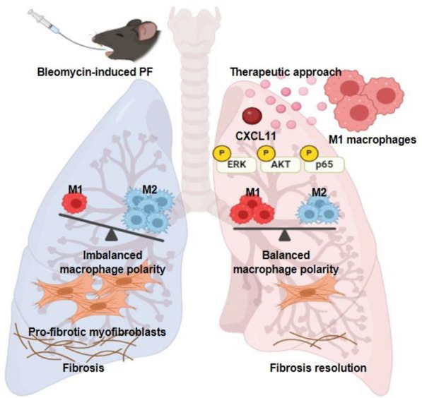
Background: In understanding the pathophysiology of pulmonary fibrosis (PF), macrophage plasticity has been implicated with a crucial role in the fibrogenic process. Growing evidence indicates that accumulation of M2 macrophages correlates with the progression of PF, suggesting that targeted modulation of molecules that influence M2 macrophage polarization could be a promising therapeutic approach for PF. Here, we demonstrated a decisive role of C-X-C motif chemokine ligand 11 (CXCL11) in driving M1 macrophage polarization to alleviate PF in the bleomycin-induced murine model.

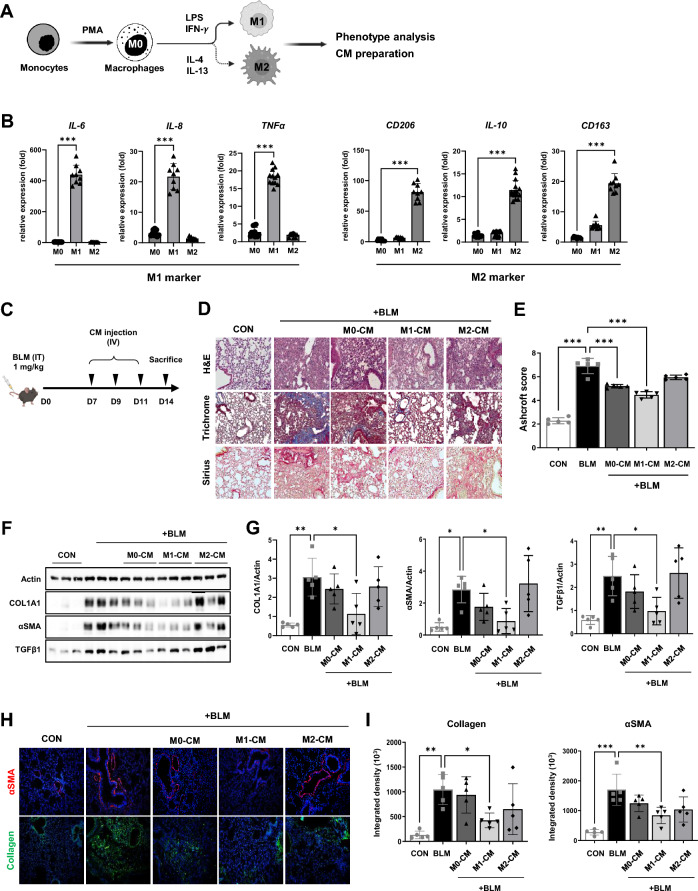
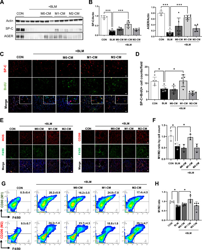
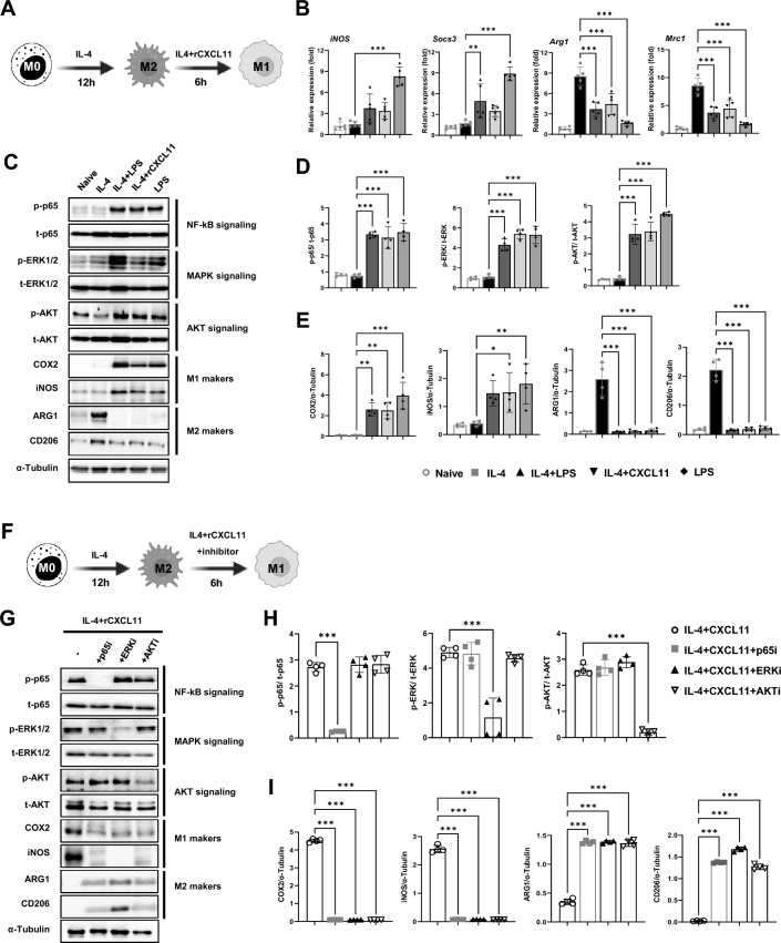
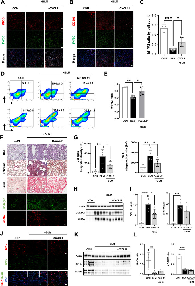
Results: We intravenously administered secretome derived from naïve (M0) and polarized macrophages (M1 and M2) into PF mice and found that lung fibrosis was effectively reversed in only the M1-treated group, with modulation of the M1/M2 ratio toward the ratio of the control group. These findings suggest that the factors secreted from M1 macrophages contribute to alleviating PF by targeting macrophages and reshaping the immunofibrotic environment in a paracrine manner. Secretome analysis of macrophages identified CXCL11 as an M1-specific chemokine, and administration of recombinant CXCL11 effectively improved fibrosis with the reduction of M2 macrophages in vivo. Furthermore, a mechanistic in vitro study revealed that CXCL11 reprogrammed macrophages from M2 to M1 through the activation of pERK, pAKT, and p65 signaling.

Conclusions: Collectively, we demonstrate an unprecedented role for M1 macrophage-derived CXCL11 as an inducer of M1 macrophage polarization to revert the fibrogenic process in mice with PF, which may provide a clinically meaningful benefit.

Keywords: CXCL11; Macrophage; Polarization; Pulmonary fibrosis.


Conflict of interest statement

Declarations Ethics approval and consent to participate The experiment was approved by the Institutional Animal Care and Use Committee (IACUC), Kangwon National University (IACUC No. KW-220509-1). Consent for publication Not applicable. Competing interests The authors have declared that no competing interest exists.

Figures

Fig. 1
Fig. 1
Polarization of macrophages during the phase of fibrotic progression in BLM-induced mice lung tissues. A Schematic overview of study BLM-induced PF mice model. B The body weight of the mice was measured from the start of BLM administration to the time of sacrifice (days 4, 7, and 14). C, D Flow cytometry analysis of the expression of M1 (F4/80+ iNOS+) and M2 (F4/80+ CD206+) macrophages in lung tissues of BLM-induced PF mice. E Mean fluorescence intensity (MFI) of M1 and M2 markers. F, G Representative images of immunostaining for iNOS (F) CD206 (G) and F4/80 in the lung tissues of each group. Scale bars, 20 μm. H The ratio of M1 and M2 macrophages was analyzed by counting the number of M1 and M2 positive cells in at least three different fields of lung tissues of each group. Data are presented as mean ± SD. *P < 0.05, **P < 0.01
Fig. 2
Fig. 2
Administration of M1-CM reverts BLM-induced PF in mice. A Schematic overview of macrophage differentiation and activation. B The expression levels of IL6, IL8, and TNFα (M1 markers) and CD206, IL10, and CD163 (M2 markers) were validated by qPCR. Data are presented as mean ± SD. C Schematic overview for testing the efficacy of the CM collected from naïve and polarized macrophage in the PF mice model. D Representative images of hematoxylin and eosin (HE), Masson’s trichrome, and Sirius Red staining in lung tissue from each group. Scale bars, 50 μm. E The Ashcroft score indicates the severity of fibrosis. F Western blotting for COL1A1, αSMA, TGFβ1, and Actin as a loading control in the lung homogenates of each group. G The graph shows the relative intensity of COL1A1, αSMA, and TGFβ1 in the lung tissue of the indicated groups. H Representative images of immunostaining for αSMA and Collagen in the lung tissue from each group. Scale bars, 50 μm. I Quantification of Collagen and αSMA positive areas by Image J 1.51j. Data are presented as mean ± SD. *P < 0.05, **P < 0.01., ***P < 0.001
Fig. 3
Fig. 3
M1 macrophage-CM promotes proliferation of AECs. A, B Western blotting (A) and subsequent quantification of SP-C (type 2 AEC marker) and AGER (type 1 AEC marker) (B) in whole-lung homogenates of mice from the indicated groups. Actin was used as a loading control. C Representative images of immunostaining for colocalization of BrdU and SP-C from lung tissues from each experimental group. Scale bars, 20 μm. D The SP-C + BrdU + cells were counted in at least 3 different fields of lung tissues of each group. E Representative images of immunostaining for iNOS, CD206, and F4/80 in the lung tissues of each group. Scale bars, 20 μm. F The ratio of M1 and M2 macrophages was analyzed by counting the number of M1 and M2 positive cells in at least three different fields of lung tissues of each group. G Flow cytometry analysis of the expression of M1 (F4/80+ CD80+) and M2 (F4/80+ CD206+) macrophages in lung tissues from each group. H Flow cytometry analysis of the M1/M2 ratio in each group. Data are presented as mean ± SD. *P < 0.05, ***P < 0.001
Fig. 4
Fig. 4
Cytokine analysis of CM from M0, M1, and M2 macrophages. A Comparative analysis of cytokine secretion in CM from naïve and polarized macrophage-CM. Each cytokine was detected in duplicate. Target cytokines are indicated using square frames with numbers. B CXCL9, CXCL11, and PTX3 proteins (indicated by arrows in a panel) were highly detected in M1-CM compared to M0- and M2-CM. C Quantification shows the mean signal intensity
Fig. 5
Fig. 5
M1 macrophage-derived CXCL11 promotes M2 to M1 phenotype polarization mediated through the p65, ERK1/2, and AKT pathway in vitro. A A schematic diagram illustrating the experimental procedures to examine the effect of CXCL11 on macrophage polarity. B Real-time RT-PCR analyzed the relative mRNA levels of M1 (iNos and Socs3)- and M2 (Arg1 and Mrc1)-related factors. C Western blotting for phosphorylation of p65, ERK1/2, AKT and COX2, iNOS, ARG1, and CD206 protein in BMDM after CXCL11 treatment. α-Tubulin was used as a loading control. D, E The graph shows the relative intensity of phosphorylation of p65, ERK1/2, and AKT (D) and COX2, iNOS, ARG1, and CD206 (E). F A schematic diagram illustrating the experimental procedures to examine the pathway of CXCL11 on macrophage polarity. G Western blotting for phosphorylation of p65, ERK1/2, AKT and COX2, iNOS, ARG1, and CD206 protein in BMDM after CXCL11 treatment with inhibitors. α-Tubulin was used as a loading control. H, I The graph shows the relative intensity of phosphorylation of p65, ERK1/2, and AKT (H) and COX2, iNOS, ARG1, and CD206 (I)
Fig. 6
Fig. 6
CXCL11 exhibits anti-fibrotic activity through the conversion of M2 macrophages to M1 macrophages. A, B Representative images of immunostaining for iNOS, CD206, and F4/80 in the lung tissue from each group. Scale bars, 20 μm. C The ratio of M1 and M2 macrophages was analyzed by counting the number of M1 and M2 positive cells in at least three different fields of lung tissues of each group. D Flow cytometry analysis of the expression of M1 (F4/80+ CD80+) and M2 (F4/80+ CD206+) macrophages in lung tissues from each group. E Flow cytometry analysis of the M1/M2 ratio in each group. F Representative images of HE, Masson’s trichrome, Sirius red staining, and immunostaining for Collagen and αSMA in lung tissue from each experimental group. Scale bars, 100 μm. G Quantification of Collagen and αSMA positive areas by Image J 1.51j. H Western blotting for COL1A1, αSMA, and Actin as a loading control in the lung homogenates of each group. I The graph shows the relative intensity of COL1A1 and αSMA in the lung tissue of the indicated groups. J Representative images of immunostaining for colocalization of BrdU and SP-C from lung tissues from each experimental group. Scale bars, 20 μm. K Western blot analysis of SP-C, AGER, and Actin as a loading control in the lung homogenates of each group of mice. L Subsequent quantification of SP-C and AGER in lung tissue of the indicated groups. Data are presented as mean ± SD. *P < 0.05, **P < 0.01., ***P < 0.001
Fig. 7
Fig. 7
A schematic illustration of M1 macrophage-derived CXCL11 in BLM-induced PF in mice. M1 macrophage-derived CXCL11 to modulate M1 macrophage polarization for reverting the fibrogenic process in mice with PF

References

    1. Das A, Sinha M, Datta S, Abas M, Chaffee S, Sen CK, et al. Monocyte and macrophage plasticity in tissue repair and regeneration. Am J Pathol. 2015;185(10):2596–606. - PMC - PubMed
    1. Deng L, Jian Z, Xu T, Li F, Deng H, Zhou Y, et al. Macrophage polarization: An important candidate regulator for lung diseases. Molecules. 2023;28(5):2379. - PMC - PubMed
    1. Wang H, Gao Y, Wang L, Yu Y, Zhang J, Liu C, et al. Lung specific homing of diphenyleneiodonium chloride improves pulmonary fibrosis by inhibiting macrophage M2 metabolic program. J Adv Res. 2023;44:213–25. - PMC - PubMed
    1. Cheng P, Li S, Chen H. Macrophages in lung injury, repair, and fibrosis. Cells. 2021;10(2):436. - PMC - PubMed
    1. Zhang F, Ayaub EA, Wang B, Puchulu-Campanella E, Li YH, Hettiarachchi SU, et al. Reprogramming of profibrotic macrophages for treatment of bleomycin-induced pulmonary fibrosis. EMBO Mol Med. 2020;12(8): e12034. - PMC - PubMed

LinkOut - more resources