. 2024 Nov 16;81(1):456.
doi: 10.1007/s00018-024-05489-5.

rTM reprograms macrophages via the HIF-1α/METTL3/PFKM axis to protect mice against sepsis

Affiliations

rTM reprograms macrophages via the HIF-1α/METTL3/PFKM axis to protect mice against sepsis

Chen Yao et al. Cell Mol Life Sci. .

Abstract

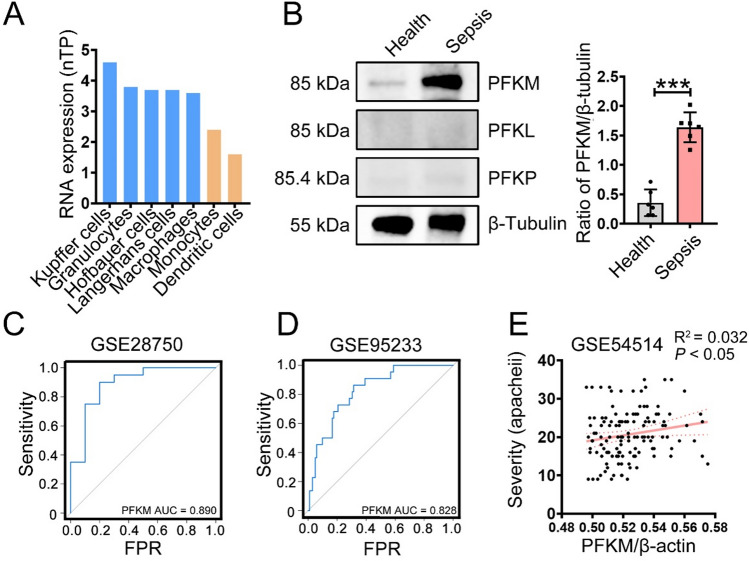
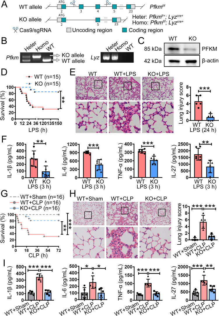
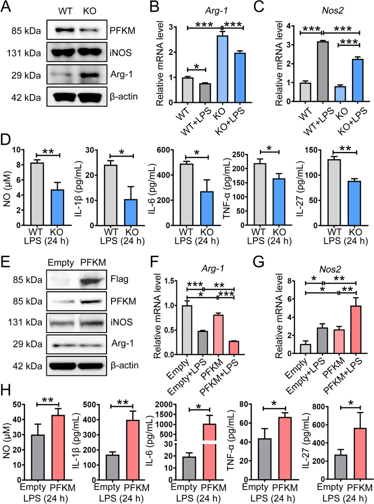
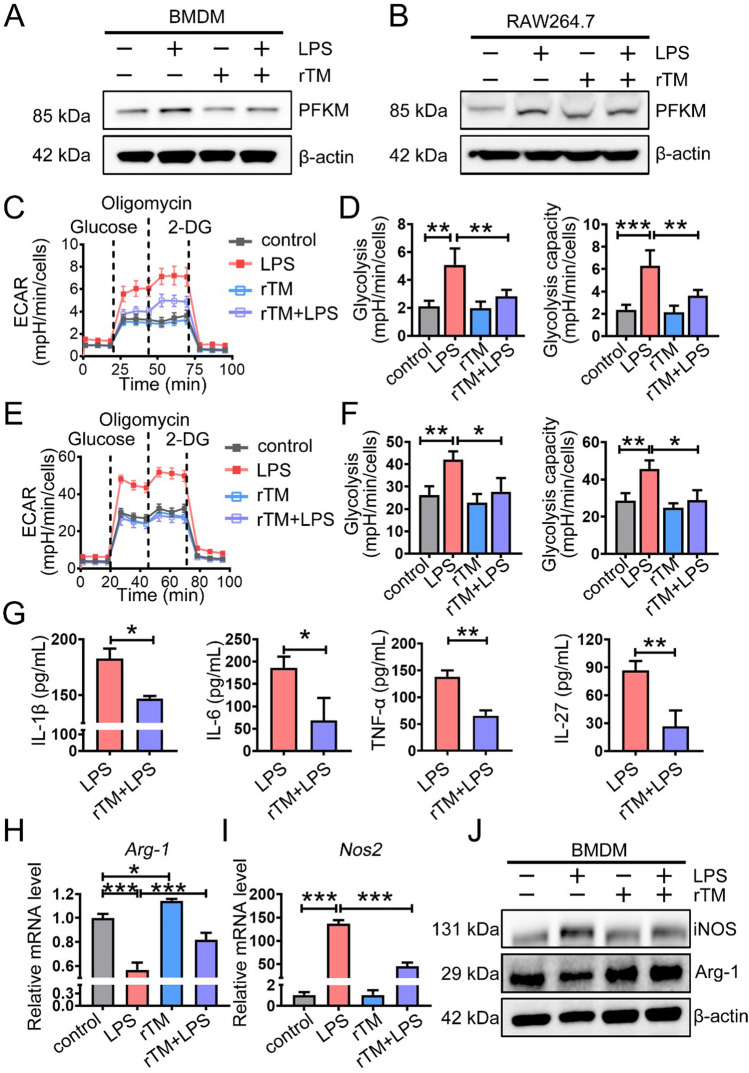
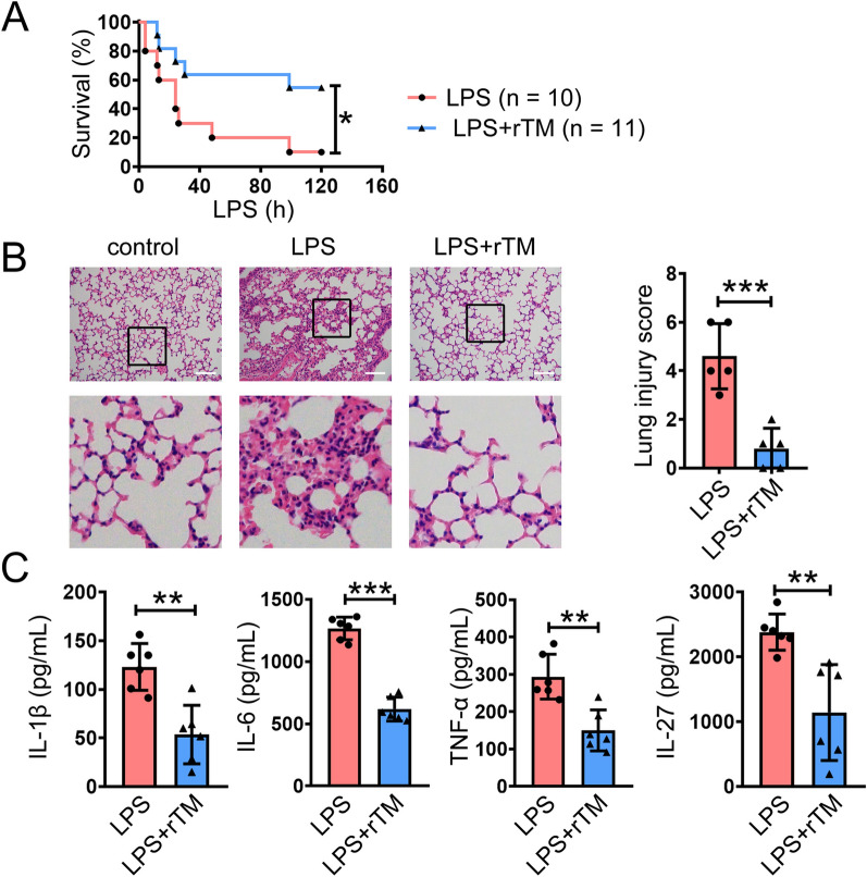
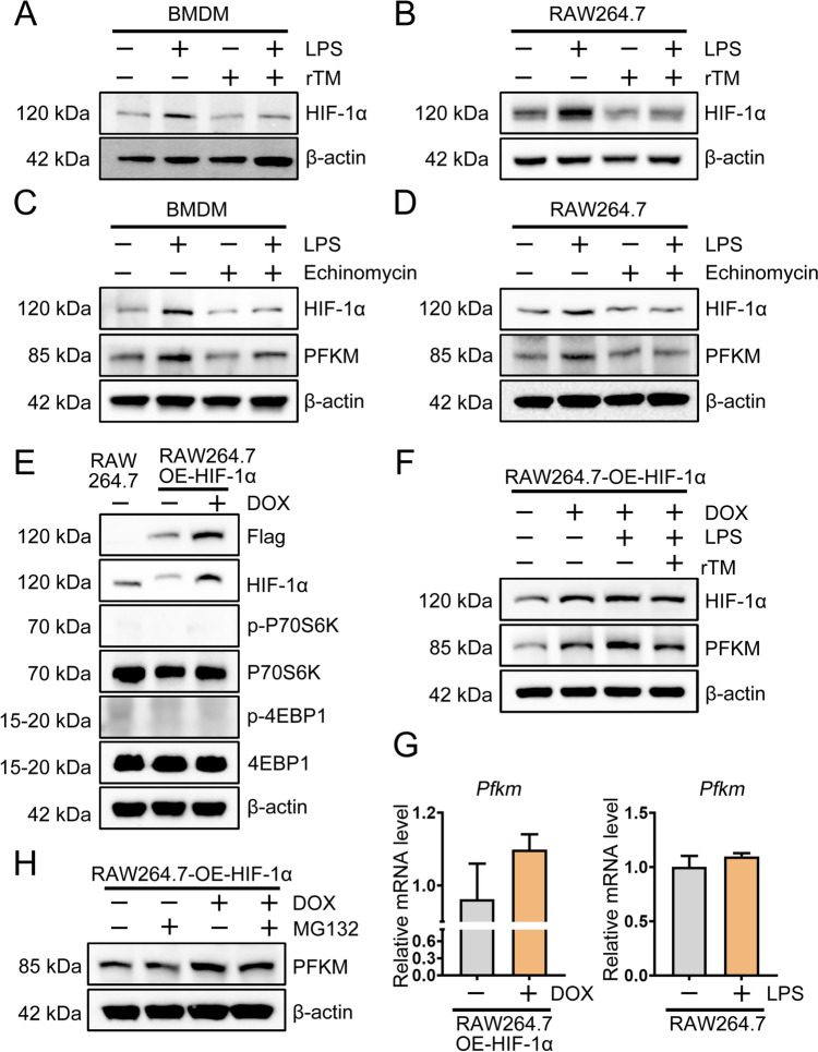
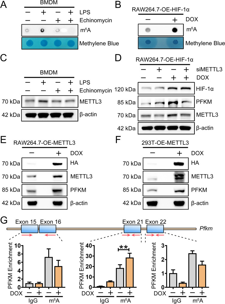
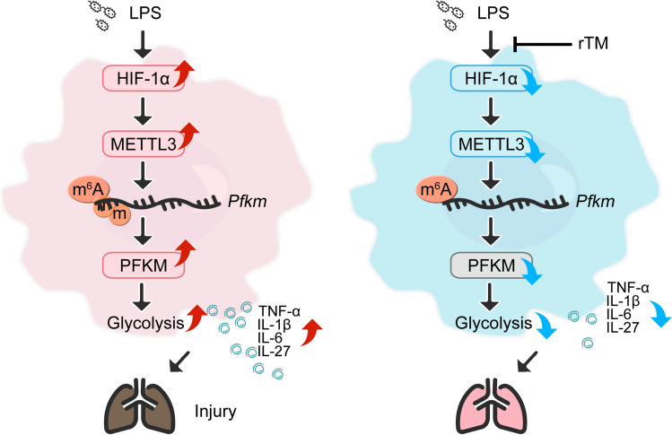
The metabolic reprogramming of macrophages is a potential therapeutic strategy for sepsis treatment, but the mechanism underlying this reprogramming remains unclear. Since glycolysis can drive macrophage phenotype switching, the rate-limiting enzymes in glycolysis may be key to treating sepsis. Here, we found that, compared with other isoenzymes, the expression of 6-phosphofructokinase, muscle type (PFKM) was the most upregulated in monocytes from septic patients. Recombinant thrombomodulin (rTM) treatment downregulated the protein expression of PFKM in macrophages. Both rTM treatment and Pfkm knockout protected mice from sepsis and reduced the production of the proinflammatory cytokines IL-1β, IL-6, TNF-α, and IL-27, whereas PFKM overexpression increased the production of these cytokines. Mechanistically, rTM treatment inhibited glycolysis in macrophages by decreasing PFKM expression in a hypoxia-inducible factor-1α (HIF-1α)-dependent manner. HIF-1α overexpression increased methyltransferase-like 3 (METTL3) expression, elevated the m6A level on Pfkm, and upregulated the protein expression of PFKM. METTL3 silence attenuated HIF-1α-mediated PFKM expression. These findings provide insight into the underlying mechanism of macrophage reprogramming for the treatment of sepsis.

Keywords: 6-phosphofructokinase; Glycolysis; Macrophages; Methyltransferase-like 3 (METTL3); Muscle type (PFKM); Sepsis; Thrombomodulin.


Conflict of interest statement

Declarations Conflict of interest The authors have no relevant financial or nonfinancial interests to disclose. Ethics approval and consent to participate The study was approved by the Ethics Committee of Xuzhou Medical University (ethical approval number: XYFY2022-KL442-01). All animal studies and all experimental protocols were approved by the Institutional Animal Care and Use Committee of Xuzhou Medical University. Consent for publication All the authors approved the submission of the manuscript to this journal.

Figures

Fig. 1
Fig. 1
PFKM is upregulated in sepsis and is positively correlated with the severity of sepsis. A The RNA expression of PFKM in different myeloid cells from the HPA database. B PBMCs (the CD14+ subset) were isolated from the peripheral blood of septic patients and healthy volunteers. The levels of the proteins were analyzed via Western blotting. C–D The ROC curves of PFKMs from the GSE28750 and GSE95233 cohorts. FPR, False Positive Rate. E The correlation between the PFKM score and sepsis severity in the GSE54514 cohort. The data are shown as the mean ± SD. ***P < 0.01
Fig. 2
Fig. 2
PFKM deficiency protected mice against sepsis. A Diagram of Pfkm knockout mice constructed by CRISPR/Cas9 technology. B Mouse genotypes were identified via agarose gel electrophoresis. C Western blotting of the expression of the indicated proteins in BMDMs. D WT and KO mice were intraperitoneally injected with LPS (20 mg/kg), and the survival rate was recorded daily. E Mice were intraperitoneally injected with LPS (20 mg/kg) and sacrificed at 24 h (n = 5). H&E staining of representative sections. Scale bars, 50 µm. F Mice were intraperitoneally injected with LPS (20 mg/kg) and sacrificed at 3 h, after which cytokine levels in the serum were determined via cytometric bead array (CBA) analysis (n = 6). (G-I) Sepsis was induced in mice with CLP, and G the survival rate was recorded. (H-I) Mice were sacrificed at 24 h (n = 5). H Representative H&E staining of sections. Scale bars, 50 µm. I The production of inflammatory factors in the serum was determined via ELISA. The data are shown as the mean ± SD. *P < 0.05, **P < 0.01, ***P < 0.001
Fig. 3
Fig. 3
PFKM promotes the polarization of macrophages toward a proinflammatory phenotype. AD BMDMs were generated from WT or KO mice. A The protein levels of PFKM, iNOS, and Arg-1 were examined via Western blotting. BC BMDMs were treated with LPS (100 ng/mL) for 3 h, and the mRNA expression levels of B Arg-1 and C Nos2 were examined via real-time RT-PCR. D BMDMs were treated with LPS (100 ng/mL) for 24 h, after which the production of inflammatory factors was determined via ELISA. EH RAW264.7 cells overexpressing PFKM were generated, and cells transfected with empty lentivirus were used as controls. E Western blotting was used to detect the protein expression levels of PFKM, iNOS, and Arg-1. FG The mRNA expression levels of F Arg-1 and G Nos2 were examined via real-time RT-PCR. H Cells were treated with LPS (100 ng/mL) for 24 h, after which the production of inflammatory factors was determined via ELISA. The data are shown as the mean ± SD. *P < 0.05, **P < 0.01, and ***P < 0.001
Fig. 4
Fig. 4
rTM treatment attenuated the proinflammatory phenotype of macrophages by regulating glycolysis. A, CD, GI BMDMs or B, EF RAW264.7 cells were treated with LPS (100 ng/mL), rTM (2 μg/mL), or a combination of LPS (100 ng/mL) and rTM (2 μg/mL). AB The protein level of PFKM in A BMDMs or B RAW264.7 cells was examined by Western blotting analysis at 3 h poststimulation. CF The ECAR of CD BMDMs and EF RAW264.7 cells was determined via a Seahorse extracellular flux analyzer at 24 h poststimulation. G BMDMs were treated with LPS (100 ng/mL) for 24 h, after which the production of inflammatory factors was determined via ELISA. The mRNA expression levels of H Arg-1 and I Nos2 were examined by real-time RT-PCR. (J) The protein levels of Arg-1 and iNOS in BMDMs were examined by Western blotting. The data are shown as the mean ± SD. *P < 0.05, **P < 0.01, ***P < 0.001
Fig. 5
Fig. 5
rTM protected mice against LPS-induced sepsis. A–C Mice were intraperitoneally injected with rTM (1 μg/kg) or normal saline 30 min before LPS injection (20 mg/kg), and (A) the survival rate was recorded daily. B Representative H&E staining of sections and the lung injury score at 24 h after LPS injection (n = 5). Scale bars, 50 µm. C Cytokine levels in the serum were determined by cytometric bead array (CBA) analysis at 3 h (n = 6). The data are shown as the mean ± SD. *P < 0.05, **P < 0.01, ***P < 0.001
Fig. 6
Fig. 6
rTM downregulates the expression of PFKM via HIF-1α. A BMDMs or B RAW264.7 cells were treated with LPS (100 ng/mL), rTM (2 μg/mL), or the combination of LPS (100 ng/mL) and rTM (2 μg/mL) for 3 h. The protein level of HIF-1α was analyzed via Western blotting. C BMDMs or D RAW264.7 cells were pretreated with or without echinomycin (1 nmol/L) and then treated with LPS (100 ng/mL) for 3 h. The protein levels of HIF-1α and PFKM were analyzed via Western blotting. E RAW264.7 cells overexpressing HIF-1α were generated with a Tet-On system (RAW264.7-OE-HIF-1α), the cells were treated with DOX (125 ng/mL), and the levels of the indicated proteins were analyzed via Western blotting. F RAW264.7-OE-HIF-1α cells were treated with LPS (100 ng/mL) and rTM (2 μg/mL) for 3 h, and the protein levels of HIF-1α and PFKM were analyzed via Western blotting. H RAW264.7-OE-HIF-1α cells were treated with MG132 (5 μmol/L) for 6 h, and the protein levels of HIF-1α and PFKM were analyzed via Western blotting. G The mRNA expression of Pfkm was examined by real-time RT-PCR. The data are shown as the mean ± SD
Fig. 7
Fig. 7
HIF-1α mediates the m6A level on Pfkm via METTL3. A, C BMDMs were pretreated with echinomycin (1 nmol/L) for 13 h and then treated with LPS (100 ng/mL) for 3 h. (A) The m6A levels were analyzed by dot blot. B RAW264.7-OE-HIF-1α cells were treated with DOX (125 ng/mL) for 48 h, and the m6A levels were analyzed via dot blot. C The protein level of METTL3 was analyzed via Western blotting. D METTL3 expression was disrupted by siRNA in RAW264.7-OE-HIF-1α cells, and the protein expression of HIF-1α, PFKM, and METTL3 was analyzed via Western blotting. E RAW264.7 cells and F 293 T cells overexpressing METTL3 were generated with the Tet-On system. The cells were treated with DOX (125 ng/mL), and the levels of the indicated proteins were analyzed via Western blotting. G MeRIP-RT-PCR was used to assess the relative expression of PFKM mRNA with m6A methylation in RAW264.7-OE-HIF-1α cells. IgG was used as a negative control. The data are shown as the mean ± SD. *P < 0.05
Fig. 8
Fig. 8
rTM inhibited the HIF-1α/METTL3/PFKM axis to reprogram macrophages toward an anti-inflammatory phenotype and protect mice against sepsis

References

    1. Singer M, Deutschman CS, Seymour CW, Shankar-Hari M, Annane D, Bauer M et al (2016) The third international consensus definitions for sepsis and septic shock (Sepsis-3). JAMA 315(8):801–810 - PMC - PubMed
    1. Gander-Bui HTT, Schläfli J, Baumgartner J, Walthert S, Genitsch V, van Geest G et al (2023) Targeted removal of macrophage-secreted interleukin-1 receptor antagonist protects against lethal Candida albicans sepsis. Immunity 56(8):1743-1760.e9 - PubMed
    1. Pan Y, Li J, Xia X, Wang J, Jiang Q, Yang J et al (2022) β-glucan-coupled superparamagnetic iron oxide nanoparticles induce trained immunity to protect mice against sepsis. Theranostics 12(2):675–688 - PMC - PubMed
    1. Peng J, Li J, Huang J, Xu P, Huang H, Liu Y et al (2019) p300/CBP inhibitor A-485 alleviates acute liver injury by regulating macrophage activation and polarization. Theranostics 9(26):8344–8361 - PMC - PubMed
    1. Seman BG, Vance JK, Rawson TW, Witt MR, Huckaby AB, Povroznik JM et al (2020) Elevated levels of interleukin-27 in early life compromise protective immunity in a mouse model of gram-negative neonatal sepsis. Infect Immun 88(3):e00828-e919 - PMC - PubMed

Substances

LinkOut - more resources