. 2025 May;82(5):871-881.
doi: 10.1016/j.jhep.2024.10.048. Epub 2024 Nov 15.

PNPLA3(148M) is a gain-of-function mutation that promotes hepatic steatosis by inhibiting ATGL-mediated triglyceride hydrolysis

Affiliations

PNPLA3(148M) is a gain-of-function mutation that promotes hepatic steatosis by inhibiting ATGL-mediated triglyceride hydrolysis

Yang Wang et al. J Hepatol. 2025 May.

Abstract

Background & aims: PNPLA3(148M) (patatin-like phospholipase domain-containing protein 3) is the most impactful genetic risk factor for steatotic liver disease. A key unresolved issue is whether PNPLA3(148M) confers a loss- or gain-of-function. Here we test the hypothesis that PNPLA3 causes steatosis by sequestering ABHD5 (α/β hydrolase domain-containing protein 5), the cofactor of ATGL (adipose TG lipase), thus limiting mobilization of hepatic triglyceride (TG).

Methods: We quantified and compared the physical interactions between ABHD5 and PNPLA3/ATGL in cultured hepatocytes using NanoBiT complementation assays and immunocytochemistry. Recombinant proteins purified from human cells were used to compare TG hydrolytic activities of PNPLA3 and ATGL in the presence or absence of ABHD5. Adenoviruses and adeno-associated viruses were used to express PNPLA3 in liver-specific Atgl-/- mice and to express ABHD5 in livers of Pnpla3M/M mice, respectively.

Results: ABHD5 interacted preferentially with PNPLA3 relative to ATGL in cultured hepatocytes. No differences were seen in the strength of the interactions between ABHD5 with PNPLA3(WT) and PNPLA3(148M). In contrast to prior findings, we found that PNPLA3, like ATGL, is activated by ABHD5 in in vitro assays using purified proteins. PNPLA3(148M)-associated inhibition of TG hydrolysis required that ATGL be expressed and that PNPLA3 be located on lipid droplets. Finally, overexpression of ABHD5 reversed the hepatic steatosis in Pnpla3M/M mice.

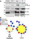
Conclusions: These findings support the premise that PNPLA3(148M) is a gain-of-function mutation that promotes hepatic steatosis by accumulating on lipid droplets and inhibiting ATGL-mediated lipolysis in an ABHD5-dependent manner. Our results predict that reducing, rather than increasing, PNPLA3 expression will be the best strategy to treat PNPLA3(148M)-associated steatotic liver disease.

Impact and implications: Steatotic liver disease (SLD) is a common complex disorder associated with both environmental and genetic risk factors. PNPLA3(148M) is the most impactful genetic risk factor for SLD and yet its pathogenic mechanism remains controversial. Herein, we provide evidence that PNPLA3(148M) promotes triglyceride (TG) accumulation by sequestering ABHD5, thus limiting its availability to activate ATGL. Although the substitution of methionine for isoleucine reduces the TG hydrolase activity of PNPLA3, the loss of enzymatic function is not directly related to the steatotic effect of the variant. It is the resulting accumulation of PNPLA3 on LDs that confers a gain-of-function by interfering with ATGL-mediated TG hydrolysis. These findings have implications for the design of potential PNPLA3(148M)-based therapies. Reducing, rather than increasing, PNPLA3 levels is predicted to reverse steatosis in susceptible individuals.

Keywords: Steatotic liver disease; lipid droplets; lipolysis; triglyceride hydrolase.


Conflict of interest statement

Conflict of interest Helen Hobbs is on the Corporate Board of Pfizer, Inc. Please refer to the accompanying ICMJE disclosure forms for further details.

Figures
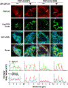
Fig. 1.
Fig. 1.. Relative strength of interactions between ABHD5 and ATGL, PNPLA3(WT or 148M) in HuH7 cells.
(A) Schematic of NanoBiT protein-protein interaction assay. Small-BiT (SmBiT, S) was fused to the N-terminus of PNPLA3-V5 (S-PNPLA3) or ATGL-V5 (S-ATGL) and Large-BiT (LgBiT, L) was fused to the C-terminus of ABHD5 (ABHD5-L). Physical proximity due to protein-protein interactions between ABHD5 and PNPLA3 or ABHD5 and ATGL results in S and L forming a functional luciferase, which catalyzes conversion of furimazine to produce a luminescent signal in live cells detected by EnVision Multilabel Plate Readers. (B) NanoBiT protein-protein interaction assays were performed in HuH7 cells expressing ABHD5-L and the indicated recombinant proteins. S-PRKACA (protein kinase cAMP-activated catalytic subunit alpha), a soluble cytoplasmic protein, was used as a negative control. Immunoblot analysis of the transfected cells is shown. CANX: calnexin; loading control. (C) Cellular luminescent signals were normalized to levels of the SmBiT-linked proteins, where levels of S-HSD17B13 and ABHD5-L pair were set to 1 (right). Data represent median [25th% - 75th%] of data pooled from 3 experiments using box-and-whisker plot in a natural logarithmic scale; p values of natural logarithms of relative luminescence were determined by one-way ANOVA followed by Tukey’s multiple comparisons test; †p < 0.0001. (D) NanoBiT-tagged protein pairs were co-expressed with proteins without NanoBiT tags to assess competitive binding to ABHD5-L. The competitor proteins were of a higher molecular weight due to addition of 3×HA, 3×FLAG and 1×V5 tags (~8.1 kDa) to the C-terminus. The ratios of the plasmids encoding the two NanoBiT components and competitors were 1:1:0.5 or 1:1:1. The asterisk indicates a non-specific signal. (E) Luminescence signals were quantified and normalized to levels of SmBiT-linked protein. The normalized luminescence signals were compared to indicated SmBiT-linked protein and ABHD5-L pair with no competitors (set at 100%). Data represent mean ± SD of technical replicates; p values were determined by one-way ANOVA followed by Sidak’s multiple comparisons test; †p < 0.0001. The experiments were repeated three times and the results were similar.
Fig. 2.
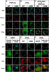
Fig. 2.. Recruitment of LD binding defective ABHD5 (ABHD5-LBD1 and ABHD5-LBD2) to LDs by PNPLA3 and ATGL in ATGL−/− QBI-293A cells.
(A) Schematics of human ABHD5(WT), ABHD5(3W3A) [ABHD5-LBD1] and ABHD5(Δ30) [ABHD5-LBD2]. The first 30 residues (gray box) are required for LD binding. (B) ABHD5-WT and the LBD variants with a myc epitope tag at the C-terminus were expressed in ATGL−/− QBI-293A cells grown in medium supplemented with oleate (200 μM). Immunofluorescence was performed using a rabbit anti-myc polyclonal antibody to detect ABHD5 (green). LDs were stained using LipidTOX Deep Red (cyan). White dashes outline the transfected cells. (C) ABHD5-WT-myc or ABHD5-LBD1-myc were co-expressed with PNPLA3(47A)-V5 or ATGL(47A)-V5 in ATGL−/− cells. LDs were stained using LipidTOX Deep Red (cyan). ABHD5 (green), PNPLA3 and ATGL (red) were visualized using anti-myc and anti-V5 primary antibodies and Alexa Fluor-conjugated secondary antibodies. Merge: ABHD5 plus PNPLA3 or ATGL. Vector: pcDNA3.1(+). White dashes outline the transfected cells. (D) The proportion of ABHD5 on LDs was quantitated using ImageJ. Data represent mean ± SD; n=5 cells/group; p values were determined using 2-way ANOVA followed by Tukey’s multiple comparisons test to compare within groups; † p<0.0001. Experiments were repeated twice, and results were similar.
Fig. 3.
Fig. 3.. ATGL inhibition by PNPLA3 requires localization of PNPLA3 to LDs.
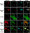
(A) Schematic of mutations in the LD binding motif (yellow, residues 347–403) to generate the LD-binding defective (LBD) variant of PNPLA3. Valine (V) substitutions are shown in red. Human PNPLA3 structure was predicted by AlphaFold 2 (left): Yellow: putative LD binding region; Cyan: patatin-like domain; Magenta spheres: catalytic dyad (Ser47 and Asp166); Black sphere: Ile148; Grey: low confidence regions of the structure. Low confidence C-terminal region (residue 409 to 481) have been removed for clarity. (right) Scheme of human PNPLA3 colored in rainbow according to pLDDT residue pLDDT confidence: lowest pLDDT (blue) to highest pLDDT (red). (B) PNPLA3(WT) or PNPLA3(148M) +/− LBD mutations were expressed in QBI-293A cells grown in medium supplemented with oleate (200 μM). (left) Immunofluorescence was performed to show localization of PNPLA3 (red) using anti-PNPLA3 primary antibody and Alexa 555-conjugated anti-mouse secondary antibody. LDs were stained using monodansylpentane (MDH, cyan). White dashes outline transfected cells. (right) Quantification of proportion of different forms of PNPLA3 localized to LD (n=5–7 cells/group). (C) Human ATGL (green) was co-expressed with PNPLA3(WT) or PNPLA3(148M) +/− LBD mutations (red) and processed as described in Panel B, except that ATGL was visualized using an anti-ATGL primary antibody. (right) Quantification of LD area in cells co-expressing target proteins (n=3 cells/group). Data are represented as mean ± SD; p values were determined by one-way ANOVA followed by Dunnett’s multiple comparisons test; **p < 0.01, ***p< 0.001, †p<0.0001. Experiment was repeated and the results were similar.
Fig. 4.
Fig. 4.. In vitro TG hydrolase activities of purified PNPLA3 and ATGL in the presence or absence of ABHD5.
(A) Coomassie stain of recombinant human ATGL, PNPLA3(WT), PNPLA3(148M), and ABHD5 purified from mammalian HEK293S GnTI cells. ATGL, PNPLA3(WT) or PNPLA3(148M) (5 μg) and ABHD5 (3.5 μg) were subjected to SDS–PAGE. (B and C) Basal and ABHD5-activated TG hydrolysis of ATGL, PNPLA3(WT) and (148M) via (B) thin layer chromatography (TLC) and (C) gas chromatography-mass spectrometry (GC-MS). A total of 5 μg of purified ATGL, PNPLA3(WT), PNPLA3(148M) +/− an equimolar amount of ABHD5 were incubated for 1 h with lipid emulsions containing 6.68 mM triolein. Lipids were extracted and fractionated by TLC. Standards for the lipids were loaded in lane 1. (C) Free fatty acids (FA) were derivatized in tri-ethylamine (1%) and pentafluorobenzyl bromide (1%) in acetone and then quantified by GC-MS. Numbers above the bars represent values of means in each group. (D) Competition between PNPLA3 and ATGL for ABHD5 in vitro. Various molar ratios (from 0.5X to 3X relative to ATGL) of PNPLA3(WT) or (148M) were added to mixtures containing equimolar amount of ATGL and ABHD5. The protein mixtures were incubated with 25 μl of emulsion containing 6.68 mM triolein for 1 h at 37°C. The released FAs were quantified using GC-MS. Data represent mean ± SD; n=3 biological replicates; p values were determined by two-tailed t-tests with Welch’s correction or one-way ANOVA followed by Dunnett’s multiple comparisons test. *p<0.05, **p<0.01, ***p<0.001, †p<0.0001. This experiment was repeated twice and the results were similar.
Fig. 5.
Fig. 5.. ATGL is required for PNPLA3(148M)-mediated hepatic steatosis
(A) Protein (left) and transcript levels (right) of LD proteins in Ls-Atgl−/− mice. LDs were purified from chow-fed Atglf/f and Ls-Atgl−/− female mice (8–10 weeks, n=4 mice/group) and immunoblot analysis was performed (5 μg of protein) using the indicated antibodies (left). Total RNA was extracted from the livers and mRNA levels were measured using quantitative real-time PCR and normalized to levels of CANX mRNA. The mRNA levels in Atglf/f mice were set as 1. The experiment was repeated in male mice fed a high sucrose diet and the results were similar. (B) Schematic of experimental design. Chow-fed Atglf/f and Ls-Atgl−/− male mice (14–17 weeks, n=3–6 mice/group) were infected with adenovirus [1.5×10¹¹ genomic copies (GC)] with no insert (RR5) or expressing human PNPLA3(WT or 148M)-V5. After 3 days, diets were synchronized and mice killed at the end of the last feeding period. (C) Immunoblot analysis of pooled hepatic LD proteins (1 μg) in infected mice (left). Protein levels were quantified using LI-COR after normalization to levels of HSD17B13 (right). (D) Relative levels of hepatic mRNAs encoding LD proteins in adenovirus-infected mice. Atglf/f mice treated with adenovirus expressing no insertion (Ad-RR5) were arbitrarily set to 1. h: human; m: mouse. (E) Hepatic TG levels were measured using an enzymatic assay and normalized to tissue weight. Data represent mean ± SD; p values were determined by one-way ANOVA followed by Tukey’s multiple comparisons test; *p<0.05, ***p<0.001. The experiment was repeated twice and the results were similar.
Fig. 6.
Fig. 6.. AAV-mediated hepatic expression of mouse ABHD5 rescued steatosis in Pnpla3M/M mice
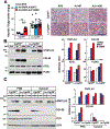
(A) Schematic of experimental design. Pnpla3+/+ and Pnpla3M/M female mice (12–15 weeks, n=4–5 mice/group) were infected with AAV expressing GFP or ABHD5-FLAG×3 [ABHD5#, 1.25×10¹¹ genomic copies (GC)]. Mice were fed a high-fructose diet for 4 weeks. Diets were synchronized by 18-h fasting and 6-h refeeding for 3 days before the mice were sacrificed after last feeding cycle. (B) (left) LD proteins (1.5 μg) were subjected to immunoblotting and (right) levels of indicated proteins were quantified using LI-COR and normalized to levels of PLIN2. (C) Relative mRNA levels of indicated LD associated genes. The mRNA levels in Pnpla3+/+ mice infected with AAV-GFP were set as 1. (D) Hepatic TG levels were measured as described in legend to Fig. 4. Data are represented as mean ± SD; n=4–5 mice/group; p values were determined by 2-way ANOVA followed by Tukey’s multiple comparisons test; *p<0.05, **p<0.01. The experiment was repeated in female mice and the results were similar.
Fig. 7.
Fig. 7.

Similar articles

Cited by

References

    1. Cohen JC, Horton JD, Hobbs HH. Human fatty liver disease: old questions and new insights. Science 2011;332:1519–1523. - PMC - PubMed
    1. Linden D, Romeo S. Therapeutic opportunities for the treatment of NASH with genetically validated targets. J Hepatol 2023;79:1056–1064. - PubMed
    1. Rinella ME, Lazarus JV, Ratziu V, et al. A multisociety Delphi consensus statement on new fatty liver disease nomenclature. Hepatology 2023;78:1966–1986. - PMC - PubMed
    1. Romeo S, Kozlitina J, Xing C, et al. Genetic variation in PNPLA3 confers susceptibility to nonalcoholic fatty liver disease. Nat Genet 2008;40:1461–1465. - PMC - PubMed
    1. Tian C, Stokowski RP, Kershenobich D, et al. Variant in PNPLA3 is associated with alcoholic liver disease. Nat Genet 2010;42:21–23. - PubMed

MeSH terms

LinkOut - more resources