. 2024 Nov 16;14(1):28308.
doi: 10.1038/s41598-024-79623-5.

Exploring the Endothelin-1 pathway in chronic thromboembolic pulmonary hypertension microvasculopathy

Affiliations

Exploring the Endothelin-1 pathway in chronic thromboembolic pulmonary hypertension microvasculopathy

Benchenouf Feriel et al. Sci Rep. .

Abstract

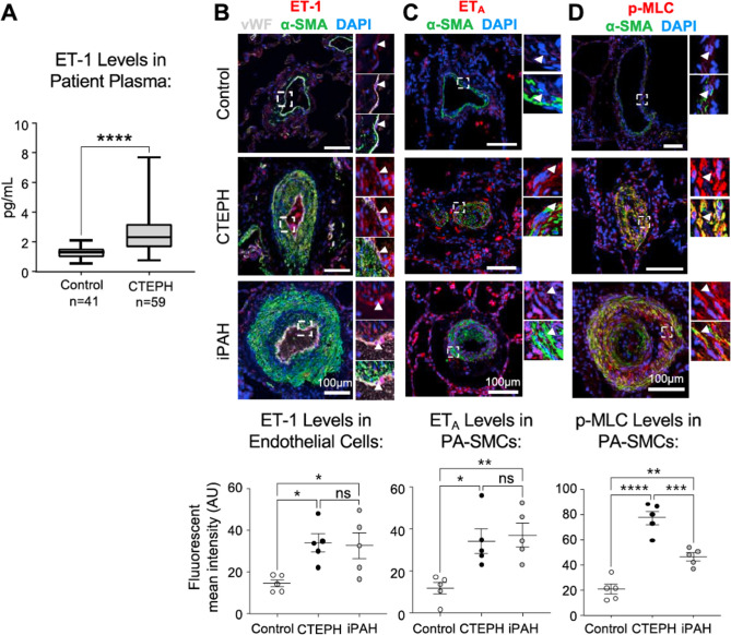
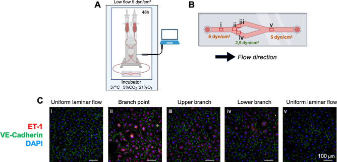
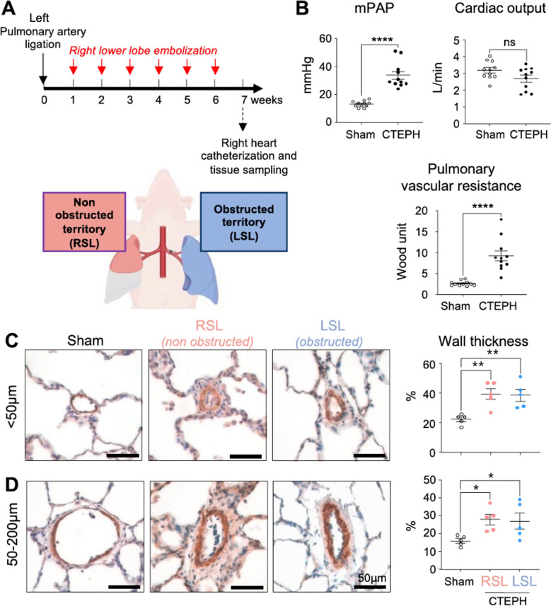
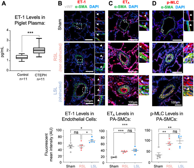
Targeted vasopeptide therapies have significantly advanced the management of pulmonary arterial hypertension (PAH). However, due to insufficient preclinical evidence regarding the involvement of the endothelin-1 (ET-1) pathway in chronic thromboembolic pulmonary hypertension (CTEPH) pathophysiology, the potential of ET-1 receptor antagonism in treating CTEPH remains uncertain. In this study, we investigated the role of the ET-1 pathway in CTEPH microvasculopathy using a multifaceted approach. Plasma ET-1 levels were measured in a cohort of 59 CTEPH patients and 41 healthy controls. Additionally, we evaluated the expression of key ET-1 pathway members in pulmonary explants from CTEPH, idiopathic PAH, and control patients. We used an in vitro system to test the hypothesis that the turbulent flow, observed near the vascular obstructions pathognomonic of CTEPH, enhances ET-1 expression. Our findings were further validated in vivo using a CTEPH piglet model that contains distinct regions representing pre- and post-thrombus lung territories. We found a twofold increase in circulating ET-1 levels in CTEPH patients compared to healthy subjects. Pulmonary explants from CTEPH patients exhibited pronounced overexpression of ET-1, endothelin receptor A (ETA), and phosphorylated myosin light chain (p-MLC) in muscularized pulmonary microvessels, suggesting heightened vascular contraction. In vitro experiments showed that turbulent flow facilitates ET-1 secretion compared to laminar flow regions. Additionally, in the CTEPH piglet model, elevated plasma ET-1 levels were observed compared to controls. Immunofluorescence and confocal microscopy analyses confirmed increased ETA and p-MLC in remodeled arteries from both pulmonary territories. However, ET-1 protein elevation was exclusively observed in the obstructed territory. These findings collectively indicate impaired vascular tone in microvessels of CTEPH patients and the CTEPH piglet model. Furthermore, our data implicates the ET-1 pathway in microvasculopathy, with turbulent flow playing a pathological role. These insights underscore the potential utility of ET-1 receptor antagonists as a promising therapeutic approach for CTEPH treatment.

Keywords: Chronic thromboembolic pulmonary hypertension; Endothelin receptor antagonist; Endothelin-1; Microvasculopathy; Pulmonary vasculature; Therapeutic target.


Conflict of interest statement

Declarations Competing interests Over the last three years, C.G. reports grants from Acceleron Pharma (Cambridge, MA, USA), a wholly-owned subsidiary of Merck & Co., Inc. (Rahway, NJ, USA), MSD, Corteria, Structure therapeutics, Diagonal Therapeutics, Gossamer, outside the submitted work. M.H. reports grants and personal fees from Acceleron, Aerovate, Altavant, AOP Orphan, Bayer, Chiesi, Ferrer, Janssen, Merck, MorphogenIX and United Therapeutics, outside the submitted work. All the other authors declare that there is no conflict of interest regarding the publication of this original article.

Figures

Fig. 1
Fig. 1
The Endothelin-1 (ET-1) Pathway Demonstrates Heightened Expression in Both the Plasma and Lung Microvessels of Chronic Thromboembolic Pulmonary Hypertension (CTEPH) Patients. (A) The concentration of ET-1 in venous plasma is elevated in CTEPH patients (n = 59) compared to control subjects (n = 41). (B) Co-immunofluorescent staining for ET-1 (red), von Willebrand factor (vWF, white), alpha-smooth muscle actin (α-SMA, green), and DAPI (blue) within lung microvessels from control subjects, CTEPH patients, and idiopathic pulmonary arterial hypertension (iPAH) patients, and the quantification of ET-1 fluorescent mean intensity (FMI) in the pulmonary endothelium (n = 5). (C) Co-immunofluorescent staining for ET-1 receptor A (ETA) (red), α-SMA (green), and DAPI (blue) in controls, CTEPH, and iPAH lungs, along with quantification of ETA FMI in pulmonary artery smooth muscle cells (PA-SMCs) (n = 5). (D) Co-immunofluorescent staining for phospho-myosin light chain (p-MLC) (red), α-SMA (green), and DAPI (blue) in controls, CTEPH, and iPAH lungs, along with quantification of p-MLC FMI in PA-SMCs (n = 5). All data are presented as mean with standard error of the mean (SEM). Statistical significance is denoted as * for P < 0.05, ** P < 0.01, *** P < 0.001, and **** P < 0.0001 and “ns” for not significant. The scale bar in all staining images is set at 100 µm.
Fig. 2
Fig. 2
Human Pulmonary Endothelial Cells (PMECs) Exhibit Increased ET-1 Expression in Regions of Disturbed Flow. (A) A schematic representation of the experimental setup for cell culture under flow within a Y-shaped ibidi plate. (B) The diagram indicates the expected shear stress distribution and highlights the image locations (i-v) for subsequent analyses. This schematic was generated using Biorender. (C) Immunofluorescent staining of PMECs at specified regions within the plate, showing ET-1 expression (red), VE-cadherin (green) and DAPI (blue). The scale bar in all staining images is set at 100 µm.
Fig. 3
Fig. 3
The CTEPH Piglet Model Demonstrates Elevated Pulmonary Vascular Resistance, Mean Pulmonary Artery Pressure (mPAP), and Pulmonary Vascular Wall Thickness Compared to Sham Controls. (A) A diagram illustrating the CTEPH piglet model with labeled territories. This schematic was generated using Biorender. (B) The mPAP, cardiac output (CO), and pulmonary vascular resistance (PVR) of the CTEPH piglet model compared to sham controls (n = 11). (C) Immunohistochemistry staining for α–smooth muscle actin and quantification of wall thickness in pulmonary arteries with diameters of < 50 µm in lungs of the CTEPH piglet model and sham controls (n = 5). (D) Immunohistochemistry staining for α–smooth muscle actin and quantification of wall thickness in pulmonary arteries with diameters of 50–200 µm in lungs of the CTEPH piglet model and sham controls (n = 5). All data are presented as the mean ± SEM. Statistical significance is denoted as * P < 0.05, **P < 0.05, ****P < 0.0001, and “ns” for not significant. Scale bars 50 µm. LSL: left superior lobe; RSL: right superior lobe.
Fig. 4
Fig. 4
The Endothelin-1 (ET-1) Pathway Demonstrates Increased Expression in both the Plasma and Lung Microvessels of the CTEPH Piglet Model. (A) The concentration of ET-1 in venous plasma in the CTEPH piglet model compared to sham controls (n = 11). (B) Co-immunofluorescent staining for ET-1 (red), von Willebrand factor (vWF, white), alpha-smooth muscle actin (α-SMA, green), and DAPI (blue) within lung microvessels from the sham controls, non-obstructed (RSL), and obstructed (LSL) territories, along with quantification of fluorescent mean intensity (FMI) in the pulmonary endothelium (n = 5). (C) Co-immunofluorescent staining for ET-1 receptor A (ETA) (red), a-SMA (green), and DAPI (blue) in sham lungs, and the RSL and LSL territories, along with quantification of FMI in pulmonary artery smooth muscle cells (PA-SMCs) (n = 5). (D) Co-immunofluorescent staining for phospho-myosin light chain (p-MLC) (red), α-SMA (green), and DAPI (blue) in sham lungs, and the RSL and LSL territories, along with quantification of FMI in PA- SMCs (n = 5). All data are presented as mean values with standard error of the mean (SEM). Statistical significance is denoted as * for P < 0.05, **P < 0.01, and ***P < 0.001. The scale bar in all staining images is set at 100 µm. LSL: left superior lobe; RSL: right superior lobe.
Fig. 5
Fig. 5
Study diagram: The obstruction of a vascular territory by the persistence of a fibro-thrombotic sequestrum leads to local disruptions in blood flow and removes a brake on ET-1 synthesis in endothelial cells surrounding this lesion. Subsequently, this ET-1 is released into circulation, exerting a paracrine action on its ETA receptor. This receptor is crucial for inducing vasoconstriction through the phosphorylation of myosin light chain (p-MLC) and promoting the proliferation of vascular smooth muscle cells (PA-SMCs). The abnormal overexpression of ETA by these downstream microvessels creates a vicious cycle that promotes the accumulation of resident pulmonary vascular cells in the walls, leading to a reduction in pulmonary vascular lumens in these unobstructed areas. Furthermore, the pulmonary endothelium of vessels downstream from the obstructed zones also contributes to the elevation of ET-1 levels in CTEPH, thereby contributing to CTEPH microvasculopathy.

References

    1. Kim, N. H. et al. Chronic thromboembolic pulmonary disease. Eur Respir J10.1183/13993003.01294-2024 (2024). - PMC - PubMed
    1. Humbert, M. et al. 2022 ESC/ERS Guidelines for the diagnosis and treatment of pulmonary hypertension. Eur Respir J61, 10.1183/13993003.00879-2022 (2023). - PubMed
    1. Dorfmuller, P. et al. Microvascular disease in chronic thromboembolic pulmonary hypertension: a role for pulmonary veins and systemic vasculature. Eur Respir J44, 1275–1288. 10.1183/09031936.00169113 (2014). - PubMed
    1. Guignabert, C. et al. Pathology and pathobiology of pulmonary hypertension: current insights and future directions. Eur Respir J10.1183/13993003.01095-2024 (2024). - PMC - PubMed
    1. Reesink, H. J. et al. Hemodynamic and clinical correlates of endothelin-1 in chronic thromboembolic pulmonary hypertension. Circ J70, 1058–1063. 10.1253/circj.70.1058 (2006). - PubMed

LinkOut - more resources