. 2024 Nov 16;7(1):1525.
doi: 10.1038/s42003-024-07240-z.

Mitochondrial ATP synthesis is essential for efficient gametogenesis in Plasmodium falciparum

Affiliations

Mitochondrial ATP synthesis is essential for efficient gametogenesis in Plasmodium falciparum

Penny C Sparkes et al. Commun Biol. .

Abstract

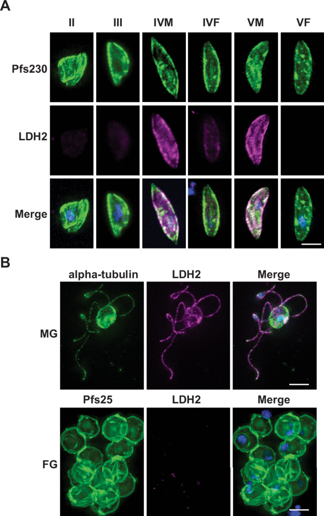
Plasmodium male and female gametocytes are the gatekeepers of human-to-mosquito transmission, therefore essential for propagation of malaria within a population. Whilst dormant in humans, their divergent roles during transmission become apparent soon after mosquito feeding with a rapid transformation into gametes - males forming eight motile sperm-like cells aiming to fertilise a single female gamete. Little is known about how the parasite fuels this abrupt change, and the potential role played by their large and elaborate cristate mitochondrion. Using a sex-specific antibody and functional mitochondrial labelling, we show that the male gametocyte mitochondrion is less active than that of female gametocytes and more sensitive to antimalarials targeting mitochondrial energy metabolism. Rather than a vestigial organelle discarded during male gametogenesis, we demonstrate that mitochondrial ATP synthesis is essential for its completion. Additionally, using a genetically encoded ratiometric ATP sensor, we show that gametocytes can maintain cytoplasmic ATP homeostasis in the absence of mitochondrial respiration, indicating the essentiality of the gametocyte mitochondrion for transmission alone. Together, this reveals how gametocytes responsively balance the conflicting demands of a dormant and active lifestyle, highlighting the mitochondria as a rich source of transmission-blocking targets for future drug development.


Conflict of interest statement

Competing interests The authors declare no competing interests.

Figures

Fig. 1
Fig. 1. Anti-LDH2 is a specific marker for P. falciparum male gametocytes and gametes.
A Immunostaining with anti-LDH2 (magenta) showed specific staining of a subpopulation of Pfs230-positive (green) stage IV and stage V gametocytes. B Male gametes stained with anti-alpha tubulin II (green) also were LDH2-positive, whilst Pfs25-positive (green) female gametes were LDH2-negative. Merged images also show DAPI (blue). Scale bars = 5 µM.
Fig. 2
Fig. 2. Contrasting the differences between nuclear organisation of male and female gametocytes using quantitative imaging.
A Anti-LDH2 (green) was used to discriminate between male (LDH2-positive) and female (LDH2-negative) gametocytes colabelled with MitoTrackerTM CMXRos (magenta) and DAPI (blue). Cell regions of interest (ROI) were manually traced, and nuclear ROI subsequently automatically traced using thresholding of DAPI staining intensity. Scale bar = 3 µM. B Using the cell ROI, mean anti-LDH2 staining per gametocyte was calculated and plotted against the calculated nuclear contour of the corresponding nucleus. Two distinct populations of cells with low LDH2 immunoreactivity (females) and high LDH2 immunoreactivity (males) were observed (n = 139 gametocytes). C A variety of nuclear parameters were calculated and compared between populations of male and female gametocytes. n = 3 independent experiments of 133, 68, and 139 gametocytes measured. Unpaired Student’s T-test: ns = p > 0.05; *p > 0.05; **p < 0.01; ***p < 0.001. Degrees of freedom (df) = 4. Error bars denote standard error of the mean (SEM).
Fig. 3
Fig. 3. Contrasting the differences between mitochondrial organisation of male and female gametocytes using quantitative imaging.
A Anti-LDH2 (green) was used to discriminate between male (LDH2-positive) and female (LDH2-negative) gametocytes co-labelled with MitoTrackerTM CMXRos (magenta) and DAPI (blue). Scale bar = 3 µM. B The same dataset used to study gametocyte nuclear morphology was reanalysed similarly using MitoTracker staining to generate mitochondrial ROIs for each cell. A variety of mitochondrial parameters were calculated and compared between populations of male and female gametocytes. n = 4 independent experiments of 133, 111, 48, and 147 gametocytes measured. Unpaired Student’s T-test: ns = p > 0.05. df = 6. Error bars denote standard deviation. C Labelling for 25 min with a non-saturating concentration of MitoTracker (12.5 nM) was used to compare mitochondrial activity of male and female gametocytes (n = 5 independent experiments of 47, 47 49, 59, and 35 gametocytes measured). Data presented here is normalised to the mitochondrial activity of the female gametocytes for each replicate, black bar indicates the mean of the replicate. A statistical comparison (Ratio paired t-test) was performed before normalisation and showed that the difference in activity between gametocyte sex over all replicates was significant (p = 0.002. df = 4). On average, male gametocytes accumulated 44.4% less MitoTracker than females.
Fig. 4
Fig. 4. The effect of mitochondrial inhibitors on male and female gametocyte mitochondrial activity and ability to form gametes.
The mitochondrial activity of male and female gametocytes treated with inhibitors for 24 h (A, C, E, G) were calculated following a 25 min incubation with 12.5 nM MitoTracker, fixing and staining with anti-LDH2. Male gametocytes were identified by LDH2 immunoreactivity. Mitchondrial activity was calculated by quantyifying the background-corrected amount of MitoTracker fluorescence within the gametocyte (n = 70–294 gametocytes per replicate, 3–5 independent experiments). Graphed data presented here is individual cells for all replicates that have been within-replicate normalised to the mean value of female DMSO. Black bar denotes the mean value. Statistical tests show unpaired Student’s T-test of normalised values. Tests performed against male gametocyte data were normalised to the female gametocyte DMSO control of each replicate and vice versa for the tests for female data. Stars and number above data indicate statistical significance and p value compared to the DMSO control (**** = p ≤ 0.0001). Absence of a star indicates not significant change. df = 8, 8, 4, 8 respectively for atovaquone, ELQ-300, DSM-265 and oligomycin A. The functional viability of male and female gametocytes was quantified in the Pf DGFA (B, D, F, H) (n = 3 independent experiments. Datapoints of each replicate are plotted with black line showing the calculated 4 parameter dose response curve used to determine the IC50 value). Mitochondrial inhibitors used: (A, B) Atovaquone; (C, D) ELQ-300; (E, F) DSM-265; (G, H) Oligomycin A.
Fig. 5
Fig. 5. Investigating the dependency of exflagellation on human serum and glucose.
A Mature gametocytes were washed in culture medium containing varying concentrations of human serum, with or without the standard RPMI glucose concentration of 2 g/L. Then, gametogenesis was triggered in condition-matched exflagellation medium. Exflagellation was then quantified 20 min later by automated microscopy. Data is normalised to the control conditions of glucose plus 10% human serum. n = 4 independent experiments. Error bars denote standard error of the mean. B Mature gametocytes were washed in culture medium containing a fixed concentration of 0.1% human serum and varying concentrations of glucose (0.1–0 g/L). Then, gametogenesis was triggered in condition-matched exflagellation medium. Exflagellation was then quantified 20 min later by automated microscopy. Data is normalised to the control conditions of glucose plus 10% human serum. n = 3 independent experiments. Datapoints of each replicate are plotted with the orange line showing the calculated 4 parameter dose response curve used to determine the IC50 value.
Fig. 6
Fig. 6. The quantitative and morphological differences in gametogenesis in the presence of mitochondrial inhibitor oligomycin A.
A Gametogenesis was triggered in ookinete medium containing 0.1% human serum, with (2 g/L “Glucose”) or without glucose (“No glucose”), or glucose and 5 µM oligomycin A (“Glucose + OA”). Exflagellation was counted 20 min later using automated microscopy to capture exflagellation centres and automated image analysis for quantification. Data presented here is the mean of five independent experiments normalised to the exflagellation activity of the “Glucose” sample for each replicate. However statistical comparison (Ratio paired t-test) was performed before normalisation and showed that the difference in activity between “Glucose” and “Glucose + OA” samples is statistically significant (p = 0.007. n = 5. df = 4). Error bars denote standard error of the mean. B Parallel samples of the cells were fixed 20 min post induction of gametogenesis and stained with anti-alpha tubulin and DAPI to visualise microtubule and DNA morphology. Bar = 5 µm.
Fig. 7
Fig. 7. Ratiometric measurement of cytoplasmic ATP using a FRET-based sensor ATeam1.03YEMK.
Gametocytes were washed in culture medium three times with or without glucose and with or without 5 µM oligomycin A. Finally, gametocytes were then pelleted in phosphate buffered saline (PBS) containing matched glucose/oligomycin A conditions and imaged over time at 37 °C. Imaging commenced at 3 min post treatment to account for processing time and time taken for cells to settle onto the slide for imaging. Dotted line indicates the FRET value recorded for gametocytes incubated in PBS alone for 120 min. Closed orange circles indicate gametocytes incubated with 2 g/L glucose; open orange circles indicate gametocytes incubated without glucose. Closed and open blue circles indicate gametocytes incubated with and without glucose respectively in the presence of 5 µM oligomycin A. Data represents the mean of 6–9 independent replicates performed pairwise with all possible combinations of treatment. Error bars denote the standard error of the mean (SEM).
Fig. 8
Fig. 8. A model showing the potential contribution of mitochondrial ATP production to male gametocytes and male gametogenesis.
Under glucose-replete conditions, gametocytes can maintain intracellular ATP homeostasis even when mitochondrial ATP production is inhibited. However, male gametogenesis cannot complete without available glucose or mitochondrial ATP production.

References

    1. World Malaria Report 2023. https://www.who.int/teams/global-malaria-programme/reports/world-malaria....
    1. Venugopal, K., Hentzschel, F., Valkiūnas, G. & Marti, M. Plasmodium asexual growth and sexual development in the haematopoietic niche of the host. Nat. Rev. Microbiol.18, 177–189 (2020). - PMC - PubMed
    1. Billker, O. et al. Identification of xanthurenic acid as the putative inducer of malaria development in the mosquito. Nature392, 289–292 (1998). - PubMed
    1. Bousema, T. et al. Revisiting the circulation time of Plasmodium falciparum gametocytes: molecular detection methods to estimate the duration of gametocyte carriage and the effect of gametocytocidal drugs. Malar. J.9, 136 (2010). - PMC - PubMed
    1. Smalley, M. E. & Sinden, R. E. Plasmodium falciparum gametocytes: their longevity and infectivity. Parasitology74, 1–8 (1977). - PubMed

LinkOut - more resources