. 2024 Nov 16;24(1):400.
doi: 10.1186/s12906-024-04689-7.

Tongguanteng injection exerts anti-osteosarcoma effects through the ER stress-associated IRE1/CHOP pathway

Affiliations

Tongguanteng injection exerts anti-osteosarcoma effects through the ER stress-associated IRE1/CHOP pathway

Xiao-Chuan Xue et al. BMC Complement Med Ther. .

Abstract

Background: In China, Tongguanteng injection (TGT) is widely used in the treatment or adjuvant treatment of various types of cancer. However, the effect and mechanism of TGT in osteosarcoma is not clear.

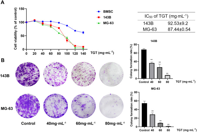
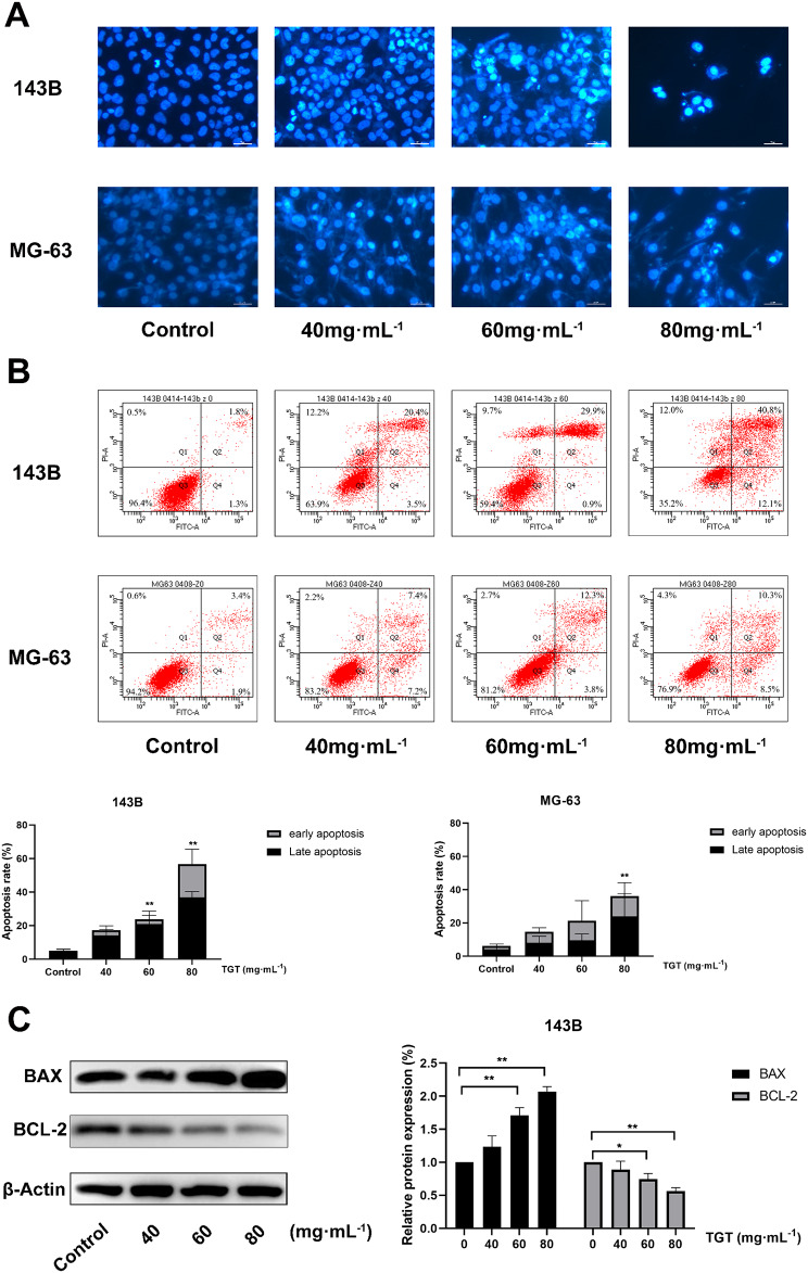
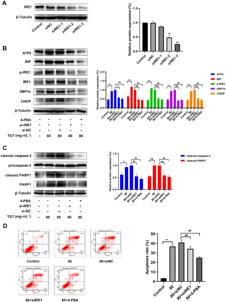
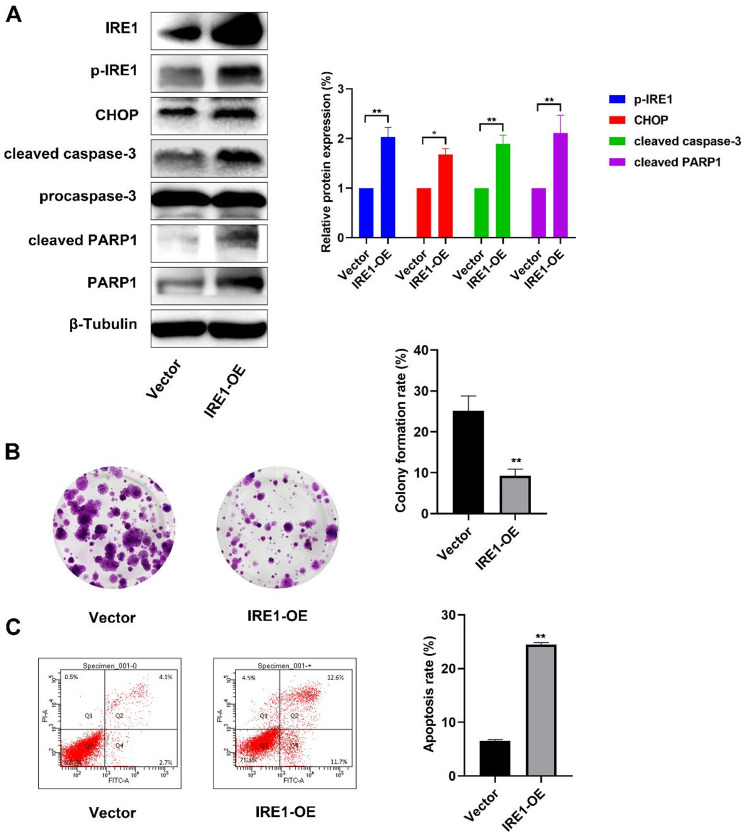
Methods: The 143B and MG-63 cells were treated with different concentrations of TGT. Cell proliferation, migration, invasion and apoptosis were detected using CCK8 assay, transwell assay and flow cytometry. Differentially expressed genes (DEGs) were screened using RNA sequencing (RNA-seq). The identified mRNA and protein expression associated with the IRE1/CHOP pathway was validated by RT-PCR and western blot assay. To explore the underlying mechanisms, 4-phenylbutyric acid (4-PBA) was selected as a specific endoplasmic reticulum (ER) stress inhibitor. Small interfering RNA (siRNA) or pEX-3-ERN1 plasmid was transfected into 143B cells to silence or overexpress IRE1, respectively. The potential downstream proteins, including CHOP, and apoptosis associated proteins, caspase-3 and PARP1 were determined. Furthermore, the effect of TGT was demonstrated in 143B cell tumor-bearing mice in vivo. H&E staining, TUNEL staining and immunohistochemistry were conducted in tumor tissues obtained from the xenograft mouse model.

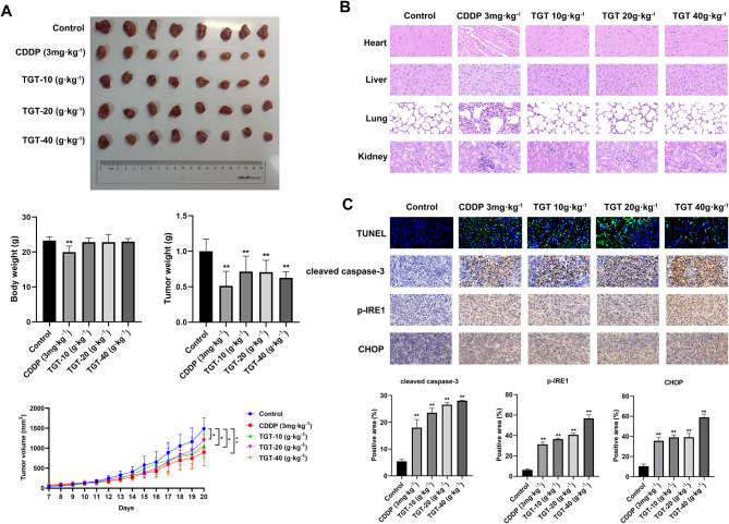
Results: TGT was shown to dramatically suppress the proliferation, migration and invasion, and induce apoptosis of osteosarcoma 143B and MG-63 cells in vitro. The identified DEGs included HSPA5 (encoding BiP) and ERN1 (encoding the IRE1 protein), as well as apoptosis-associated gene DDIT3 (encoding the CHOP protein). The term "IRE1-mediated unfolded protein response" was screened to be the most enriched biological process GO term. The expression of ER stress-associated proteins including ATF6, BiP, p-IRE1, XBP1s and CHOP, as well as apoptosis-associated cleaved caspase-3 and cleaved PARP1 proteins, was significantly upregulated by TGT treatment in osteosarcoma 143B cells, suggesting that TGT might promote the apoptosis of osteosarcoma 143B cells through the IRE1/CHOP pathway. Furthermore, knocking down IRE1 with si-IRE1 or inhibiting of ER stress with 4-PBA suppressed the expression of ATF6, BiP, XBP1s and CHOP induced by TGT, as well as the expression of cleaved caspase-3 and cleaved PARP1. On the contrary, overexpressing IRE1 promoted CHOP expression and induced osteosarcoma cell apoptosis. Consistent with in vitro results, TGT dramatically inhibited the tumor growth and promoted the expression of p-IRE1 and CHOP in tumor-bearing mice.

Conclusion: The findings suggest that TGT exerts an anti-osteosarcoma effect in vitro and in vivo. The underlying mechanism might be associated with the activation of IRE1/CHOP pathway in ER stress. Our findings suggest that targeting IRE1/CHOP pathway might be a potential novel approach for osteosarcoma treatment.

Keywords: Apoptosis; ER stress; IRE1/CHOP pathway; Osteosarcoma; Tongguanteng injection.


Conflict of interest statement

Declarations Ethics approval and consent to participate The animal researches were accredited by the Animal Welfare Ethics Committee of Shanghai Sixth People’s Hospital (No. SYXK 2018-0028). All animal experiments were conducted rigorously in compliance with Provision and General Recommendation of Chinese Experimental Animals Administration Legislation. Consent for publication Not applicable. Competing interests The authors declare no competing interests.

Figures

Fig. 1
Fig. 1
TGT attenuates the growth of osteosarcoma cells. A 143B, MG-63 and BMSC cells were treated with TGT at different concentrations for 24 h. The cell viability was evaluated and the IC50 value was calculated. B After treatment with TGT (40, 60, or 80 mg·mL− 1) for 24 h, the colony formation ability of the 143B and MG-63 cells was detected. Data are expressed as the mean ± SD (n = 3). **p < 0.01, vs. the control group
Fig. 2
Fig. 2
TGT inhibits the migration and invasion of osteosarcoma cells. A TGT suppressed the lateral migration capacity of 143B and MG-63 cells in the scratch test. Scale bar = 100 μm. B TGT inhibited the longitudinal migration of osteosarcoma cells according to the transwell assay. Scale bar = 50 μm. C TGT suppressed the invasion ability of osteosarcoma cells according to the transwell assay. Scale bar = 50 μm. Data are presented as the mean ± SD (n = 3). **p < 0.01, vs. the control group
Fig. 3
Fig. 3
TGT promotes apoptosis in osteosarcoma cells. A TGT induced the apoptosis of 143B and MG-63 cells, as determined by a Hoechst staining assay. Scale bar = 25 μm. B TGT increased the apoptosis rate of osteosarcoma cells according to the flow cytometry assay. C The expression of BAX protein was enhanced and BCL-2 protein was reduced by TGT treatment, as detected by western blot experiment. Data are expressed as the mean ± SD (n = 3). *p < 0.05, **p < 0.01, vs. the control group
Fig. 4
Fig. 4
RNA-seq analysis of 143B cells treated with TGT for 24 h. A Volcano plot of DEGs with q-value < 0.05 and |log2 FC|>1 as threshold in the TGT 40 and 80 mg·mL− 1 group. B Heatmap and q-PCR validation of the ER stress-associated genes and randomly selected DEGs. C GO and KEGG analysis of genes categorized into profile 15. Data are denoted as the mean ± SD (n = 3). *p < 0.05, **p < 0.01, vs. the control group
Fig. 5
Fig. 5
TGT triggers the ER stress response pathway and activates apoptotic proteins in 143B cells. A After TGT treatment, the expression levels of ATF6, BiP, p-IRE1, XBP1s and CHOP were enhanced, as shown by western blot experiment. B The expression of cleaved caspase-3 and cleaved PARP1 was enhanced by TGT treatment, as detected by western blot experiment. Data are displayed as the mean ± SD (n = 3). *p < 0.05, **p < 0.01, vs. the control group
Fig. 6
Fig. 6
Knocking down of IRE1 or inhibiting of ER stress suppresses the 143B cell apoptosis. A The knockdown efficiency of three siIRE1s was measured via a western blot experiment. B The expression levels of proteins involved the regulation of ER stress, including ATF6, BiP, p-IRE1, XBP1s and CHOP, were significantly decreased according to western blot analysis. C The expression levels of cleaved caspase-3 and cleaved PARP1 were significantly reduced by knockdown of IRE1, as detected by western blot experiment. D The apoptosis rate was significantly decreased by IRE1 knockdown and 4-PBA treatment, as determined via flow cytometry analysis. Data are displayed as the mean ± SD (n = 3). #p < 0.05, ##p < 0.01, vs. the control group; *p < 0.05, **p < 0.01, vs. the TGT alone group
Fig. 7
Fig. 7
IRE1 overexpression enhances CHOP expression and apoptosis in 143B cells. A Western blot analysis was used to evaluate the expression levels of IRE1, CHOP, cleaved caspase-3 and cleaved PARP1 in 143B cells transfected with the IRE1-OE plasmid. B The colony formation capacity of 143B cells was decreased upon IRE1 overexpression. C The apoptosis level of the 143B cells was reduced upon IRE1 overexpression, as determined by flow cytometry. Data are displayed as the mean ± SD (n = 3). *p < 0.05, **p < 0.01, vs. the control group
Fig. 8
Fig. 8
TGT inhibits osteosarcoma growth in vivo through the IRE1/CHOP pathway. A Tumor morphology, body weight and tumor volume curve were displayed (mean ± SD, n = 8). B Representative H&E staining images of the heart, liver, lung and kidney tissues from mice in each group (400×, n = 3, Scale bar = 20 μm). C TGT-induced cell apoptosis (TUNEL staining, 400×) and enhanced cleaved caspase-3, p-IRE1 and CHOP levels (IHC staining, 400×) in the xenograft model (mean ± SD, n = 3, Scale bar = 20 μm). *p < 0.05, **p < 0.01, vs. the control group
Fig. 9
Fig. 9
TGT inhibits the growth of osteosarcoma through the ER stress-associated IRE1/CHOP pathway

References

    1. Meltzer PS, Helman LJ. New horizons in the treatment of osteosarcoma. N Engl J Med. 2021;385(22):2066–76. - PubMed
    1. Beird HC, Bielack SS, Flanagan AM, et al. Osteosarcoma Nat Rev Dis Primers. 2022;8(1):77. - PubMed
    1. Shoaib Z, Fan TM, Irudayaraj JMK. Osteosarcoma mechanobiology and therapeutic targets. Br J Pharmacol. 2022;179(2):201–17. - PMC - PubMed
    1. Hudson MM, Bhatia S, Casillas J, et al. Long-term follow-up care for childhood, adolescent, and young adult cancer survivors. Pediatrics. 2021;148(3):e2021053127. - PMC - PubMed
    1. Chen X, Cubillos-Ruiz JR. Endoplasmic reticulum stress signals in the tumour and its microenvironment. Nat Rev Cancer. 2021;21(2):71–88. - PMC - PubMed

MeSH terms

LinkOut - more resources