

Changes in choroidal thickness quantified by Optical Coherence Tomography across cognitive impairment: data from the NORFACE cohort
- PMID: 39550564
- PMCID: PMC11568571
- DOI: 10.1186/s13195-024-01616-3
Changes in choroidal thickness quantified by Optical Coherence Tomography across cognitive impairment: data from the NORFACE cohort
Abstract
Background: Optical coherence tomography (OCT) enables high-resolution imaging of ocular structures in health and disease. Choroid thickness (CT) is a key vascular retinal parameter that can be assessed by OCT and might be relevant in the evaluation of the vascular component of cognitive decline. We aimed to investigate CT changes in a large cohort of individuals cognitive unimpaired (CU), with mild cognitive impairment due to Alzheimer's (MCI-AD), mild cognitive impairment due to cerebrovascular disease (MCI-Va), Alzheimer's disease dementia (ADD), and vascular dementia (VaD).
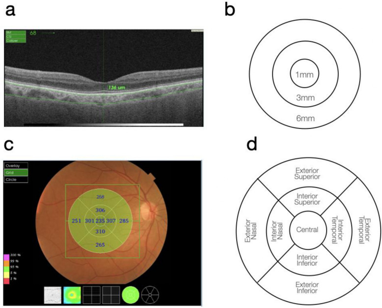
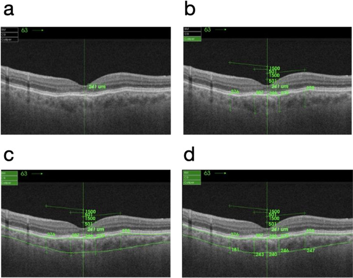
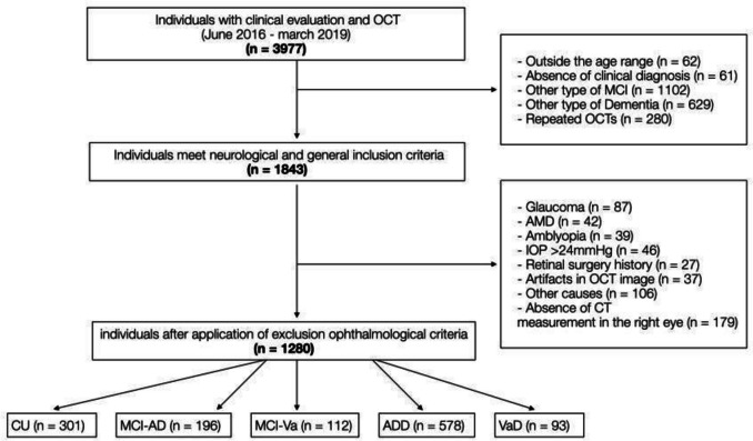
Methods: Clinical, demographical, ophthalmological and OCT data from the Neuro-ophthalmological Research at Fundació ACE (NORFACE) project were analyzed. CT was assessed in the macula across nine Early Treatment Diabetic Retinopathy Study (ETDRS) quadrants, average thickness, total volume, and subfoveal choroidal thickness. Differences of CT among the five diagnostic groups were assessed in a multivariate regression model, adjusting for demographic and cardiovascular risk factors and OCT image quality. A comparison between manual and automatic CT measurements in a subset of participants was also performed.
Results: The study cohort comprised 1,280 participants: 301 CU, 196 MCI-AD, 112 MCI-Va, 578 ADD, and 93 VaD. CT was significantly increased in individuals with cognitive impairment compared to those CU, particularly in the VaD and MCI-Va groups and in the peripheral ETDRS regions. No significant differences were found in inner superior, center and subfoveal choroidal thickness. The interaction of sex and diagnosis had no effect in differentiating CT. Mini-Mental State Examination (MMSE) scores were not correlated to CT. Manual and automated CT measurements showed good reliability.
Discussion: Our findings indicated that peripheral choroidal thickening, especially in patients with cerebrovascular disease, may serve as a potential choroidal biomarker for cognitive decline and suggest different pathogenic pathways in AD and VaD. Further research is required to explore CT as a reliable ocular biomarker for cognitive impairment.
Keywords: Alzheimer's disease; Biomarkers; Choroidal thickness; NORFACE cohort; Optical coherence tomography; Vascular dementia.
© 2024. The Author(s).
Conflict of interest statement
Figures

References
-
- American Psychiatric Association. American Psychiatric Association. Diagnostic and Statistical Manual of Mental Disorders. 5th ed. Text Revision. Arlington: American Psychiatric Publishing; 2022. ISBN: 978–0890425756. Diagnostic and Statistical Manual of Mental Disorders. 2022.
-
- Schneider JA, Arvanitakis Z, Bang W, Bennett DA. Mixed brain pathologies account for most dementia cases in community-dwelling older persons. Neurology. 2007 [cited 2024 Sep 8];69:2197–204. Available from: https://pubmed.ncbi.nlm.nih.gov/17568013/. - PubMed
-
- McKhann GM, Knopman DS, Chertkow H, Hyman BT, Jack CR, Kawas CH, et al. The diagnosis of dementia due to Alzheimer’s disease: Recommendations from the National Institute on Aging-Alzheimer’s Association workgroups on diagnostic guidelines for Alzheimer’s disease. Alzheimer’s & Dementia. 2011;7:263–9. - DOI - PMC - PubMed
MeSH terms
LinkOut - more resources
Full Text Sources
Medical
Miscellaneous
