. 2024 Nov 17;12(11):e009860.
doi: 10.1136/jitc-2024-009860.

Inhibition of Cbl-b restores effector functions of human intratumoral NK cells

Affiliations

Inhibition of Cbl-b restores effector functions of human intratumoral NK cells

Sofia Tundo et al. J Immunother Cancer. .

Abstract

Background: T cell-based immunotherapies including immune checkpoint blockade and chimeric antigen receptor T cells can induce durable responses in patients with cancer. However, clinical efficacy is limited due to the ability of cancer cells to evade immune surveillance. While T cells have been the primary focus of immunotherapy, recent research has highlighted the importance of natural killer (NK) cells in directly recognizing and eliminating tumor cells and playing a key role in the set-up of an effective adaptive immune response. The remarkable potential of NK cells for cancer immunotherapy is demonstrated by their ability to broadly identify stressed cells, irrespective of the presence of neoantigens, and their ability to fight tumors that have lost their major histocompatibility complex class I (MHC I) expression due to acquired resistance mechanisms.However, like T cells, NK cells can become dysfunctional within the tumor microenvironment. Strategies to enhance and reinvigorate NK cell activity hold potential for bolstering cancer immunotherapy.

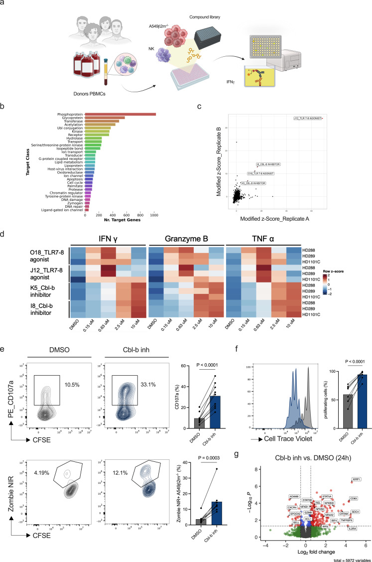
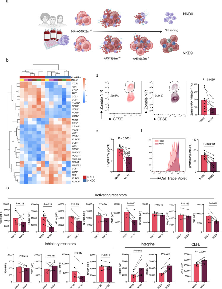
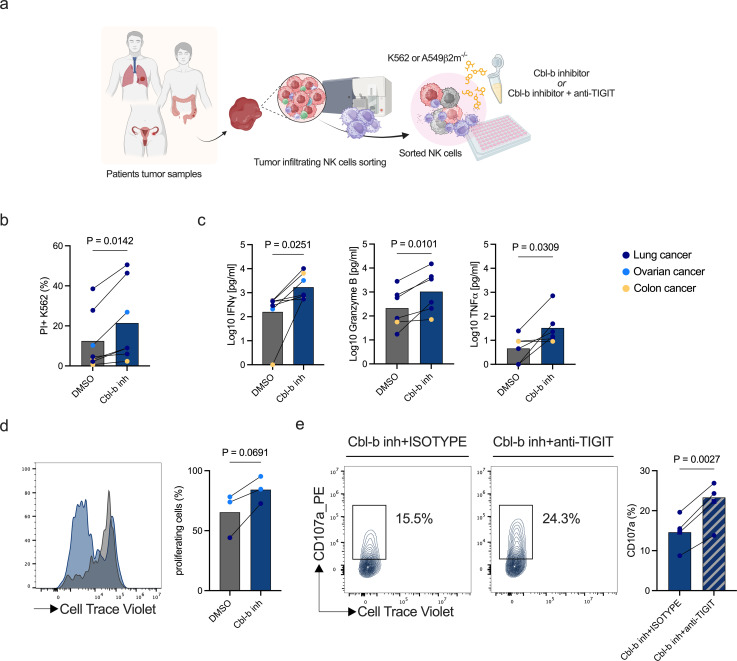
Methods: In this study, we conducted a high-throughput screen to identify molecules that could enhance primary human NK cell function. After compound validation, we investigated the effect of the top performing compounds on dysfunctional NK cells that were generated by a newly developed in vitro platform. Functional activity of NK cells was investigated using compounds alone and in combination with checkpoint inhibitor blockade. The findings were validated on patient-derived intratumoral dysfunctional NK cells from different cancer types.

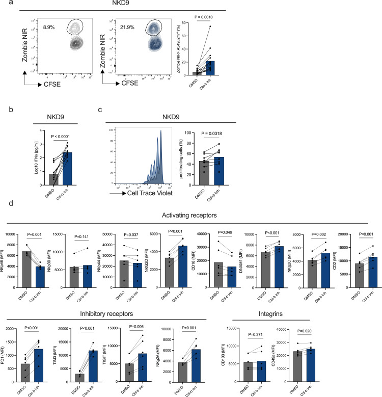
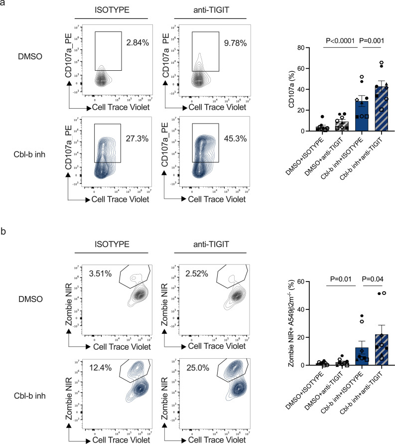
Results: The screening approach led to the identification of a Casitas B-lineage lymphoma (Cbl-b) inhibitor enhancing the activity of primary human NK cells. Furthermore, the Cbl-b inhibitor was able to reinvigorate the activity of in vitro generated and patient-derived dysfunctional NK cells. Finally, Cbl-b inhibition combined with T-cell immunoreceptor with Ig and ITIM domains (TIGIT) blockade further increased the cytotoxic potential and reinvigoration of both in vitro generated and patient-derived intratumoral dysfunctional NK cells.

Conclusions: These findings underscore the relevance of Cbl-b inhibition in overcoming NK cell dysfunctionality with the potential to complement existing immunotherapies and improve outcomes for patients with cancer.

Keywords: Immune Checkpoint Inhibitor; Immunotherapy; Intratumoral; Natural Killer - NK.


Conflict of interest statement

Competing interests: ARom and OG reports personal fees from Hoffmann-La Roche during the conduct of the study and personal fees from Hoffmann-La Roche outside the submitted work. AZ received consulting/advisor fees from Bristol-Myers Squibb, Merck Sharp & Dohme, Hoffmann–La Roche, NBE Therapeutics, Engimmune, and maintains further non-commercial research agreements with Hoffmann–La Roche, T3 Pharma, Bright Peak Therapeutics, AstraZeneca.

Figures

Figure 1
Figure 1. A small molecule library screening approach identifies Cbl-b inhibitors enhancing the activity of primary human NK cells. (a) Experimental design of the small molecule library screening approach on primary human NK cells in co-culture with A549 β2m−/− (created with BioRender.com). NK cells were enriched from healthy donors’ PBMCs and primed with a suboptimal concentration of IL-15 (0.2 ng/mL). Following compound treatment, NK cells were co-cultured with A549 β2m−/− overnight. Screening hits were identified by measurement of IFN-γ secretion from supernatants. (b) Small molecule screening compounds target classes. (c) Volcano plot representing screening hits based on modified Z score value. Top four screening hits were identified based on a threshold of normalized modified Z score ≥3. (d) Heatmap of IFN-γ, granzyme B, and TNF-α concentrations for screening hits validation. Primary human NK cells from healthy donors PBMCs (n=3) were treated in dose response with the top four screening compounds and cytokines measured from the supernatants. (e) Representative flow cytometry plots for NK cell degranulation (CD107a) and NK cell cytotoxicity. NK cells from healthy donors PBMCs (n=9) were primed with a suboptimal concentration of IL-15 (0.2 ng/mL) and co-cultured with A549 β2m−/− (CFSE+) at an E:T ratio of 2:1 for 6 hours (p values are from paired t-test). (f) CellTrace Violet dilution and statistics of primary human NK cells after treatment with Cbl-b inhibitor (p values are from paired t-test). (g) Volcano plot showing differentially expressed proteins on primary human NK cells after overnight treatment with Cbl-b inhibitor in the presence of a suboptimal concentration of 0.2 ng/mL IL-15 (n=6; gray=NS, green=log2 FC, blue=adjusted p value, red=log2 FC and adjusted p value. For statistical analyses see “Material and methods”). Cbl-b, Casitas B-lineage lymphoma; CFSE, carboxyfluorescein succinimidyl ester; DMSO, dimethylsulfoxide; E:T, effector to target; IFN, interferon; IL, interleukin; NK, natural killer; PBMCs, peripheral mononuclear cells; TLR, toll-like receptor; TNF, tumor necrosis factor.
Figure 2
Figure 2. In vitro model for the generation of dysfunctional NK cells. (a) Schematic representation of the in vitro model (created with BioRender.com). Primary human NK cells were enriched from healthy donors PBMCs and co-cultured for 9 days after single (NKD0) or continuous (NKD9) exposure to A549 ß2m−/− NK cells were then sorted to be used in functional assays. (b) Heatmap of selected differentially expressed genes associated with NK cell functionality between NKD0 and NKD9 (n=6). Volcano plot with differentially expressed genes in online supplemental figure S4a. (c) Multicolor extracellular staining of in vitro generated NK cells from healthy donors PBMCs (n=6) and Cbl-b intracellular staining of in vitro generated NK cells (n=6). Cbl-b expression is represented as delta isotype MFI (p values are from paired t-test). (d) Representative flow cytometry plots of cytotoxicity of in vitro generated NK cells (n=10). NK-mediated cytotoxicity is represented as the percentage of dead (zombie NIR+) target cancer cells (CFSE+). Percentage of cytotoxicity was normalized by cancer cell viability (p values are from paired t-test). (e) Interferon-γ concentration of in vitro generated NK cells following restimulation with target cells (n=6) (p values are from paired t-test). (f) Flow cytometry histogram and statistics of CellTrace Violet dilution of in vitro generated NK cells (n=7, p values are from paired t-test with Benjamini-Hochberg correction). Cbl-b, Casitas B-lineage lymphoma; CFSE, carboxyfluorescein succinimidyl ester; MFI, mean fluorescence intensity; NK, natural killer; PBMCs, peripheral mononuclear cells; PD-1, programmed cell death protein; TIGIT, T-cell immunoreceptor with Ig and ITIM domains; TIM3, T-cell immunoglobulin and mucin domain 3.
Figure 3
Figure 3. Cbl-b inhibitor reinvigorates the activity of in vitro generated dysfunctional NK cells. In vitro generated dysfunctional NK cells (NKD9) from healthy donors PBMCs (n=6) were sorted and re-challenged with fresh A549 ß2m−/− (CFSE+) at a 2:1 E:T ratio overnight in the presence of DMSO or Cbl-b inhibitor (3 µM). (a) Cytotoxicity of in vitro generated dysfunctional NK cells (NKD9) following Cbl-b inhibitor treatment by flow cytometry. In vitro generated dysfunctional NK cells (NKD9) from healthy donors PBMCs (n=6) were sorted and re-challenged with fresh A549 ß2m−/− (CFSE+) in the presence of DMSO or Cbl-b inhibitor (3 µM) (p values are from paired t-test). (b) IFN-γ concentration of in vitro generated dysfunctional NK cells (NKD9) after Cbl-b inhibitor treatment (p values are from paired t-test). (c) Flow cytometry histogram and statistics of CellTrace Violet dilution of in vitro generated dysfunctional NK cells after Cbl-b inhibitor (3 µM) treatment (n=7). (P values are from paired t-test). (d) Multicolor extracellular staining of in vitro generated dysfunctional NK cells following Cbl-b inhibitor treatment (3 µM) (n=6, p values are from paired t-test with Benjamini-Hochberg correction). Cbl-b, Casitas B-lineage lymphoma; CFSE, carboxyfluorescein succinimidyl ester; E:T, effector to target; MFI, mean fluorescence intensity; NK, natural killer; PBMCs, peripheral mononuclear cells; PD-1, programmed cell death protein; TIGIT, T-cell immunoreceptor with Ig and ITIM domains; TIM3, T-cell immunoglobulin and mucin domain 3.
Figure 4
Figure 4. Combination of Cbl-b inhibitor with TIGIT checkpoint blockade results in increased reinvigoration of dysfunctional NK cells. (a) Degranulation (CD107a) of in vitro generated dysfunctional NK cells (NKD9) following Cbl-b inhibitor and TIGIT blockade (n=8, for p values see Material and methods). (b) Cytotoxicity of in vitro generated NK cells following Cbl-b inhibitor treatment and TIGIT blockade treatment (n=8, for p values see “Material and methods”). Cbl-b, Casitas B-lineage lymphoma; DMSO, dimethylsulfoxide; NK, natural killer; TIGIT, T-cell immunoreceptor with Ig and ITIM domains.
Figure 5
Figure 5. Cbl-b inhibitor enhances the activity of intratumoral NK cells from cancer patient samples. (a) Schematic representing experimental design to assess the effect of Cbl-b inhibitor compound on tumor-infiltrating NK cells. Tumor-infiltrating NK cells were previously sorted from patients’ tumor samples and treated overnight with Cbl-b inhibitor in the presence of target cells (K562 or A549 ß2m−/−) (created with BioRender.com). (b) Flow cytometry-based cytotoxicity of Cbl-b inhibitor treated tumor-infiltrating NK cells (n=7 tumor digests, p values are from paired t-test). (c) IFN-γ, granzyme B and TNF-ɑ supernatant concentrations of Cbl-b inhibitor treated tumor-infiltrating NK cells from patients’ samples (n=6 tumor digests, p values are from paired t-test). (d) Histogram and statistics of flow cytometry CellTrace Violet dilution of patients tumor-infiltrating NK cells treated with Cbl-b inhibitor (n=3 tumor digests, p values are from paired t-test). (e) Degranulation (CD107a) of Cbl-b inhibitor treated intratumoral NK cells in combination with anti-TIGIT antibody (n=3 lung adenocarcinoma digests, n=1 lung adenocarcinoma pleural effusion, p values are from paired t-test). Cbl-b, Casitas B-lineage lymphoma; CFSE, carboxyfluorescein succinimidyl ester; DMSO, dimethylsulfoxide; IFN, interferon; NK, natural killer; TIGIT, T-cell immunoreceptor with Ig and ITIM domains; TNF, tumor necrosis factor.

References

    1. Tawbi HA, Schadendorf D, Lipson EJ, et al. Relatlimab and Nivolumab versus Nivolumab in Untreated Advanced Melanoma. N Engl J Med. 2022;386:24–34. doi: 10.1056/NEJMoa2109970. - DOI - PMC - PubMed
    1. Cho BC, Abreu DR, Hussein M, et al. Tiragolumab plus atezolizumab versus placebo plus atezolizumab as a first-line treatment for PD-L1-selected non-small-cell lung cancer (CITYSCAPE): primary and follow-up analyses of a randomised, double-blind, phase 2 study. Lancet Oncol. 2022;23:781–92. doi: 10.1016/S1470-2045(22)00226-1. - DOI - PubMed
    1. Sharma P, Goswami S, Raychaudhuri D, et al. Immune checkpoint therapy—current perspectives and future directions. Cell. 2023;186:1652–69. doi: 10.1016/j.cell.2023.03.006. - DOI - PubMed
    1. Postow MA, Chesney J, Pavlick AC, et al. Nivolumab and ipilimumab versus ipilimumab in untreated melanoma. N Engl J Med. 2015;372:2006–17. doi: 10.1056/NEJMoa1414428. - DOI - PMC - PubMed
    1. Larkin J, Chiarion-Sileni V, Gonzalez R, et al. Five-Year Survival with Combined Nivolumab and Ipilimumab in Advanced Melanoma. N Engl J Med. 2019;381:1535–46. doi: 10.1056/NEJMoa1910836. - DOI - PubMed

MeSH terms

Substances

LinkOut - more resources