. 2024 Nov 17;22(1):718.
doi: 10.1186/s12951-024-02897-4.

Exploring the anti-inflammatory effects of curcumin encapsulated within ferritin nanocages: a comprehensive in vivo and in vitro study in Alzheimer's disease

Affiliations

Exploring the anti-inflammatory effects of curcumin encapsulated within ferritin nanocages: a comprehensive in vivo and in vitro study in Alzheimer's disease

Carlo Morasso et al. J Nanobiotechnology. .

Abstract

Background: The global demographic shift towards an aging population is generating a rise in neurodegenerative conditions, with Alzheimer's disease (AD) as the most prominent problem. In this landscape, the use of natural supplements has garnered attention for their potential in dementia prevention. Curcumin (Cur), derived from Curcuma longa, has demonstrated promising pharmacological effects against AD by reducing the levels of inflammatory mediators. However, its clinical efficacy is hindered by poor solubility and bioavailability. Our study introduces the use of H-Ferritin nanocages (HFn) as a nanoformulation vehicle for Cur, aiming to enhance its therapeutic potential for AD. In this work, we characterized a nanoformulation of Cur in HFn (HFn-CUR) by evaluating its safety, stability, and its transport across the blood-brain barrier (BBB) in vitro. Moreover, we evaluated the efficacy of HFn-CUR by transcriptomic analysis of peripheral blood mononuclear cells (PBMCs) from both AD patients and healthy controls (HC), and by using the well-established 5xFAD mouse model of AD.

Results: Our data show that HFn-CUR exhibits improved water dispersibility, is non-toxic, and can traverse the BBB. Regarding its activity on PBMCs from AD patients, HFn-CUR enhances cellular responses to inflammation and reduces RAGE-mediated stress. Studies on an AD mouse model demonstrate that HFn-CUR exhibits mild beneficial effects on cognitive performance. Moreover, it effectively reduces microgliosis and astrogliosis and in vivo in mouse, suggesting potential neuroprotective benefits.

Conclusions: Our data suggest that HFn-CUR is safe and effective in reducing inflammation in both in vitro and in vivo models of AD, supporting the need for further experiments to define its optimal use.

Keywords: 5xFAD mice; Alzheimer’s disease; Blood-brain barrier; Curcumin; H-Ferritin; Nanoparticles; Neuroinflammation; Toxicity.


Conflict of interest statement

Declarations Ethics approval and consent to participate All procedures performed in studies involving human participants were in accordance with the ethical standards of the institutional and/or national research committee and with the Helsinki declaration and its later amendments or comparable ethical standards. The study design was examined by the IRBs of the enrolling institutions. The study protocol to obtain blood from patients was approved by the Ethical Committee of the IRCCS Mondino Foundation (Pavia, Italy) (Code: 20170001758). Informed consent was obtained from all subjects involved in the study. Consent for publication Not applicable. Competing interests The authors declare no competing interests.

Figures

Fig. 1
Fig. 1
A) Comparison of curcumin dispersion: On the left, water-dispersed Cur settles at the bottom of the flask, on the right, well-dispersed HFn-CUR. B) TEM images depict HFn-CUR, affirming the structural integrity of the HFn. C) Time-release profile of Cur from HFn-CUR: Black squares represent release at neutral pH, while white squares denote release in slightly acidic conditions
Fig. 2
Fig. 2
A) Concentration of Cur measured in the lower compartment of BBB transwells after 3- and 7-hours incubation with HFn-CUR or free Cur equally dosed at 0.1 mg/mL. B) Percent uptake of HFn-CUR or free Cur by RBMEC cells. Reported values are the mean or at least 3 replicates. Statistical analysis was done by unpaired t-test: * p < 0.05; ** p < 0.01; ***p < 0.0005 HFn-CUR vs. free CUR; §§ p < 0.01 3 vs. 7 h
Fig. 3
Fig. 3
Expression profiles of differently expressed genes in AD and healthy controls. In panel A), differentially expressed (DE) mRNAs between HFnCurAD vs. ntAD are shown, while in panel B) DE HFnCurAD vs. HFnCurHC are shown by Heatmap. All comparisons are given between the disease state and the control samples. We considered as differentially expressed only genes showing |log2(disease sample/healthy donor) | ≥1 and a False Discovery Rate ≤ 0.1
Fig. 4
Fig. 4
GO Pathway analysis. Molecular Function and Biological process analysis of deregulated genes for HFnCurAD vs. ntAD (A, B) and HFnCurAD vs. HFnCurHC (C, D)
Fig. 5
Fig. 5
Morris water maze. Although saline-treated WT mice showed excellent location to the correct quadrant (A), saline-treated TG mice only learned the correct quadrant by day 6 (B) whereas HFn-CUR-treated TG mice learned this by day 5 (C). When testing memory during probe trials, again, the ability to focus upon the correct quadrant was consistently best in saline-treated WT mice and HFn-CUR-treated TG mice, followed by saline-treated TG mice during probe testing of spatial memory (D) with a similar pattern observed during reversal memory testing (E). A: 2-way ANOVA, day x quadrant interaction: WT saline F (2.3, 34.0) = 7.6, p < 0.002; B: 2-way ANOVA, day x quadrant interaction: TG saline F (3.7, 33.6) = 5.4, p < 0.003; C: 2-way ANOVA, day × quadrant interaction: HFn-Cur TG F (2.2, 21.5) = 9.3, p < 0.002); D: 2-way ANOVA effect of quadrant WT saline versus TG saline F (1.7, 39.9) = 10.6; 2-way ANOVA effect of quadrant TG saline versus TG HFn-Cur F (2.1, 39.7) = 11.5, p < 0.0001; E: reversal probe test, 2-way ANOVA effect of quadrant WT saline versus TG saline F (2.3, 54.1) = 20.1 p < 0.0001; 2-way ANOVA effect of quadrant TG saline versus TG HFn-Cur F (2.2, 42.7) = 21.7 p < 0.0001). Symbols are of group mean ± sem. A, B, C: * p < 0.05, **p < 0.01, ***p < 0.001 only when correct quadrant occupation (NE, denoted by box) was significantly different to all three other quadrants. G, H: *p < 0.05, **p < 0.01, ***p < 0.001, ***p < 0.0001 compared with same-group, correct quadrant. Correct quadrant denoted by box (E: SW; E: NE)
Fig. 6
Fig. 6
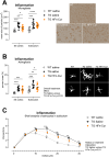
Inflammation. Microgliosis (A): Treatment with HFn-CUR reduced microglial size in frontal somatosensory cortex but not in subiculum. Photomicrographs depict somatosensory cortex of a saline-treated WT (top), saline-treated TG (lower left) and HFn-CUR-treated TG (lower right) mouse. Scalebar = 100 μm. Frontal cortex: TG saline 95% CI: 112–142, TG HFn-Cur 95% CI: 93–116. * p < 0.05, **p < 0.01, ***p < 0.001. Astrocytosis (B): HFn-CUR treatment resulted in an overall reduction in GFAP-positive astrocytes compared with saline-treated TG mice, with post-hoc testing revealing a specific effect in subiculum (B). Saline-treated TG 95% CI 7.5–11.5, HFn-CUR-treated TG 95% CI 4.6–8.5. ** p < 0.01, *** p < 0.0001. Morphological analysis of astrocytes in subiculum revealed a partial normalisation of astrocyte morphology in HFn-CUR-treated TG mice (C). Black and white images show representative astrocytes used for morphological analysis (saline-treated WT: left column, saline-treated TG: middle column; HFn-CUR-treated TG: right column). Each image is 80 μm x 80 μm. Symbols in graphs are of individual mice together with group mean ± sem (A,B) or of group mean ± sem (C, where there are no error bars, they are obscured behind symbols). ** p < 0.01, *** p < 0.001 compared with WT saline

References

    1. Golde TE, Levey AI. Immunotherapies for Alzheimer’s disease. Science. 2023;382:1242–4. - PubMed
    1. Rodrigue KM, Kennedy KM, Devous MD, Rieck JR, Hebrank AC, Diaz-Arrastia R, et al. β-Amyloid burden in healthy aging: Regional distribution and cognitive consequences. Neurology. 2012;78:387–95. - PMC - PubMed
    1. Hampel H, Hardy J, Blennow K, Chen C, Perry G, Kim SH, et al. The Amyloid-β pathway in Alzheimer’s Disease. Mol Psychiatry. 2021;26:5481–503. - PMC - PubMed
    1. Mir RH, Shah AJ, Mohi-ud-din R, Pottoo FH, Dar MA, Jachak SM, et al. Natural anti-inflammatory compounds as drug candidates in Alzheimer’s Disease. Curr Med Chem. 2021;28:4799–825. - PubMed
    1. Gagliardi S, Morasso C, Stivaktakis P, Pandini C, Tinelli V, Tsatsakis A, et al. Curcumin formulations and trials: what’s New in Neurological diseases. Molecules. 2020;25:5389. - PMC - PubMed