. 2024 Nov 17;10(1):472.
doi: 10.1038/s41420-024-02242-6.

DDX3 is critical for female fertility via translational control in oogenesis

Affiliations

DDX3 is critical for female fertility via translational control in oogenesis

Shang-Yu Tsai et al. Cell Death Discov. .

Abstract

DEAD-box RNA helicase 3 (DDX3) and its homologs play a vital role in translation initiation by unwinding secondary structures of selected mRNAs. The human DDX3 gene is located on the sex chromosomes, so there are DDX3X and DDX3Y. DDX3X is ubiquitously expressed in almost all tissues and critical for embryonic development, whereas DDX3Y is only expressed in the testis and essential for male fertility. Drosophila belle (bel) is the single ortholog of DDX3, and mutations in bel cause male and female infertility. Using Drosophila bel mutants and Ddx3x conditional knockout (cKO) mice, we confirmed the pivotal role of DDX3 in female fertility and ovarian development. Drosophila bel mutants exhibited female infertility and immature egg chambers. Consistently, oocyte-specific Ddx3x knockout in mice resulted in female infertility and impaired oogenesis. We further found that immature egg chambers in Drosophila bel mutants and impaired follicular development in oocyte-specific Ddx3x cKO mice were caused by excessive apoptosis. We also identified a set of DDX3 target genes involved in oocyte meiosis and maturation and demonstrated that DDX3 is involved in their translation in human cells. Our results suggest that DDX3 is critical for female fertility via translational control in oogenesis.


Conflict of interest statement

Competing interests The authors declare no competing interests. Ethics approval All animal studies were carried out in accordance with the recommendations in the Guide for the Care and Use of Laboratory Animals of the NIH. The experimental procedures using mice were reviewed and approved by the Institutional Animal Care and Use Committee at Chang Gung University, Taiwan (IACUC:CGU110-208).

Figures

Fig. 1
Fig. 1. Mutations in Drosophila bel cause female infertility and ovarian agenesis.
A Adult females were lysed in 1× RIPA Lysis Buffer and subjected to western blot analysis using antibodies against DDX3 and α-tubulin proteins. Detection of α-tubulin served as a loading control. B Three virgin females were mated with two w1118 males for 5 days. Fecundity was assessed by the number of eggs laid within 24 h. Fertility was evaluated by the number of offspring after 19 days of ovulation. The strain w1118 served as a control. Data are shown as mean and standard deviation (n = 10/group). C Adult females were fed diets containing yeast for 3 days and then dissected to collect their ovaries. Body length and ovary size were measured and recorded under a dissecting microscope. Data are shown as mean and standard deviation (n = 10/group). Statistical significance was determined using the Student’s t-test (ns not significant; **p < 0.01; ****p < 0.0001).
Fig. 2
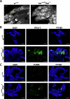
Fig. 2. Immature egg chambers and apoptosis in Drosophila belneo30/bel6 mutants.
Adult females (w1118 and belneo30/bel6) were fed diets containing yeast for 3 days and then dissected to collect their ovaries. Ovaries were fixed with 4% formaldehyde and washed with 1× PBS containing 0.3% Triton X-100. A DAPI staining shows the presence of nuclei in Drosophila ovaries. NCs: nurse cells. B Ovaries were permeabilized with 1% Triton X-100. Immunofluorescence staining was performed using antibodies against cleaved Dcp-1 (cDcp-1), commonly used as a marker of apoptosis in Drosophila. DAPI staining shows the presence of nuclei in ovaries. C Ovaries were permeabilized with 1% Triton X-100. TUNEL assay was performed to detect apoptotic DNA fragmentation in Drosophila ovaries. DAPI staining shows the presence of nuclei in ovaries. Scale bars, 100 μm.
Fig. 3
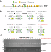
Fig. 3. Oocyte-specific Ddx3x cKO mice are generated and genotyped by PCR analysis.
A The loxP site (ATAACTTCGTATAATGTATGCTATACGAAGTTAT), followed by an ApaI restriction site (GGGCCC), was inserted into intron 1 and intron 10 of the Ddx3x gene using a CRISPR/Cas9-based gene editing strategy. The length of a PCR product with a loxP site will increase by 40 bp, and it can be cut into two fragments by the ApaI restriction enzyme. B Diagram of breeding strategy for generation of Cre-loxP-mediated oocyte-specific Ddx3x cKO mice. C Homozygous Ddx3xloxP/loxP female mice were detected by PCR product (636 bp) compared to wild-type mice (596 bp). Zp3-Cre mice were detected by PCR product (~300 bp). Among the 13 female mice, 7 were oocyte-specific Ddx3x cKO mice (Ddx3xloxP/loxP; Zp3-Cre) (red circle).
Fig. 4
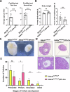
Fig. 4. Female infertility and impaired ovarian development in oocyte-specific Ddx3x cKO mice.
A Ten-week-old Ddx3xloxP/loxP female mice (n = 9) and Ddx3xloxP/loxP; Zp3-Cre female mice (n = 10) were mated with wild-type C57BL/6 male mice for 10 days (1 female:1 male). Females were then separated from the males and allowed to rest for 21 days, the average gestation period in mice. The number of pups was counted after birth. Fertility tests were carried out twice. Data are shown as mean ± SD. B Ten-week-old Ddx3xloxP/loxP female mice (n = 6) and Ddx3xloxP/loxP; Zp3-Cre female mice (n = 6) were dissected to collect their uteruses (with ovaries). The weight of the body and uterus (with ovaries) was measured and recorded. The ratio of uterus weight to body weight of mice was calculated. Data are shown as mean ± SD. C Experimental mice are as described in (B). Mouse ovaries were observed under a dissecting microscope. Representative images of mouse ovaries were shown. D Experimental mice are as described in (B). Mouse ovaries were fixed in formalin, embedded in paraffin, and then sectioned at a thickness of 5 μm. Ovarian sections were stained with hematoxylin and eosin (H&E). Representative images of H&E-stained ovarian sections were shown. E Experimental mice were prepared and treated as described in (D). The number of follicles in the ovarian sections at different stages (primordial, primary, secondary, and antral) was counted. The bar graph shows the number of follicles at different stages in the ovaries of oocyte-specific Ddx3x cKO mice (Ddx3xloxP/loxP; Zp3-Cre) compared to control mice (Ddx3xloxP/loxP). Data are shown as mean and standard deviation (n = 6/group). Statistical significance was determined using the Student’s t-test (ns not significant; *p < 0.05; **p < 0.01; ***p < 0.001).
Fig. 5
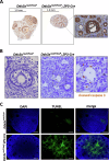
Fig. 5. Impaired oogenesis and extensive apoptosis occur in the ovaries of oocyte-specific Ddx3x cKO mice.
10-week-old Ddx3xloxP/loxP female mice (n = 6) and Ddx3xloxP/loxP; Zp3-Cre female mice (n = 6) were dissected to collect their ovaries. Mouse ovaries were fixed in formalin, embedded in paraffin, and then sectioned at a thickness of 5 μm. A Ovarian sections were processed for immunohistochemistry (IHC) to stain endogenous mouse Ddx3x. Representative images of IHC-stained ovarian sections were shown. An antral follicle and nearby degenerated oocytes were enlarged in Ddx3xloxP/loxP; Zp3-Cre ovarian sections. B Ovarian sections were processed for immunohistochemistry (IHC) to stain cleaved caspase 3. Representative images of IHC-stained ovarian sections were shown. Brown signals were frequently observed in Ddx3xloxP/loxP; Zp3-Cre ovarian sections. C TUNEL assay was performed to detect apoptotic DNA fragmentation in mouse ovarian sections. DAPI staining shows the presence of nuclei in mouse ovaries. Scale bars, 100 μm.
Fig. 6
Fig. 6. DDX3 regulates the protein expression of candidate target genes involved in oogenesis in HeLa cells.
HeLa cells were transduced with the empty lentiviral vector (pLKO.1) or the pLKO.1 vector expressing DDX3 shRNAs (shDDX3-1 and shDDX3-2). Cells were harvested for analysis at day 3 post-transduction. A Western blot analysis was performed using antibodies against DDX3, APC1, cyclin E1, p38 MAPK, B56β, PP2Aβ, PKACα, FBXW11, Rac1, GNAS, and α-tubulin proteins. Detection of α-tubulin served as a loading control. B The expression of APC1, CCNE1, P38γ, B56β, PP2Aβ, PKACα, FBXW11, RAC1, P38β, GNAS, DDX3, and GAPDH mRNAs were detected by quantitative real-time RT-PCR. The bar graph shows the relative mRNA levels normalized to GAPDH as mean and standard deviation from three independent experiments. Statistical significance was tested by one-way ANOVA (ns not significant; **p < 0.01; ***p < 0.001; ****p < 0.0001).
Fig. 7
Fig. 7. DDX3 regulates translation of candidate target genes involved in oogenesis in 293T cells.
293T cells were transduced with the empty lentiviral vector (pLKO.1) or the pLKO.1 vector expressing shDDX3-1. Cells were harvested for analysis at day 3 post-transduction. A Western blot analysis was performed using antibodies against DDX3 and α-tubulin proteins. Detection of α-tubulin served as a loading control. B Cytoplasmic extracts were loaded on a linear 15-40% sucrose gradient ultracentrifugation. Total RNA was extracted from each fraction for analysis. Polysome profile analysis and quantitative real-time RT-PCR were performed to assess translational efficiency of APC1, CCNE1, P38γ, B56β, PP2Aβ, PKACα, FBXW11, RAC1, P38β, GNAS, and GAPDH mRNAs in DDX3 knockdown 293T cells (shDDX3-1) compared to control cells (pLKO.1). Detection of GAPDH mRNA served as a negative control. The bar graph shows the changes in translational efficiency as mean and standard deviation from three independent experiments. Statistical significance was determined using the Student’s t-test (ns not significant; *p < 0.05; **p < 0.01; ***p < 0.001).
Fig. 8
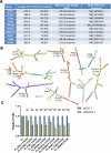
Fig. 8. DDX3 facilitates translation of candidate target mRNAs that contain complex 5′ UTRs.
A The 5′ UTR sequences of APC1, CCNE1, P38γ, B56β, PP2Aβ, PKACα, FBXW11, RAC1, P38β, and GNAS mRNAs obtained from the NCBI Reference Sequence (RefSeq) database were analyzed. The length, GC content, and minimum free energy (MFE) of the 5′ UTR sequences were indicated. B Secondary structures within the 5′ UTRs were predicted using the RNAfold web server. The nucleotides are colored according to their probabilities in the structure. C 293T cells were transduced with the empty lentiviral vector (pLKO.1) or the pLKO.1 vector expressing shDDX3-1. After 48 h, 293T cells were co-transfected with firefly luciferase (Fluc) reporters containing the 5′ UTRs of APC1, CCNE1, P38γ, B56β, PP2Aβ, PKACα, FBXW11, RAC1, P38β, or GNAS mRNAs in combination with the control pRL-SV40 vector encoding the Renilla luciferase (Rluc). 293T cells were lysed for analysis at 24 h post transfection. The Fluc activity was normalized for each transfectant to that of the Rluc control. The bar graph shows the relative Fluc/Rluc activities in DDX3 knockdown 293T cells (shDDX3-1) compared to control cells (pLKO.1). Data are shown as mean and standard deviation from three independent experiments. Statistical significance was determined using the Student’s t-test (**p < 0.01; ***p < 0.001; ****p < 0.0001).

Similar articles

Cited by

References

    1. Chuang RY, Weaver PL, Liu Z, Chang TH. Requirement of the DEAD-Box protein ded1p for messenger RNA translation. Science. 1997;275:1468–71. - PubMed
    1. Soto-Rifo R, Rubilar PS, Limousin T, de Breyne S, Decimo D, Ohlmann T. DEAD-box protein DDX3 associates with eIF4F to promote translation of selected mRNAs. EMBO J. 2012;31:3745–56. - PMC - PubMed
    1. Lai MC, Lee YH, Tarn WY. The DEAD-box RNA helicase DDX3 associates with export messenger ribonucleoproteins as well as tip-associated protein and participates in translational control. Mol Biol Cell. 2008;19:3847–58. - PMC - PubMed
    1. Lee CS, Dias AP, Jedrychowski M, Patel AH, Hsu JL, Reed R. Human DDX3 functions in translation and interacts with the translation initiation factor eIF3. Nucleic Acids Res. 2008;36:4708–18. - PMC - PubMed
    1. Calviello L, Venkataramanan S, Rogowski KJ, Wyler E, Wilkins K, Tejura M, et al. DDX3 depletion represses translation of mRNAs with complex 5′ UTRs. Nucleic Acids Res. 2021;49:5336–50. - PMC - PubMed

LinkOut - more resources