

Comparative efficacy of anteroposterior and lateral X-ray based deep learning in the detection of osteoporotic vertebral compression fracture
- PMID: 39551876
- PMCID: PMC11570669
- DOI: 10.1038/s41598-024-79610-w
Comparative efficacy of anteroposterior and lateral X-ray based deep learning in the detection of osteoporotic vertebral compression fracture
Abstract
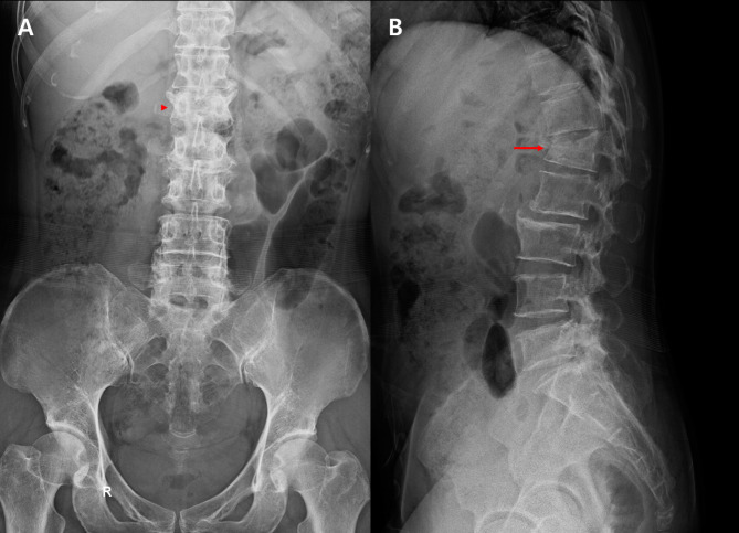
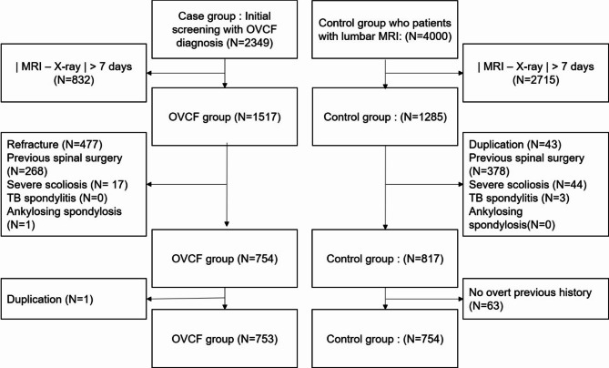
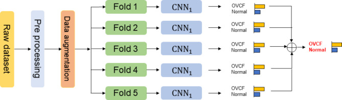
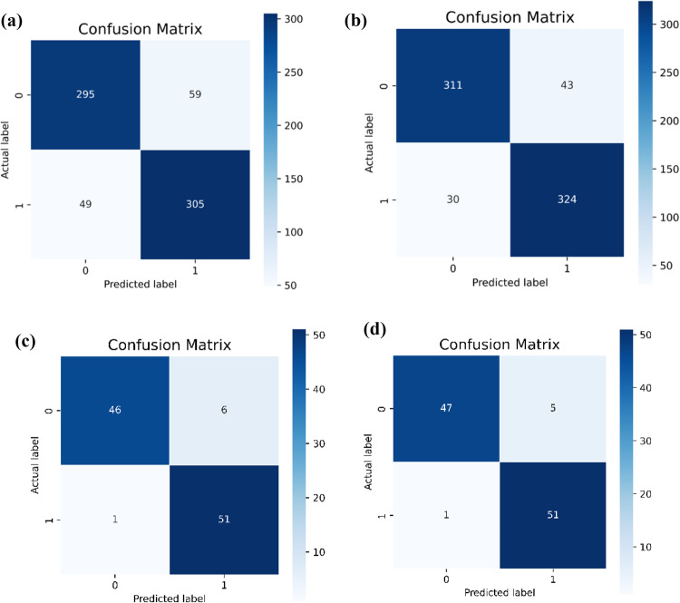
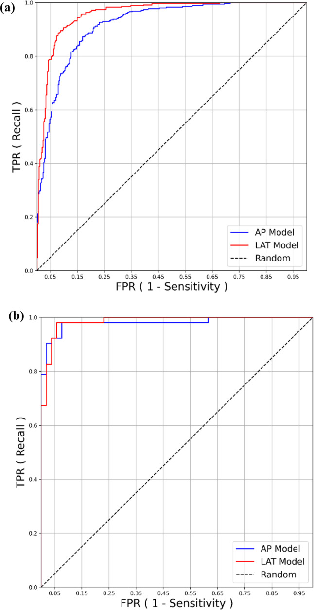
Magnetic resonance imaging remains the gold standard for diagnosing osteoporotic vertebral compression fractures (OVCF), but the use of X-ray imaging, particularly anteroposterior (AP) and lateral views, is prevalent due to its accessibility and cost-effectiveness. We aim to assess whether the performance of AP images-based deep learning is comparable compared to those using lateral images. This retrospective study analyzed X-ray images from two tertiary teaching hospitals, involving 1,507 patients for the training and internal test, and 104 patients for the external test. The EfficientNet-B5-based algorithms were employed to classify OVCF and non-OVCF group. The model was trained with a 1:1 balanced dataset and validated through 5-fold cross validation. Performance outcomes were compared with the area under receiver operating characteristic (AUROC) curve. Out of a total of 1,507 patients, 799 were included in the training dataset and 708 were included in the internal test dataset. The training and internal test datasets were matched 1:1 as OVCF and non-OVCF patients. The DL model showed comparable classifying performance with internal test data (N = 708, AUROC for AP, 0.915; AUROC for lateral, 0.953) and external test data (N = 104, AUROC for AP, 0.982; AUROC for lateral, 0979), respectively. The other performances including F1 score and accuracy were also comparable. Especially, The AUROC of AP and lateral x-ray image-based DL was not significantly different (p for DeLong test = 0.604). The EfficientNet-B5 algorithms using AP X-ray images shows comparable efficacy for classifying OVCF and non-OVCF compared to lateral images.
Keywords: Anteroposterior; Deep learning; Diagnostic accuracy; Lateral views; Machine learning in radiology; Osteoporotic vertebral compression fractures; X-ray imaging.
© 2024. The Author(s).
Conflict of interest statement
Figures
Similar articles
-
Constructing a Deep Learning Radiomics Model Based on X-ray Images and Clinical Data for Predicting and Distinguishing Acute and Chronic Osteoporotic Vertebral Fractures: A Multicenter Study.Acad Radiol. 2024 May;31(5):2011-2026. doi: 10.1016/j.acra.2023.10.061. Epub 2023 Nov 27. Acad Radiol. 2024. PMID: 38016821
-
Development and validation of a radiomics-based model for predicting osteoporosis in patients with lumbar compression fractures.Spine J. 2024 Sep;24(9):1625-1634. doi: 10.1016/j.spinee.2024.04.016. Epub 2024 Apr 26. Spine J. 2024. PMID: 38679078
-
Can a Deep-learning Model for the Automated Detection of Vertebral Fractures Approach the Performance Level of Human Subspecialists?Clin Orthop Relat Res. 2021 Jul 1;479(7):1598-1612. doi: 10.1097/CORR.0000000000001685. Clin Orthop Relat Res. 2021. PMID: 33651768 Free PMC article.
-
Exploring deep learning radiomics for classifying osteoporotic vertebral fractures in X-ray images.Front Endocrinol (Lausanne). 2024 Mar 28;15:1370838. doi: 10.3389/fendo.2024.1370838. eCollection 2024. Front Endocrinol (Lausanne). 2024. PMID: 38606087 Free PMC article.
-
Artificial intelligence in risk prediction and diagnosis of vertebral fractures.Sci Rep. 2024 Dec 19;14(1):30560. doi: 10.1038/s41598-024-75628-2. Sci Rep. 2024. PMID: 39702597 Free PMC article.
Cited by
-
AI system for diagnosing mucosa-associated lymphoid tissue lymphoma and diffuse large B cell lymphoma using ImageNet and hematoxylin and eosin-stained specimens.PNAS Nexus. 2025 Apr 30;4(5):pgaf137. doi: 10.1093/pnasnexus/pgaf137. eCollection 2025 May. PNAS Nexus. 2025. PMID: 40365164 Free PMC article.
-
[Analysis of demographic and clinical characteristics of 744 inpatients with osteoporotic vertebral compression fractures].Zhongguo Xiu Fu Chong Jian Wai Ke Za Zhi. 2025 Mar 15;39(3):354-361. doi: 10.7507/1002-1892.202411068. Zhongguo Xiu Fu Chong Jian Wai Ke Za Zhi. 2025. PMID: 40101912 Free PMC article. Chinese.
References
-
- Nicolaes, J. et al. Detection of vertebral fractures in CT using 3D Convolutional Neural Networks. arXiv preprint arXiv:1911.01816 (2019).
Publication types
MeSH terms
Grants and funding
LinkOut - more resources
Full Text Sources
Medical
