. 2024 Dec 31;13(1):2429846.
doi: 10.1080/2162402X.2024.2429846. Epub 2024 Nov 17.

Systemic administration of a viral nanoparticle neoadjuvant prevents lung metastasis development through emergency myelopoiesis

Affiliations

Systemic administration of a viral nanoparticle neoadjuvant prevents lung metastasis development through emergency myelopoiesis

Léa Bourguignon et al. Oncoimmunology. .

Abstract

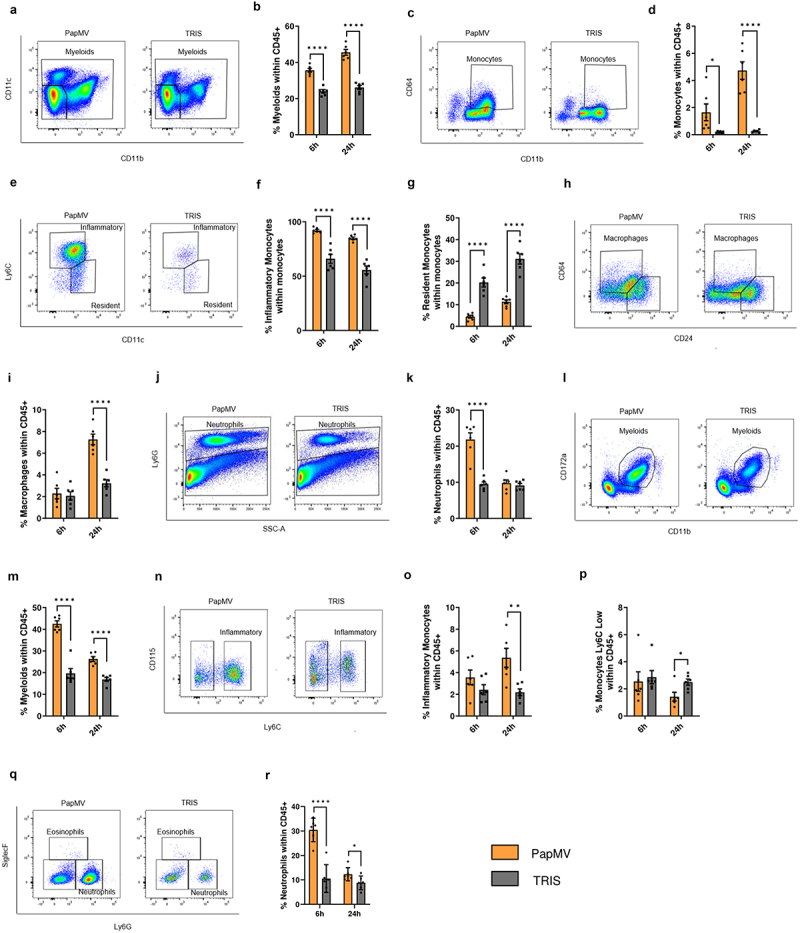
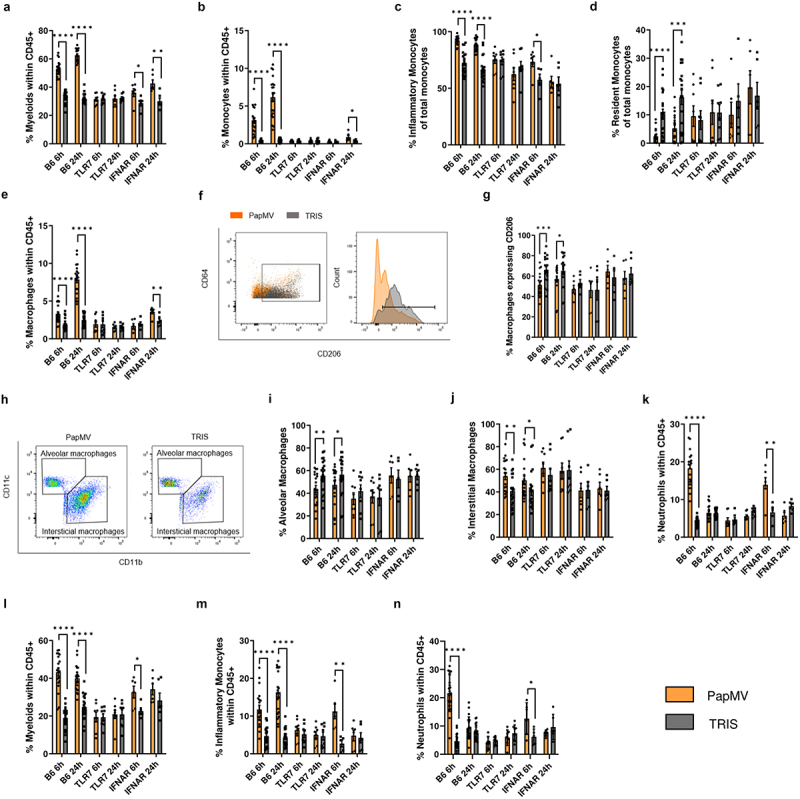
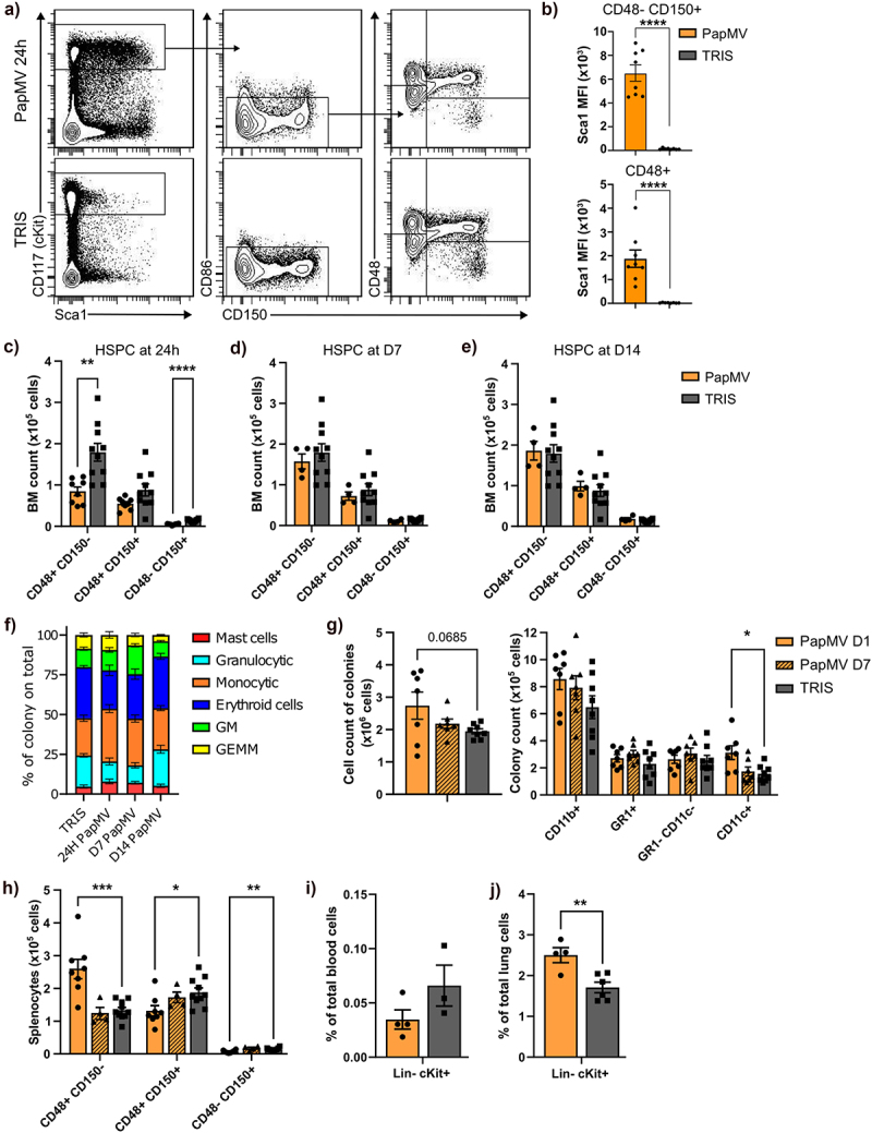
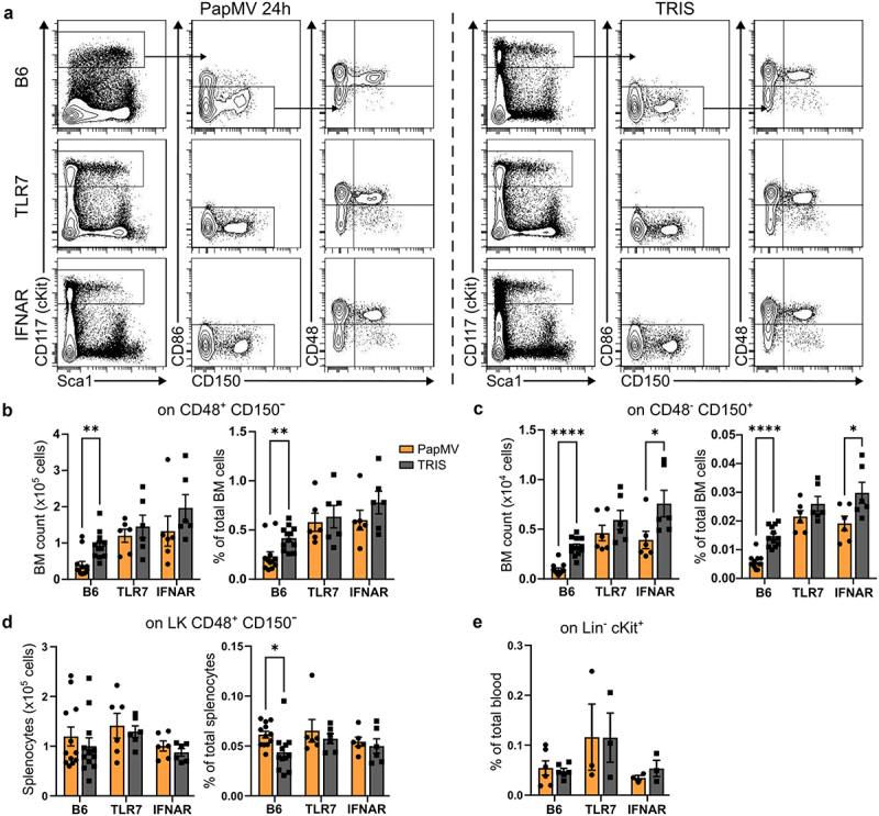
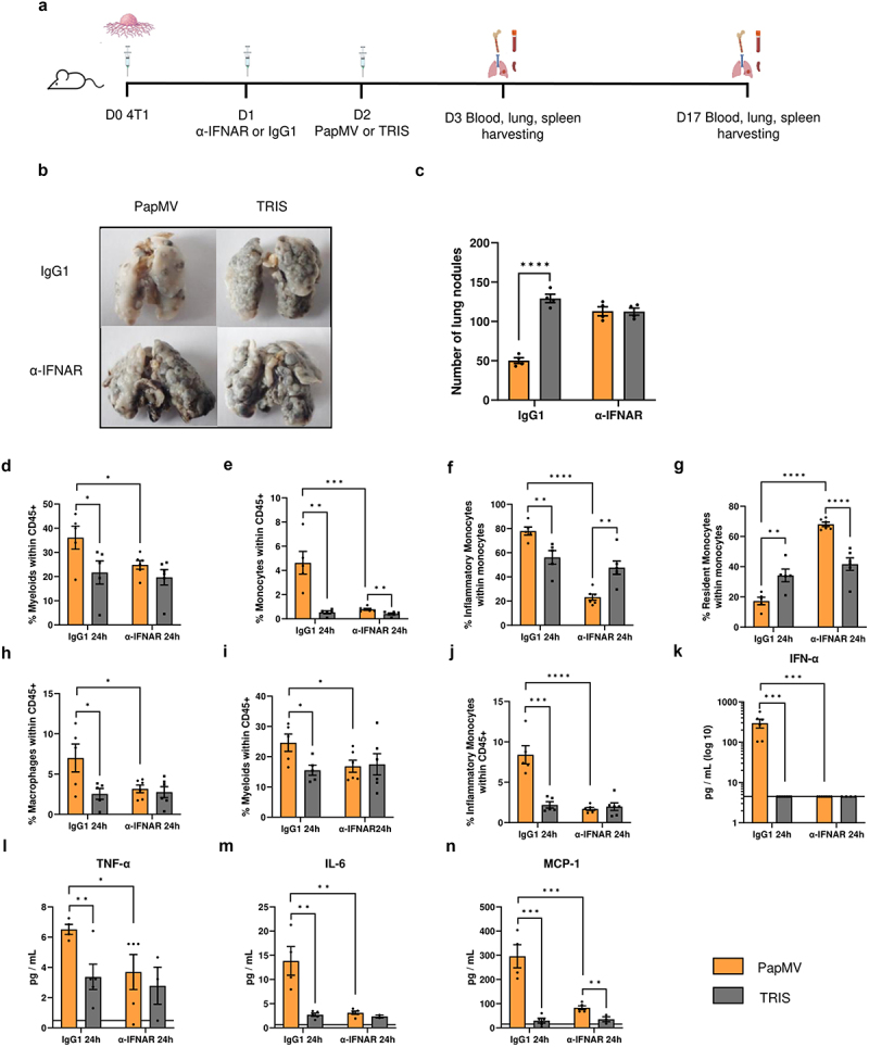
Cancer presents a significant public health concern, particularly in the context of metastatic disease. Surgical removal of primary tumors, while essential, can inadvertently heighten the risk of metastasis. Thus, there is a critical need for innovative neoadjuvant therapies capable of curtailing metastatic progression before or immediately following tumor resection. Addressing this imperative, the papaya mosaic virus nanoparticle (PapMV) has demonstrated potent immunostimulatory capabilities against both viruses and tumors, effectively hindering their proliferation. Our study reveals that PapMV exerts a protective effect against lung metastasis when administered systemically prior to tumor implantation or during the early stages of metastasis in various mouse models of cancer. This anti-tumor effect is initiated by the recruitment of myeloid cells in the lungs. These cells adopt a pro-inflammatory profile, secreting cytokines such as IFN-α, thus fostering a tumor microenvironment inhospitable to tumor progression. Crucially, this protective mechanism hinges on the presence of macrophages before treatment. TLR7 and IFN-I signaling pathways also play pivotal roles in this process. Furthermore, our findings demonstrate that PapMV triggers the activation of the bone marrow emergency response, which accounts for the influx of myeloid cells into the lungs. This study unveils a novel aspect of PapMV's functionality. By bolstering the immune system, PapMV confers robust protection against metastasis at an early stage of disease progression. This discovery holds promise for therapeutic intervention, particularly as a preemptive measure prior to or just after surgical intervention.

Keywords: Cancer immunotherapy; emergency myelopoiesis; metastasis prevention; neoadjuvant therapy; virus-based nanoparticle.


Conflict of interest statement

No potential conflict of interest was reported by the author(s).

Figures

Figure 1.
Figure 1.
Systemic PapMV administration reduces metastasis implantation in the lungs. (a) Injection schedule for metastasis implantation model in Balb/c mice (created with BioRender.com). Balb/c mice were injected IV with PapMV or TRIS 6 h before (top) or 2 days after (bottom) IV inoculation with 4T1 or CT26 cells. (b) Lungs were harvested 17 days post injection of 3×10⁵ 4T1 cells and (c) nodules were enumerated by visual inspection. (d) Lungs were harvested 20 days post injection of 3×10⁵ CT26 cells and (e) nodules enumerated as above. Values are means ± SEM and compiled from two independent experiments (n = 6 for 6h before, n = 3 for 2 days after).
Figure 2.
Figure 2.
Systemic PapMV administration induces pro-inflammatory myeloid cell recruitment to the lungs. Balb/c mice were injected IV with PapMV or TRIS and euthanized 6h and 24h post-injection. (a-k) lungs and (l-r) blood were harvested for flow cytometric cell labeling. In lungs, proportions of (a-b) myeloid cells, (c-d) total monocytes, (e-f-g) monocyte subtypes, (h-i) macrophages, and (j-k) neutrophils were determined based the cell population indicated on the Y axis. In blood, proportions of (l-m) myeloid cells, (n-o-p) monocyte subtypes, and (q-r) neutrophils were determined based on total CD45+ immune cells. A, C, E, H and N represent dot plots at 6h post-injection. J, L, and Q represent dot plots at 24h post-injection. Values are means ± SEM and compiled from three independent experiments (n = 6).
Figure 3.
Figure 3.
Myeloid cell infiltration into the lungs is dependent on TLR7 and partially dependent on IFN-I signaling. B6, TLR7, and IFNAR mice were injected IV with PapMV or TRIS and euthanized 6h and 24h post-injection. (a-k) lungs and (l-n) blood were harvested for flow cytometric cell labeling. In lungs, proportions of (a) myeloid cells, (b) total monocytes, (c-d) monocyte subtypes, (e) total macrophages, (f-g) M2 macrophages, (h-i) alveolar macrophages, (h-j) interstitial macrophages, and (k) neutrophils were determined based the cell population indicated on the Y axis. In blood, proportions of (l) myeloid cells, (m) inflammatory monocytes, and (n) neutrophils were determined based on total CD45+ immune cells. Values are means ± SEM and compiled from three or four independent experiments (n = 20 for B6, n = 8 for TLR7, and n = 6 for IFNAR).
Figure 4.
Figure 4.
Systemic PapMV injection promotes the differentiation of hematopoietic stem cells into myeloid cells. Balb/c mice were injected IV with PapMV or TRIS and were euthanized 24h, day-7 or day-14 post-injection. (a-g) bone marrow, (j) spleen, (i) blood and (j) lung were analyzed for their HSPC composition. (a) Flow cytometry analysis of bone marrow progenitors. (b) Sca1 mean fluorescence intensity (MFI) on CD48+ and CD48- CD150+ cells at 24 hours post-injection. (c-e) number of CD48+ CD150-, CD48+ CD150+ and CD48- CD150+ cells in BM at (c) 24 hours, (d) 7 days and (e) 14 days post-injection. (f) Ratio of colony types over the total having grown from 104 BM cells 24h post-injection plated in M3434 methylcellulose-based medium (GM : granulocyte/macrophage colony; GEMM : granulocyte/erythroid/macrophage/megakaryocyte colony). (g) Flow cytometry analysis of colonies from 24 hours and 7 days post-injection of BM cells. Number of total cells from colony assay (left panel) and number of CD11b+ cells and granulocytes (GR1+ CD11c- cells), monocytes (GR1- CD11c- cells) and dc-like (GR1- CD11c+) sub-populations within the CD11b+ cells (right panel). (h) Number of CD48+ CD150-, CD48+ CD150+ and CD48- CD150+ cells in splenocytes at 24 hours and 7 days post-injection. (i-j) percentage of lin- cKit+ cells over total (i) blood cells and (j) lung cells at 24 hours post-injection. Values are means ± SEM and are compiled from three independent experiments for 24 hours BM, splenocytes and blood data, and two independent experiments for colony assays (CFU and flow cytometry), and one experiment for 7 and 14 days BM data, 7 days splenocytes data and 24 hours lungs data. Blood results are from pooled mice (2 animals per n showed) to increase cell numbers. The control values are compiled data from 24 hours, 7 days and 14 days controls.
Figure 5.
Figure 5.
The production of new myeloid cells, in the bone marrow, is dependent on TLR7 and partially dependent on IFN-I signaling. (a) Flow cytometry analysis of bone marrow progenitors from representative B6, TLR7 KO and IFNAR KO mice at 24h post-PapMV-injection (left panels) and littermate mice injected with TRIS (right panels). Number (left panel) and percentage (right panel) of (b) CD48+ CD150- and (c) CD48- CD150+ cells in BM at 24 hours post-PapMV-injection. (d) Number (left panel) and percentage (right panel) of CD48+ CD150- cells in splenocytes at 24 hours post-PapMV-injection. (e) Percentage of lin- cKit+ blood cells at 24 hours post-PapMV-injection. Values are means ± SEM and are compiled from three independent experiments. Blood results are from pooled mice (2 animals per data) to increase cell numbers. The B6 data are compiled from all the IFNAR KO and TLR7 KO experiments.
Figure 6.
Figure 6.
The immunomodulatory properties of PapMV are maintained when treatment is given two days after tumor implantation. (a) Injection schedule for metastasis implantation model in Balb/c mice (created with BioRender.com). Balb/c mice were injected IV with 3×10⁵ 4T1 cells, 24 hours later they were treated with an α-ifnar or IgG1 control antibody. 24 hours later they were injected IV with PapMV or TRIS. Mice were euthanized at D3 or D17. (b) Images and (c) numbers of lung metastases at D17 following treatment with PapMV or TRIS. At D3, (d-h) lungs and (i-j) blood were harvested for flow cytometric cell labeling. In the lungs, proportions of (d) myeloid cells, (e) total monocytes, (f) inflammatory monocytes, (g) resident monocytes, and (h) macrophages were determined based the cell population indicated on the Y axis. In the blood, the proportion of (i) myeloid cells and (j) inflammatory monocytes were determined based on total CD45+ immune cells. Serum was collected 24h following PapMV treatment and (k) ifn-α, (l) tnf-α, (m) IL-6 and (n) MCP-1 concentrations were measured. Values are means ± SEM and compiled from of two independent experiments (n = 6).
Figure 7.
Figure 7.
Role of macrophages in the anti-metastatic properties of PapMV. (a) Injection schedule for the metastasis implantation model in Balb/c mice (created with BioRender.com). Balb/c mice were injected IV with 3×10⁵ 4T1 cells. After 24 hours, they received IP administration of control or clodronate containing liposomes. Subsequently, 24 hours later, they were injected IV with PapMV or TRIS. Mice were euthanized at 6 hours, 24 hours, or on day 17. At 6 hours and 24 hours (b-d) lungs and (e, f) blood were harvested for flow cytometric cell labeling. In the lungs, proportions of (b) monocytes and (c-d) monocyte subtypes were determined based the cell population indicated on the Y axis. In the blood, proportions of (e) inflammatory monocytes and (f) Ly6C low monocytes were determined based on total CD45+ immune cells. Serum was collected 6 hours and 24 hours following PapMV treatment and (g) ifn-α, (h) IL-6, (i) MCP-1, and (j) tnf-α concentrations were measured. (k) Lung metastases were visualized at D17 and (l) enumerated. Values are means ± SEM and complied from two independent experiments. (n = 2 for 6h, n = 4 for 24h).

References

    1. Lin D, Shen Y, Liang T.. Oncolytic virotherapy: basic principles, recent advances and future directions. Sig Transduct Target Ther. 2023;8(1):156. doi:10.1038/s41392-023-01407-6. - DOI - PMC - PubMed
    1. Shahgolzari M, Venkataraman S, Osano A, Akpa PA, Hefferon K. Plant virus nanoparticles combat cancer. Vaccines (Basel). 2023;11(8):1278. doi:10.3390/vaccines11081278. - DOI - PMC - PubMed
    1. Lebel ME, Chartrand K, Leclerc D, Lamarre A. Plant viruses as nanoparticle-based vaccines and adjuvants. Vaccines (Basel). 2015;3(3):620–14. doi:10.3390/vaccines3030620. - DOI - PMC - PubMed
    1. Lacasse P, Denis J, Lapointe R, Leclerc D, Lamarre A. Novel plant virus-based vaccine induces protective cytotoxic T-lymphocyte-mediated antiviral immunity through dendritic cell maturation. J Virol. 2008;82(2):785–794. doi:10.1128/JVI.01811-07. - DOI - PMC - PubMed
    1. Hanafi LA, Bolduc M, Gagné MÈL, Dufour F, Langelier Y, Boulassel M-R, Routy J-P, Leclerc D, Lapointe R. Two distinct chimeric potexviruses share antigenic cross-presentation properties of MHC class I epitopes. Vaccine. 2010;28(34):5617–5626. doi:10.1016/j.vaccine.2010.06.024. - DOI - PubMed

MeSH terms

LinkOut - more resources