Cerebrovascular Function in Sporadic and Genetic Cerebral Small Vessel Disease
- PMID: 39552538
- PMCID: PMC11831873
- DOI: 10.1002/ana.27136
Cerebrovascular Function in Sporadic and Genetic Cerebral Small Vessel Disease
Abstract
Objective: Cerebral small vessel diseases (SVDs) are associated with cerebrovascular dysfunction, such as increased blood-brain barrier leakage (permeability surface area product), vascular pulsatility, and decreased cerebrovascular reactivity (CVR). No studies assessed all 3 functions concurrently. We assessed 3 key vascular functions in sporadic and genetic SVD to determine associations with SVD severity, subtype, and interrelations.
Methods: In this prospective, cross-sectional, multicenter INVESTIGATE-SVDs study, we acquired brain magnetic resonance imaging in patients with sporadic SVD/cerebral autosomal dominant arteriopathy with subcortical infarcts and leukoencephalopathy (CADASIL), including structural, quantitative microstructural, permeability surface area product, blood plasma volume fraction, vascular pulsatility, and CVR (in response to CO2) scans. We determined vascular function and white matter hyperintensity (WMH) associations, using covariate-adjusted linear regression; normal-appearing white matter and WMH differences, interrelationships between vascular functions, using linear mixed models; and major sources of variance using principal component analyses.
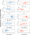
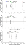
Results: We recruited 77 patients (45 sporadic/32 CADASIL) at 3 sites. In adjusted analyses, patients with worse WMH had lower CVR (B = -1.78, 95% CI -3.30, -0.27) and blood plasma volume fraction (B = -0.594, 95% CI -0.987, -0.202). CVR was worse in WMH than normal-appearing white matter (eg, CVR: B = -0.048, 95% CI -0.079, -0.017). Adjusting for WMH severity, SVD subtype had minimal influence on vascular function (eg, CVR in CADASIL vs sporadic: B = 0.0169, 95% CI -0.0247, 0.0584). Different vascular function mechanisms were not generally interrelated (eg, permeability surface area product~CVR: B = -0.85, 95% CI -4.72, 3.02). Principal component analyses identified WMH volume/quantitative microstructural metrics explained most variance in CADASIL and arterial pulsatility in sporadic SVD, but similar main variance sources.
Interpretation: Vascular function was worse with higher WMH, and in WMH than normal-appearing white matter. Sporadic SVD-CADASIL differences largely reflect disease severity. Limited vascular function interrelations may suggest disease stage-specific differences. ANN NEUROL 2025;97:483-498.
© 2024 The Author(s). Annals of Neurology published by Wiley Periodicals LLC on behalf of American Neurological Association.
Conflict of interest statement
Nothing to report.
Figures


References
-
- Wardlaw JM, Smith C, Dichgans M. Small vessel disease: mechanisms and clinical implications. Lancet Neurology 2019;18:684–696. - PubMed
Publication types
MeSH terms
Grants and funding
- 224912/Z/21/Z/WT_/Wellcome Trust/United Kingdom
- CAF/18/08, UC/CSO_/Chief Scientist Office/United Kingdom
- EXC 2145 SyNergy-ID 390857198/Deutsche Forschungsgemeinschaft
- INST 409/193-1 FUGG/Deutsche Forschungsgemeinschaft
- Scottish Funding Council through the SINAPSE collaboration
- BB/W008793/1/BB_/Biotechnology and Biological Sciences Research Council/United Kingdom
- 16 CVD 05/Fondation Leducq
- WT_/Wellcome Trust/United Kingdom
- TSALECT2015/04/Stroke Association Postdoctoral Fellowship, Garfield Weston Foundation Senior Clinical Lecture, Princess Margaret Research Development Fellowship
- SA LECT 2015/04/Stroke Association Postdoctoral Fellowship, Garfield Weston Foundation Senior Clinical Lecture, Princess Margaret Research Development Fellowship
- Edinburgh and Lothians Health Foundation
- UK Dementia Research Institute
- 666881/Horizon 2020 Framework Programme
- NHS Research Scotland
- Stiftung zur Erforschung der Vaskulären Demenz
- 252(AS-PG-14-033)/ALZS_/Alzheimer's Society/United Kingdom
- Translational Neuroscience PhD programme at the University of Edinburgh, DJG/WT_/Wellcome Trust/United Kingdom
- SA PDF 23\100007/Stroke Association Postdoctoral Fellowship, Garfield Weston Foundation Senior Clinical Lecture, Princess Margaret Research Development Fellowship
- Mrs Gladys Row Fogo Charitable Trust
- NHS Lothian Research and Development Office
- Age UK
LinkOut - more resources
Full Text Sources
Molecular Biology Databases

