

Atypical Sensory Loss Pattern in an Isolated Thalamic Stroke: A Case Report
- PMID: 39553054
- PMCID: PMC11566376
- DOI: 10.7759/cureus.71607
Atypical Sensory Loss Pattern in an Isolated Thalamic Stroke: A Case Report
Abstract
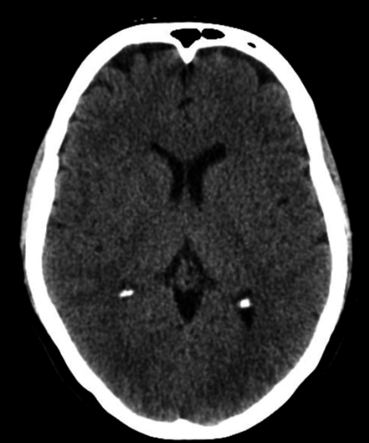
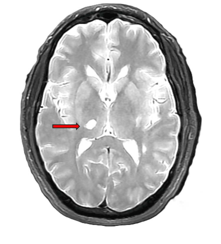
This case report discusses a 51-year-old male who presented to the emergency department (ED) with left-sided hemiparesthesia and left leg incoordination. The initial brain computed tomography (CT) scan was negative, and the follow-up brain CT three days after the onset of symptoms was also negative. Although sensitivity and specificity are not 100%, CT remains the first-line diagnostic test for detecting a cerebrovascular accident (CVA). In this unique case, CT was not sufficient. Following two negative CT scans, magnetic resonance imaging (MRI) finally revealed the cause of this patient's symptoms, an ischemic incident in the right thalamus. Thalamic strokes typically present with contralateral hemiparesis and hemisensory loss, unreactive pupils, and gaze palsy with gaze deviation away from the side of the infarct. It is unusual to see a thalamic lesion present with pure hemiparesthesia without facial involvement. This patient's clinical presentation is discussed, as well as future investigations and ways to prevent this diagnostic delay. This case demonstrates the importance of follow-up imaging based on the clinical presentation of potentially subtle imaging findings.
Keywords: atypical thalamic stroke; cerebrovascular accident; computed tomography imaging; ischemic stroke; magnetic resonance imaging; thalamic stroke.
Copyright © 2024, Tadros et al.
Conflict of interest statement
Human subjects: Consent was obtained or waived by all participants in this study. Conflicts of interest: In compliance with the ICMJE uniform disclosure form, all authors declare the following: Payment/services info: All authors have declared that no financial support was received from any organization for the submitted work. Financial relationships: All authors have declared that they have no financial relationships at present or within the previous three years with any organizations that might have an interest in the submitted work. Other relationships: All authors have declared that there are no other relationships or activities that could appear to have influenced the submitted work.
Figures
References
-
- Global burden of stroke. Katan M, Luft A. Semin Neurol. 2018;38:208–211. - PubMed
-
- Imaging of acute stroke: CT and/or MRI. Lövblad KO, Altrichter S, Pereira VM, Vargas M, Gonzalez AM, Haller S, Sztajzel R. J Neuroradiol. 2015;42:55–64. - PubMed
-
- Missed strokes using computed tomography imaging in patients with vertigo: population-based cohort study. Grewal K, Austin PC, Kapral MK, Lu H, Atzema CL. Stroke. 2015;46:108–113. - PubMed
-
- Vascular syndromes of the thalamus. Schmahmann JD. Stroke. 2003;34:2264–2278. - PubMed
-
- Delays in the diagnosis of ischaemic stroke presenting with persistent reduced level of consciousness: a systematic review. Tan S, Tang C, Ng JS, et al. J Clin Neurosci. 2023;115:14–19. - PubMed
Publication types
LinkOut - more resources
Full Text Sources