. 2024 Oct 15;14(10):4817-4829.
doi: 10.62347/JKTJ7904. eCollection 2024.

Comprehensive analysis of the oncogenic potential of eukaryotic initiation factor 3M via SAAL1 interaction in lung adenocarcinoma

Affiliations

Comprehensive analysis of the oncogenic potential of eukaryotic initiation factor 3M via SAAL1 interaction in lung adenocarcinoma

Hung-Hsing Chiang et al. Am J Cancer Res. .

Abstract

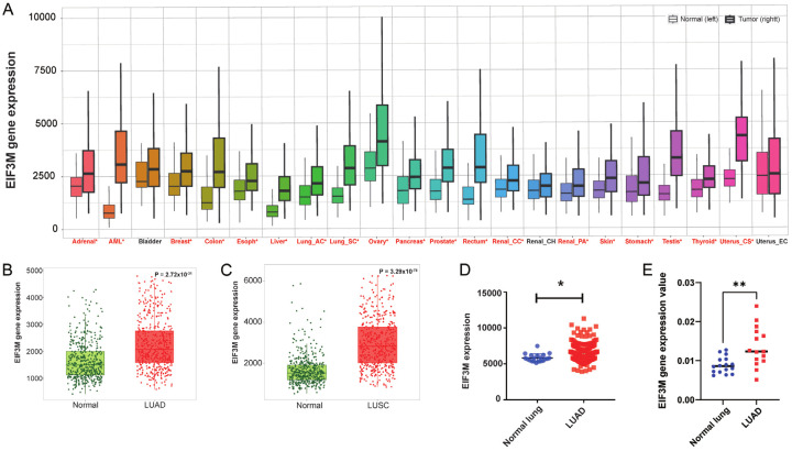
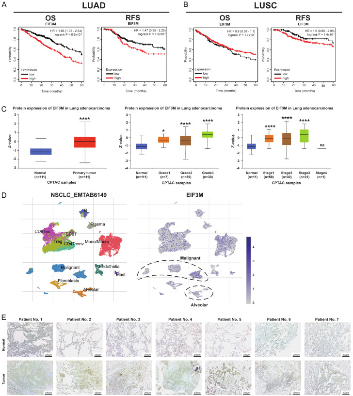
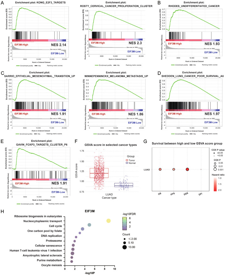
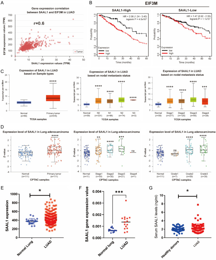
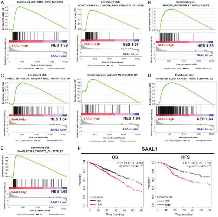
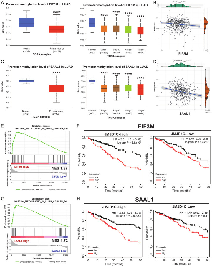
Lung adenocarcinoma (LUAD) carries a poor prognosis at advanced stages underscoring the need to elucidate the underlying molecular mechanisms driving its pathogenesis. This study aimed to investigate the roles of eukaryotic translation initiation factor 3 subunit M (EIF3M) and its associated effector, serum amyloid A-like 1 (SAAL1), in LUAD development and progression. Bioinformatic analyses such as TNMplot, The Cancer Genome Atlas (TCGA), Gene Expression Omnibus (GEO), and other public databases were used to evaluate EIF3M and SAAL1 expression levels, methylation status, clinical associations, and potential transcriptional regulators across LUAD datasets. Patient samples were analyzed for EIF3M/SAAL1 expression by qRT-PCR, immunohistochemistry, and ELISA. EIF3M and SAAL1 were overexpressed in LUAD tumor tissues compared with normal lung tissues, correlated with advanced stage, nodal metastasis, and poor survival outcomes. High EIF3M/SAAL1 levels associated with increased cell proliferation, epithelial-mesenchymal transition, metastasis, and regulatory T cell dysfunction based on gene set enrichment analysis (GSEA). Mechanistically, EIF3M/SAAL1 upregulation was linked to promoter hypomethylation, and transcriptionally regulated by JMJD1C, via hTFtarget prediction. The EIF3M/SAAL1 promote oncogenic cellular programs and immunosuppressive microenvironments that conferred unfavorable prognosis. These findings nominate EIF3M/SAAL1 as potential therapeutic targets and biomarkers in LUAD.

Keywords: EIF3M; JMJD1C; SAAL1; lung adenocarcinoma.


Conflict of interest statement

None.

Figures

Figure 1
Figure 1
Tumor cells expressed higher level of EIF3M in lung cancer. Expression levels of EIF3M in different types of cancer (A) from TNMplot. The levels of EIF3M expressed in LUAD (B) and LUSC (C) compared with adjancent normal parts. The gene expression of EIF3M in GSE31210 dataset (D) and in the KMUH cohort (E). LUAD, lung adenocarcinoma; LUSC, lung squamous cell carcinoma. *, P < 0.05; **, P < 0.01.
Figure 2
Figure 2
Higher expression of EIF3M conferred poor prognosis in LUAD. Survival analysis as overall survival (OS) and relapse-free survival (RFS) in LUAD (A) and LUSC (B) using Kaplan-Meier survival analysis. The protein expression of eIF3m in primary tumor tissue, tumor grades, and cancer stages in LUAD from CPTAC dataset (C). The expression of EIF3M in the tumor part of NSCLC_EMTAB6149 from the TISCH2 (D). Immunohistochemical staining of eIF3m from in-house LUAD patients (E). *, P < 0.05; ****, P < 0.001; na = not applicable.
Figure 3
Figure 3
Higher EIF3M expression held aggressive biologic functions in LUAD. The functions of EIF3M of LUAD disclosed by the gene set enrichment analysis (GSEA) as cell proliferation (A), cell undifferentiation (B), epithelial mesenchymal transition, and metastasis (C), poor survival (D), and regulatory T cell (E). Gene set variation analysis (GSVA) score of EIF3M between the tumor tissue and normal tissue (F) showed higher hazard ratio as OS, PFS, DSS, but DFI was not correlated with GSVA score. (G) KEGG pathway analysis using a gene set of EIF3M associated 200 genes (H). PFS, progression free survival; DSS, disease free survival; DFI, disease free interval.
Figure 4
Figure 4
SAAL1 synergized with EIF3M exerted poor survival outcome. The correlation between EIF3M and SAAL1 using TCGA cohort (A). The effects of SAAL1 on EIF3M was elucdated using cross-analysis as OS (B). The gene expression of SAAL1 expression in primary tumor parts, nodal metastasis status, and cancer stage from TCGA in LUAD (C). Protein expression of SAAL1 in LUAD in primary tumor, tumor stage, and grade from CPTAC dataset for LUAD (D). SAAL1 expression in tumor parts of LUAD from GSE31210 dataset (E) and in-house cohort (F). Serum SAAL1 levels from healthy donors and LUAD patients (G). *, P < 0.05; **, P < 0.01; ***, P < 0.005; ****P < 0.001; na = not-applicable.
Figure 5
Figure 5
Higher SAAL1 expression held poor survival in LUAD. The functions of SAAL1 through GSEA revealed assoication with cell proliferation (A), cell undifferentiation (B), epithelial mesenchymal transition and metastasis (C), poor survival (D), and Treg cells (E) in LUAD. The survival analysis of SAAL1 regarding OS and RFS (F) (HR = 1.6 and 1.92, respectively, P < 0.05).
Figure 6
Figure 6
JMJD1C regulated EIF3M and SAAL1 in LUAD. The gene expression is under the regulation of different mechanisms. Promoter methylation levels of EIF3M presented as either in normal/tumor or tumor stages (A) and the correlation between levels of methylation and EIF3M expression (B) in LUAD. The expression of SAAL1 regarding promoter methylation levels either in normal/tumor or tumor stages (C) and the correlation between levels of methylation and SAAL1 expression (D). GSEA showed the association of EIF3M promoter methylation and gene expression (E). The cross analysis revealed the impact of JMJD1C on the OS by levels of EIF3M in LUAD (F). GSEA showed the association of SAAL1 promoter methylation and gene expression (G). The cross analysis revealed the impact of JMJD1C on the OS by levels of SAAL1 in LUAD (H). ****, P < 0.001.

References

    1. Kratzer TB, Bandi P, Freedman ND, Smith RA, Travis WD, Jemal A, Siegel RL. Lung cancer statistics, 2023. Cancer. 2024;130:1330–1348. - PubMed
    1. Nicholson AG, Tsao MS, Beasley MB, Borczuk AC, Brambilla E, Cooper WA, Dacic S, Jain D, Kerr KM, Lantuejoul S, Noguchi M, Papotti M, Rekhtman N, Scagliotti G, van Schil P, Sholl L, Yatabe Y, Yoshida A, Travis WD. The 2021 WHO classification of lung tumors: impact of advances since 2015. J Thorac Oncol. 2022;17:362–387. - PubMed
    1. Xie S, Wu Z, Qi Y, Wu B, Zhu X. The metastasizing mechanisms of lung cancer: recent advances and therapeutic challenges. Biomed Pharmacother. 2021;138:111450. - PubMed
    1. Wang M, Herbst RS, Boshoff C. Toward personalized treatment approaches for non-small-cell lung cancer. Nat Med. 2021;27:1345–1356. - PubMed
    1. Teimouri H, Kochugaeva MP, Kolomeisky AB. Elucidating the correlations between cancer initiation times and lifetime cancer risks. Sci Rep. 2019;9:18940. - PMC - PubMed