. 2024 Nov 14;16(1):2419155.
doi: 10.1080/20002297.2024.2419155. eCollection 2024.

Oral administration of Porphyromonas gingivalis to mice with diet-induced obesity impairs cognitive function associated with microglial activation in the brain

Affiliations

Oral administration of Porphyromonas gingivalis to mice with diet-induced obesity impairs cognitive function associated with microglial activation in the brain

Kana Oue et al. J Oral Microbiol. .

Abstract

Objective: Both periodontal disease and obesity are risk factors for dementia, but their links to 1brain function remain unclear. In this study, we examined the effects of oral infection with a periodontal pathogen on cognitive function in a mouse model of obesity, focusing on the roles of microglia.

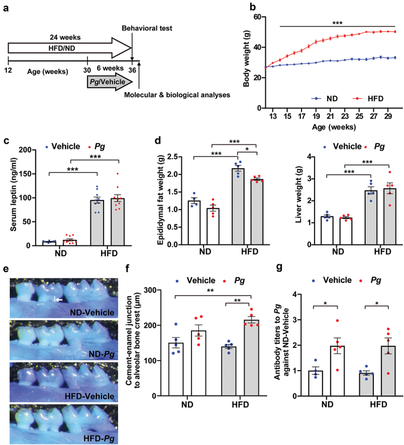
Methods: To create a mouse model of diet-induced obesity and periodontitis, male C57BL/6 J mice were first fed a high-fat diet containing 60% lipid calories for 18 weeks, beginning at 12 weeks of age, to achieve diet-induced obesity. Then, Porphyromonas gingivalis administration in the oral cavity twice weekly for 6 weeks was performed to induce periodontitis in obese mice.

Results: Obese mice orally exposed to P. gingivalis showed cognitive impairment in the novel object recognition test. Increased expression levels of inflammatory cytokines (e.g. interleukin-1β and tumor necrosis factor-α) were observed in the hippocampus of P. gingivalis-treated obese mice. Immunohistochemical analysis revealed that microglia cell body size was increased in the hippocampus and prefrontal cortex of P. gingivalis-treated obese mice, indicating microglial activation. Furthermore, depletion of microglia by PLX3397, a colony-stimulating factor 1 receptor inhibitor, ameliorated cognitive dysfunction.

Conclusion: These results suggest that microglia mediate periodontal infection-induced cognitive dysfunction in obesity.

Keywords: Periodontal disease; Porphyromonas gingivalis; cognitive dysfunction; inflammation; microglia; obesity; oral infection.


Conflict of interest statement

No potential conflict of interest was reported by the author(s).

Figures

Figure 1.
Figure 1.
Effects of long-term high-fat diet intake and oral administration of P. gingivalis in mice. (a) Timeline of experimental procedures, including administration of respective diets and oral administration of P. gingivalis (Pg). (b) Body weights of mice fed a normal diet (ND) and high-fat diet (HFD) were measured up to 30 weeks of age. Results are expressed as the mean ±S.E.M. of 10 mice per group. ***P < 0.001. Serum leptin levels (c) and weights of epididymal fat and liver (d) were analyzed in 36-week-old mice. Results are expressed as the mean ±S.E.M. of 10 (c) and five (d) mice per group. *P < 0.05, ***P < 0.001. (e, f) Alveolar bone levels of the upper jaw were analyzed. Representative images are shown (e); the distance from the alveolar bone crest of the proximal buccal root of the second molar to the cementoenamel junction (indicated by white line in e) was measured (f). Results are expressed as the mean ±S.E.M. of five mice per group. **P < 0.01. (g) Serum Pg antibody titer was measured by ELISA, and the data were expressed as a ratio to the ND-vehicle group. Results are expressed as the mean ±S.E.M. of five mice per group. **P < 0.01.
Figure 2.
Figure 2.
Effects of diet-induced obesity and periodontal infection on cognitive function. Recognition memory was analyzed by the novel object recognition test. Time spent exploring each object (a) and discrimination index (b) are shown. ND; normal diet, HFD; high-fat diet, Pg; P. gingivalis. Results are expressed as the mean ± S.E.M. of 10 mice per group. **P < 0.01, ***P < 0.001.
Figure 3.
Figure 3.
Effects of diet-induced obesity and periodontal infection on inflammation-related gene expression in the mouse hippocampus. mRNA levels were analyzed by quantitative reverse transcription-polymerase chain reaction, and the data were normalized to the level of the housekeeping gene GAPDH. ND; normal diet, HFD; high-fat diet, Pg; P. gingivalis. Results are expressed as the mean ±S.E.M. of five mice per group. *P < 0.05, **P < 0.01.
Figure 4.
Figure 4.
Microglial proliferation and enlargement in the hippocampus of P. gingivalis-treated obese mice. (a) Representative images of Iba1-positive cells in CA1, CA3, and dentate gyrus regions of the hippocampus in 36-week-old mice are shown. (b) The number of microglia and mean cell area per Iba1-labeled microglia in each region of the hippocampus are shown. The numbers of microglia were counted in each left and right hemisphere section of each mouse; 10 total sections were analyzed from five mice per group. For analysis of the cell area of microglia, five microglia per left and right hemisphere section were randomly selected from each mouse, and the values were averaged for each section. Ten total sections were analyzed from five mice per group. ND; normal diet, HFD; high-fat diet, Pg; P. gingivalis. Results are expressed as the mean ±S.E.M. *< 0.05, **P < 0.01, ***P < 0.001.
Figure 5.
Figure 5.
Microglial proliferation and enlargement in the prefrontal cortex of P. gingivalis-treated obese mice. (a) Representative images of Iba1-positive cells in the prelimbic, infralimbic, and orbitofrontal cortices in 36-week-old mice are shown. (b) The number of microglia and mean cell area per Iba1-labeled microglia in each region of the hippocampus are shown. The numbers of microglia were counted in each left and right hemisphere section of each mouse; 10 total sections were analyzed from five mice per group. For analysis of the cell area of microglia, five microglia per left and right hemisphere section were randomly selected from each mouse, and the values were averaged for each section. Ten total sections were analyzed from five mice per group. ND; normal diet, HFD; high-fat diet, Pg; P. gingivalis. Results are expressed as the mean ±S.E.M. ***P < 0.001.
Figure 6.
Figure 6.
Effects of PLX3397 on cognitive impairment in P. gingivalis-treated obese mice. To deplete microglia, PLX3397 (PLX), a CSF1 receptor inhibitor, was orally administered to 35-week-old mice for 10 days. The AIN-76A rodent diet was used as a control (for more details; see Materials and Methods). Recognition memory was analyzed using the novel object recognition test. Time spent exploring each object (a) and discrimination index (b) are shown. HFD; high-fat diet, Pg; P. gingivalis. Results are expressed as the mean ±S.E.M. of five mice per group. *P < 0.05, **P < 0.01, ***P < 0.001.

References

    1. Nichols E, Steinmetz JD, Vollset SE, et al. Estimation of the global prevalence of dementia in 2019 and forecasted prevalence in 2050: an analysis for the Global Burden of Disease Study 2019. Lancet Public Health. 2022;7(2):e105–13. - PMC - PubMed
    1. Livingston G, Huntley J, Sommerlad A, et al. Dementia prevention, intervention, and care: 2020 report of the Lancet Commission. Lancet. 2020;396(10248):413–446. - PMC - PubMed
    1. Balin BJ, Gérard HC, Arking EJ, et al. Identification and localization of Chlamydia pneumoniae in the Alzheimer’s brain. Med Microbiol Immunol. 1998;187(1):23–42. - PubMed
    1. Miklossy J, Kis A, Radenovic A, et al. Beta-amyloid deposition and Alzheimer’s type changes induced by Borrelia spirochetes. Neurobiol Aging. 2006;27(2):228–236. - PubMed
    1. Dunn N, Mullee M, Perry VH, et al. Association between dementia and infectious disease: evidence from a case-control study. Alzheimer Dis Assoc Disord. 2005;19(2):91–94. - PubMed

LinkOut - more resources