. 2024;2(1):55.
doi: 10.1038/s44298-024-00063-z. Epub 2024 Nov 14.

Potent neutralization by a RBD antibody with broad specificity for SARS-CoV-2 JN.1 and other variants

Affiliations

Potent neutralization by a RBD antibody with broad specificity for SARS-CoV-2 JN.1 and other variants

Michael S Piepenbrink et al. Npj Viruses. 2024.

Abstract

SARS-CoV-2 continues to be a public health burden, driven in-part by its continued antigenic diversification and resulting emergence of new variants. By increasing herd immunity, current vaccines have improved infection outcomes for many. However, prophylactic and treatment interventions that are not compromised by viral evolution of the Spike protein are still needed. Using a differential staining strategy with a rationally designed SARS-CoV-2 Receptor Binding Domain (RBD) - ACE2 fusion protein and a native Omicron RBD protein, we developed a recombinant human monoclonal antibody (hmAb) from a convalescent individual following SARS-CoV-2 Omicron infection. The resulting hmAb, 1301B7 potently neutralized a wide range of SARS-CoV-2 variants including the original Wuhan-1, the more recent Omicron JN.1 strain, and SARS-CoV. 1301B7 contacts the ACE2 binding site of RBD exclusively through its VH1-69 heavy chain. Broad specificity is achieved through 1301B7 binding to many conserved residues of Omicron variants including Y501 and H505. Consistent with its extensive binding epitope, 1301B7 is able to potently diminish viral burden in the upper and lower respiratory tract and protect mice from challenge with Omicron XBB1.5 and Omicron JN.1 viruses. These results suggest 1301B7 has broad potential to prevent or treat clinical SARS-CoV-2 infections and to guide development of RBD-based universal SARS-CoV-2 prophylactic vaccines and therapeutic approaches.

Keywords: Antibodies; SARS-CoV-2.


Conflict of interest statement

Competing interestsM.S.P., A.M.K., A.C., M.B., S.S., S.P., N.B.E., P.A.G., M.R.W., L.M.-S., and J.J.K. are co-inventors on patent applications that include claims related to the hmAbs described in this manuscript.

Figures

Fig. 1
Fig. 1. RBD-ACE2 stabilized fusion protein for isolation of B cells.
A Representation of stabilized WA-1 RBD-ACE2 fusion protein. B Strategy for isolation of RBD-specific B cells by flow cytometry. Initial plot gated on live, annexin V negative, CD19 + , IgD negative, HIV p24 negative, peripheral blood B cells.
Fig. 2
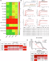
Fig. 2. SARS-CoV-2 binding and neutralization by 1301B7 hmAb.
A hmAbs were tested at 1 μg/ml in triplicate for binding to indicated protein in the presence of 8 M urea by ELISA. Average optical density (OD) at 450 nM is shown. B Binding to indicated RBD was determined by SPR. Concentrations of the RBDs evaluated are 50, 12.5, 3.125, and 0.781 nM. The 50 nM concentration was not evaluated for 1301B7-FL.1 interactions. Embedded table indicates key amino acid residues and fold difference (FD) relative KD of XBB.1.5. hmAbs were tested at increasing concentrations in triplicate for neutralization by pseudovirus-based (C) or live virus (D) neutralization assays.
Fig. 3
Fig. 3. Structure of the 1301B7Fab – EG5.1 SARS-CoV-2 Spike.
A Orthogonal views of the Cryo-EM density for the upRBD-1301B7 Fv complex with RBD cyan, 1301B7 heavy and light chains yellow and grey, respectively, and the glycan attached to CDRH3 is shown in magenta. B Ribbon diagram of the final model in the orientations shown in A. (C, D) Orthogonal views of the 1301B7 RBD binding site shown as a green surface on the EG5.1 RBD, with CDR/FR3 residues colored in green. The location of two significant JN.1 mutations L452W and V483del are shown in red. E 1301B7 CDRs/FR3 contact residues shown on the ACE2 binding surface (yellow) of RBD, showing 1301B7 does not bind to the “tip” region of RBD encircled in red on the figure. F Electron density from the 4.1 Å local-refined map corresponding to a portion of CDRH3 and the N-linked glycan attached to Asn100E. G Molecular interactions between 1301B7 and EG5.1 RBD.
Fig. 4
Fig. 4. Prophylactic activity of 1301B7 hmAb against SARS-CoV-2 XBB.1.5.
K18 hACE2 transgenic mice were treated i.n. with 1301B7 (1 mg/kg or 10 mg/kg) or isotype control hmAb (10 mg/kg), followed by infection with 105 PFU SARS-CoV-2 XBB.1.5. Body weight (A) and survival (B) were evaluated at the indicated days post-infection (n = 5 mice/group). Mice that lost >25% of their initial body weight were humanely euthanized. Error bars represent standard error of the mean (SEM) for each group of mice. Viral titers in the nasal turbinate (C) and lung (D) at 2 and 4 days p.i. were determined by plaque assay in Vero AT cells (n = 4 mice/group/day). Symbols represent individual mice, bars indicate the mean of virus titers. Dotted lines indicate limit of detection. & indicates virus not detected in any mice from that group. α indicates virus detected in only one mouse from that group. * indicates p < 0.05 as compared to isotype control hmAb as determined by t-test.
Fig. 5
Fig. 5. Prophylactic activity of 1301B7 hmAb against SARS-CoV-2 JN.1.
K18 hACE2 transgenic mice were treated i.n. with 1301B7 (2 mg/kg or 20 mg/kg) or isotype control hmAb (20 mg/kg), followed by infection with 105 PFU SARS-CoV-2 JN.1. Body weight (A) and survival (B) were evaluated at the indicated days post-infection (n = 5 mice/group). Mice that lost >25% of their initial body weight were humanely euthanized. Error bars represent standard error of the mean (SEM) for each group of mice. Viral titers in the nasal turbinate (C) and lung (D) at 2 and 4 days p.i. were determined by plaque assay in Vero AT cells (n = 4 mice/group/day). Symbols represent individual mice, bars indicate the mean of virus titers. Dotted lines indicate limit of detection. & indicates virus not detected in any mice from that group. α indicates virus detected in only one mouse for that group. * indicates p < 0.05 as compared to isotype control hmAb as determined by t-test.

Update of

References

    1. Holshue, M. L. et al. First Case of 2019 Novel Coronavirus in the United States. N. Engl. J. Med.382, 929–936 (2020). - PMC - PubMed
    1. WHO. WHO COVID-19 dashboard. 2024 [cited 2024]Available from: https://data.who.int/dashboards/covid19/deaths?n=c.
    1. Arabi, M. et al. Role of previous infection with SARS-CoV-2 in protecting against omicron reinfections and severe complications of COVID-19 compared to pre-omicron variants: a systematic review. BMC Infect. Dis.23, 432 (2023). - PMC - PubMed
    1. Planas, D. et al. Distinct evolution of SARS-CoV-2 Omicron XBB and BA.2.86/JN.1 lineages combining increased fitness and antibody evasion. Nat. Commun.15, 2254 (2024). - PMC - PubMed
    1. Kaku, Y. et al. Virological characteristics of the SARS-CoV-2 JN.1 variant. Lancet Infect. Dis.24, e82 (2024). - PubMed

LinkOut - more resources