[Preprint]. 2024 Oct 29:2024.10.27.620508.
doi: 10.1101/2024.10.27.620508.

DDO-adjuvanted influenza A virus nucleoprotein mRNA vaccine induces robust humoral and cellular type 1 immune responses and protects mice from challenge

Affiliations

DDO-adjuvanted influenza A virus nucleoprotein mRNA vaccine induces robust humoral and cellular type 1 immune responses and protects mice from challenge

Victoria Gnazzo et al. bioRxiv. .

Update in

Abstract

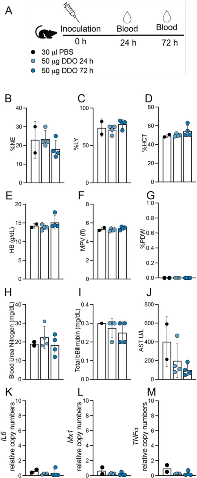
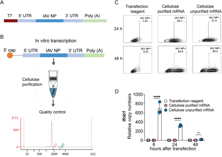
A challenge in viral vaccine development is to produce vaccines that generate both neutralizing antibodies to prevent infection and cytotoxic CD8+ T-cells that target conserved viral proteins and can eliminate infected cells to control virus spread. mRNA vaccines offer an opportunity to design vaccines based on conserved CD8-targeting epitopes, but achieving robust antigen-specific CD8+ T-cells remains a challenge. Here we tested the viral-derived oligonucleotide DDO268 as an adjuvant in the context of a model influenza A virus (IAV) nucleoprotein (NP) mRNA vaccine in C57BL/6 mice. DDO268 safely induced local type I interferon (IFN) production, stimulated dendritic cells type 1 (DC1) activation and migration to the draining lymph nodes, and improved the generation of IgG2c antibodies and antigen-specific effector Th1 CD4+ and CD8+ T-cells (IFNγ+TNFα+IL2+) when co-packaged with NP mRNA. The DDO268 adjuvanted vaccine provided enhanced protection against lethal IAV challenge and reduced the antigen dose required to achieve this protection. These results highlight the potential of DDO268 as an effective mRNA vaccine adjuvant and show that an IAV NP mRNA/DDO268 vaccine is a promising approach for generating protective immunity against conserved IAV epitopes.


Conflict of interest statement

Declaration of Competing Interest The following financial interests/personal relationships may be considered as potential competing interests: [The University of Pennsylvania and C.B.L. have a patent for Methods and Compositions for Stimulating Immune Response Using Potent Immunostimulatory RNA Motifs.].

Figures

Figure 1:
Figure 1:. Absence of systemic toxicity after DDO268 inoculation in the footpad.
(A) Study design. C57BL6 mice were inoculated s.c. with 50μg DDO in the rear footpad. (B-C) Leukogram results for percent neutrophils (%NE) and leukocytes (%LY). (D-E) Erythrogram results for percent hematocrit (%HCT) and hemoglobin concentration (g/dL) (HB g/dL). (F-G) Thrombocytes showing mean platelet volume (femtoliters)(MPV fl) and Platelet Distribution Width (% PDW). (H-J) Chemical parameters measurements: Blood Urea Nitrogen (BUN mg/dL); Total Bilirubin (mg/dL); Aspartate Aminotransferase (AST U/L). (K-M) Transcript levels of IL6, M×1 and TNFα relative to the housekeeping genes GAPDH and β-actin in liver of inoculated mice. The mean ± SD of each group is shown n = 2 (mock) and 4(treated).
Figure 2:
Figure 2:. DDO268 improves type 1 immune responses to the original Pfizer SARS-CoV2 mRNA vaccine.
C57BL6 WT and Ifnar−/− mice were immunized in the rear footpad with the Pfizer SARS-CoV-2 mRNA vaccine BNT162b2 (0.125 μg), or with BNT162b2 (0.125 μg) + DDO268 (5 μg). (A) Timeline and groups for the study design. (B) Number of total cDC1 in the draining lymph nodes 12 hours after vaccination of C57BL6 WT mice with: PBS, DDO268 (5 μg), BNT162b2 (0.125 μg), BNT162b2 (0.125 μg) + non-immunostimulatory RNA (5 μg) or BNT162b2 (0.125 μg) + DDO268 (5 μg). cDC1 were characterized as live, CD3NK1.1B220CD19MHCIIhiCD64Ly6cCD11chiXCR1+SIRPa. N = 3 mice per group (C) Ifnb1 and M×1 transcript level at the inoculation site of WT mice measured by qPCR at 2, 4, 8, and 12 hours after vaccination. (D) Number of total and activated cDC1 in the draining lymph nodes 12 hours after vaccination of WT and Ifnar−/− mice. cDC1 were characterized as Live, CD3NK1.1B220CD19 MHCIIhiCD11chi CD64Ly6c XCR1+ SIRPa, activated cDC1 are Live, CD3NK1.1B220CD19 MHCIIhiCD11chi CD64Ly6c XCR1+ SIRPa CD86+ (E) SARS-CoV-2 Spike specific IgG21 and IgG2c antibodies in vaccinated animals 60 days post vaccination (32 days post booster). (F) Number of CD8+ Tetramer+ T-cells in the spleens on day 39 after boost and specific CD8+ Tetramer+ IFNγ+ T-cells in the spleen. Number of cells shown was normalized to 500,000 live cells. In all experiments the mean ± SEM of each group is shown (n = 3– 5/group) except for (F) WT # CD8+ Tetramer+ where data was pooled from two independent experiments n= 5 mice each. *=p <0.05, **=p <0.01, ***=p <0.005 ****=p <0.001 by unpaired t-test for comparisons between two groups, and one-way ANOVA or two-way ANOVA with Bonferroni’s multiple comparison test for comparisons among three or more groups.
Figure 3:
Figure 3:. Design, characterization and functional assessment of IAV NP mRNA.
(A) pJB201.1 plasmid schematic. (B) mRNA encoding the IAV NP in vitro transcription and downstream process. Cellulose purification and quality control by Bioanalyzer. (C, D) Functional assessment of cellulose purified and unpurified mRNA. (C) Comparison of IAV NP expression in A549 cells (1×10⁶ cells) transfected with 1mg of cellulose purified or unpurified IVT NP mRNA at 24 and 48 hours post transfection. Protein detection by flow cytometry upon intracellular staining with anti-NP antibody. (D) Transcript levels of Ifnb1 relative to the housekeeping genes GAPDH and β-actin in A549 cells at 6, 24 and 48 hours after transfection with cellulose purified and unpurified IVT NP mRNA. The mean ± SD of each group is shown (n = 3/group) ****=p <0.001 by one-way ANOVA test.
Figure 4:
Figure 4:. IAV NP mRNA vaccine formulation, characterization and in vivo testing.
(A) Scheme of the different vaccine formulations tested: Empty LNP; IAV NP mRNA; IAV NP mRNA/X region; IAV NP mRNA/DDO268; IAV NP mRNA/DDO268B (B) Size distribution of nanoparticles empty or carrying IAV NP mRNA; IAV NP mRNA/X region; IAV NP mRNA/DDO268; IAV NP mRNA/DDO268B measured by Dynamic Light Scattering. (C, D) Expression of IAV NP at the (C) inoculation site (rear footpad) or (D) draining lymph nodes detected by flow cytometry upon intracellular staining with anti-NP antibody 24 hours after vaccination of C57B6 mice with 1 μg NP mRNA alone or 1 μg or NP mRNA/DDO268. Gating for immune cells (CD45+): single cells, live, CD45+ IAV NP+. Gating for non-immune cells (CD45): single cells, live, CD45 IAV NP+. Gating for myeloid cells: single cells, live, CD3 B220 CD19 NK1.1 IAV NP+. Gating for lymphoid cells: single cells, live, CD3+ B220+ CD19+ NK1.1+ IAV NP+. Gating for myeloid cells: single cells, live, CD3 B220 CD19 NK1.1 IAV NP+. The mean ± SD of each group is shown n = 3/group ns=p > 0.05 by unpaired t-test.
Figure 5:
Figure 5:. DDO268 promotes strong type 1 innate immune responses to the IAV NP mRNA vaccine.
(A) Timeline and groups for the study design. (B) Transcript levels of Ifnb1, Il6, Il1b, Ccl2, Cxcl10 and Il13 relative to the housekeeping genes GAPDH and β-actin in the footpad of C57BL6 mice 12 hours after vaccination with empty LNPs. Transcript levels of (C) Ifnb1, (D) Il6, (E) Il1b, (F) Ccl2, (G) Cxcl10 and (H) Il13 relative to the housekeeping genes GAPDH and β-actin in the footpad of C57BL6 mice 12 hours after vaccination with PBS; 0.5 μg NP mRNA or 0.5 μg NP mRNA/X region; 0.5 μg NP mRNA/DDO268; 0.5 μg NP mRNA/DDO268B (I) Transcript levels of Ifnb1 in spleen of C57BL6 mice 12 hours after vaccination with PBS; 0.5 μg NP mRNA or 0.5 μg NP mRNA/X region; 0.5 μg NP mRNA/DDO268; 0.5 μg NP mRNA/DDO268B. Transcript levels of (J) Il6, (K) M×1, and (L) TNFα in spleen of C57BL6 mice 24 hours after vaccination with PBS; 0.5 μg NP mRNA or 0.5 μg NP mRNA/X region; 0.5 μg NP mRNA/DDO268; 0.5 μg NP mRNA/DDO268B. (M) Number of cDC1 in the draining lymph nodes 12 hours after vaccination with PBS; empty LNPs; 0.5 μg NP mRNA or 0.5 μg NP mRNA/X region; 0.5 μg NP mRNA/DDO268; 0.5 μg NP mRNA/DDO268B. cDC1 were characterized as Live, CD3NK1.1B220CD19 MHCIIhiCD11chi CD64Ly6C XCR1+SIRPa. Mean ± SEM of each group is shown (n = 3/group) *=p <0.05; **=p <0.01; ***=p <0.005, by one-way ANOVA with Bonferroni’s multiple comparison test. (C-H) Two independent experiments are shown n=3 mice each. (M) Data represent two independent experiments where data was pooled with n=3 mice, n = 2/3 for PBS group.
Figure 6:
Figure 6:. DDO268 promote strong type 1 adaptive humoral and cellular immune responses to the IAV NP mRNA vaccine.
(A) Timeline and groups for the study design. C57BL6 mice were immunized twice 28 days apart with 0.5 μg NP mRNA or 0.5 μg of NP mRNA/DDO268. (B) Blood was collected 3 weeks after booster and specific NP antibodies IgG1 and IgG2c subtypes were evaluated by ELISA. Sera were serially diluted, and the orange line correspond to the cut off (normal mouse serum OD + 2DS). The pie graphs represent the ratio of IgG1 and IgG2c in mouse serum at a dilution of 1:32. (C-I) Antigen-experienced cells in the spleen were examined on day 32 after the booster immunization. (C) Representative flow cytometry plots for CD4+ and CD8+ TNFα+ IFNγ+ from the spleens of vaccinated mice. CD4+ TNFα+ IFNγ+ were identified by gating on live, singlets, CD3+ CD4+ CD8 CD11a+ cells. CD8+ TNFα+ IFNγ+ were identified by gating on live, singlets, CD3+, CD8+ CD4 CD11a+ cells. (D) Number of CD4+ IFNγ+ and CD8+ IFNγ+ T-cells in the spleens of individual mice in each vaccination group after Ionomycin/PMA restimulation. (E) Number of CD4+ TNFα+ IFNγ+ and CD8+ TNFα+ IFNγ+ T-cells in the spleens of individual mice in each vaccination group after Ionomycin/PMA restimulation. (F) Representative flow cytometry plots for CD8+ Tetramer (NP336–374)+ and CD4+ Tetramer (NP311–325)+ TNFα+ IFNγ+ T-cells. Specific CD4+ TNFα+ IFNγ+ were identified by gating on live, singlets, CD3+, CD4+ CD8, Tetramer (NP311–325)+. CD8+ TNFα+ IFNγ+ were identified by gating on live, singlets, CD3+, CD8+ CD4, Tetramer (NP336–374)+. (G) Number of tetramer specific CD4+ Tetramer (NP311–325)+ TNFα+ IFNγ+ and CD8+ Tetramer (NP336–374)+ TNFα+ IFNγ+ in the spleens of individual mice in each vaccination group after specific IAV NP restimulation. Number of cells shown was normalized to 500,000 live cells. (H) Number of spots counted as IFNγ+ after specific IAV NP restimulation (from 200,000 cells). (I) Number of spots counted as double-positives for IL2 and IFNγ after specific IAV NP restimulation (from 200,000 cells). Data corresponds to individual mice with mean ± SEM (n = 5/group). *=p <0.05, ***= p <0.005, ****= p <0.001 by one-way ANOVA with Bonferroni’s multiple comparison test.
Figure 7:
Figure 7:. DDO268 enhances protective immunity against Influenza A/PR8 following IAV NP mRNA vaccination.
(A) Timeline and groups for the study design. Vaccinated C57BL6 mice were challenged with 40 TCID50/dose of mouse passaged PR8 (H1N1) intranasally on day 39 after boost immunization. (B) Probability of survival in two independent experiments, n=5 mice each (C) weight relative to initial body weight over time in two independent experiments, n=5 mice each (D-F) Data corresponds to Experiment 2 n=3–5 (surviving animals). (D) Transcript levels of IAV NP relative to the housekeeping genes GAPDH and β-actin in the lungs of vaccinated mice at 7 days after challenge. (E) Percentage of effectors CD8+ in the lungs that are tetramer (NP336–374)+ at day 24 post challenge determined by staining and flow cytometry analysis. Effector Tetramer+ cells were identified after gating on single cells, live, CD3+, CD8+, CD44+, CD62L, CCR7, Tetramer+. (F) Percentage of CD8+ CD103+ T-cells in the lungs that are tetramer (NP336–374)+ at day 24 post challenge determined by staining and flow cytometry analysis (identified after gating on single cells, live, CD3+, CD8+, CD44+, CD103+, Tetramer(NP336–374)+. *= p <0.05 ** = p <0.01, as determined by log-rank Mantel-Cox test for survival and by one-way ANOVA with Bonferroni’s multiple comparison test.

References

    1. Freyn AW R.J., Rosado VC, Bliss CM, Pine M, Mui BL, A multi-targeting, nucleoside-modified mRNA influenza virus vaccine provides broad protection in mice, Molecular Therapies (2020). - PMC - PubMed
    1. Harrington WN K.C., Webby RJ, The evolution and future of influenza pandemic preparedness, Exp Mol Med (2021). - PMC - PubMed
    1. Ye L O.A., Ong LC, Gad HH, Hartmann R, Lycke N, Staeheli P.., Type I and Type III Interferons Differ in Their Adjuvant Activities for Influenza Vaccines, J Virol (2019). - PMC - PubMed
    1. Takeuchi A., Saito T., CD4 CTL, a Cytotoxic Subset of CD4+ T Cells, Their Differentiation and Function, Frontiers in immunology (2017). - PMC - PubMed
    1. Malyshkina A., Brüggemann A., Paschen A., Dittmer U., Cytotoxic CD4+ T cells in chronic viral infections and cancer, Frontiers in immunology (2023). - PMC - PubMed

Publication types